Figures & data
Table 1 Quadriceps and diaphragm structure and function in patients with COPD compared with controls
Figure 1 The downward spiral of disease.
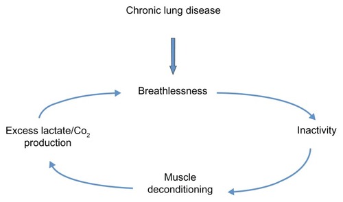
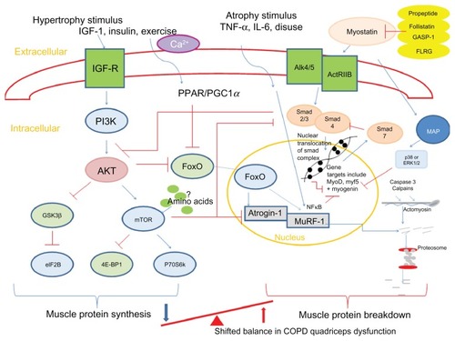
Figure 2 Summary of pathways controlling muscle protein synthesis (MPS) and muscle protein breakdown (MPB). The role of myostatin in MPS and MPB has also been included. Myostatin is held in an inactive state by its pro-peptide, follistatin, and inhibitory binding proteins – growth and differentiation factor-associated serum protein-1 (GASP-1) and follistatin-like related gene, (FLRG) as shown. Upon activation, it binds to its transmembrane receptor activin receptor type IIB (ActRIIB), which then forms homodimers with activin receptor-like kinase 4 or 5 (Alk 4/5). The SMAD signaling pathway is then activated and translocation of this transcription factor complex to the nucleus occurs, where MyoD production and therefore myoblast proliferation and fusion are blocked. Myostatin is also proposed to increase proteosomal activity in a FoxO-dependent manner. Activation of MAP kinase is mediated via myostatin either via p38 or ERK1/2, which leads to the blocking of genes involved in myogenesis.
Abbreviations: 4E-BP1, eukaryotic translation initiation factor 4E binding protein-1; Akt, protein kinase B; ERK, extracellular signal-regulated kinase; eIF2B, eukaryotic initiation factor 2B; FoxO, forkhead box class O; GSK-3β, glycogen synthase kinase-3β; IGF-1, insulin-like growth factor-1; IL-6, interleukin-6; MAP, mitogen-activated protein; mTOR, mammalian target of rapamycin; MuRF, muscle-specific RING finger protein; p70S6k, 70-kD ribosomal S6 protein kinase; PGC1α, peroxisome proliferator-activated receptor gamma co-activator 1-alpha; PI3K, phosphoinositide 3-kinase; PPAR, peroxisome proliferator-activated receptor; SMAD,; TNF-α, tumor necrosis factor-alpha; GASP-1, growth and differentiation factor-associated serum protein-1; IGF-R, insulin-like growth factor-1 receptor; Ca2+, calcium ion; NFκB, nuclear factor κB.