Figures & data
Figure 1 Synthesis of phenylthiazole acids.
Notes: Reagent and conditions: (a) CuBr2, EtOAc/CHCl3 (v:v=1:1), reflux; (b) thiourea, EtOH, reflux; (c) succinic anhydride or diglycolic anhydride or thiodiglycolic anhydride, pyridine, DMF, rt; then HCl
Abbreviations: DMF, dimethylformamide; rt, room temperature.
Abbreviations: DMF, dimethylformamide; rt, room temperature.
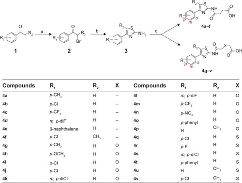
Figure 2 Fluorescence polarization (FP)-based PPARγ ligand screening assay of phenylthiazoles.
Note: Results were mean ± SD of two independent experiments.
Abbreviations: PPARγ, peroxisome proliferator-activated receptor gamma; RSG, rosiglitazone.
Abbreviations: PPARγ, peroxisome proliferator-activated receptor gamma; RSG, rosiglitazone.
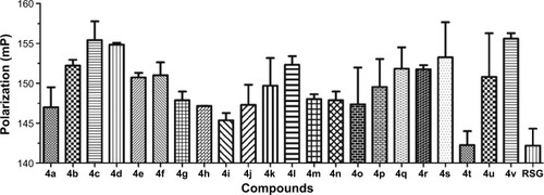
Table 1 EC50s of phenyl thiazole acids in FP-based PPARγ ligand screening assay
Table 2 The molecular properties and binding energy of selected phenylthiazole acids
Figure 3 The binding modes of agonists with PPARγ. Orange and cyans colors represented RSG and other agonists, respectively.
Notes: H-binding interactions of RSG and other agonists were shown in orange and cyans dotted line, respectively. (A) 4t and RSG, (B) 4a and RSG, (C) 4i and RSG, and (D) 4h and RSG.
Abbreviations: PPARγ, peroxisome proliferator-activated receptor gamma; RSG, rosiglitazone.
Abbreviations: PPARγ, peroxisome proliferator-activated receptor gamma; RSG, rosiglitazone.
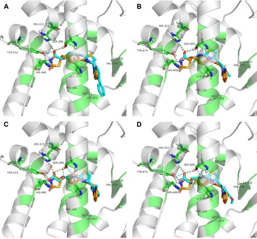
Figure 4 The RMSD of ligands from the starting structure during 25 ns MD simulations.
Notes: (A) PPARγ in complex with RSG and (B) PPARγ in complex with 4t.
Abbreviations: RMSD, root mean square deviation; MD, molecular dynamics; PPARγ, peroxisome proliferator-activated receptor gamma; RSG, rosiglitazone.
Abbreviations: RMSD, root mean square deviation; MD, molecular dynamics; PPARγ, peroxisome proliferator-activated receptor gamma; RSG, rosiglitazone.
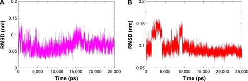
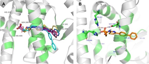
Figure 5 The binding modes of 4t.
Notes: (A) The binding modes of 4t in binding site of PPARγ obtained from Autodock docking (cyans), 21 ns (slate), 22 ns (gray), 23 ns (magenta), 24 ns (violet), and 25 ns (yellow). (B) The binding mode of 4t obtained after MD simulations.
Abbreviations: PPARγ, peroxisome proliferator-activated receptor gamma; MD, molecular dynamics.
Abbreviations: PPARγ, peroxisome proliferator-activated receptor gamma; MD, molecular dynamics.