123
Views
51
CrossRef citations to date
0
Altmetric
Review

Historical development of vaginal microbicides to prevent sexual transmission of HIV in women: from past failures to future hopes

, &
Pages 1767-1787 | Published online: 15 Jun 2017

Figures & data

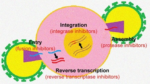
Table 1 Classification of antiretroviral drugs

Figure 1 Targeting site of antiretroviral drugs at different stages of the viral cycle.

Figure 1 Targeting site of antiretroviral drugs at different stages of the viral cycle.

Figure 2 Diagram of the action sites of different microbicides against HIV.

Figure 2 Diagram of the action sites of different microbicides against HIV.

Figure 3 Chemical structure of tenofovir (A) and tenofovir disoproxil fumarate (B).

Figure 3 Chemical structure of tenofovir (A) and tenofovir disoproxil fumarate (B).

Figure 4 Structure of a matrix type (A) and reservoir type (B) vaginal ring.

Figure 4 Structure of a matrix type (A) and reservoir type (B) vaginal ring.

Figure 5 Mechanism of drug release from sustained release tablets by in situ gelation of the polymer.

Notes: At the time of administration, the tablet is completely solid (A). In contact with the vaginal fluid, the polymer of the outer layers forms a drug-loaded gel (B). The drug reaches the vaginal environment by diffusing through the gel layer or by erosion of the gel (C).
Figure 5 Mechanism of drug release from sustained release tablets by in situ gelation of the polymer.

Figure 6 Mechanism of drug release from osmotic release tablets.

Figure 6 Mechanism of drug release from osmotic release tablets.

Figure 7 Structure of a drug-loaded polymer nanoparticle.

Figure 7 Structure of a drug-loaded polymer nanoparticle.

Figure 8 Diagram of the nanofiber manufacturing process by electrospinning.

Figure 8 Diagram of the nanofiber manufacturing process by electrospinning.

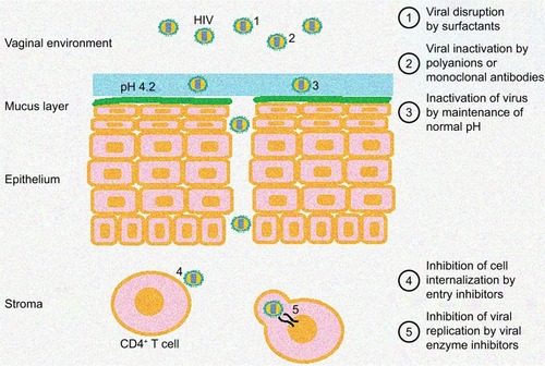
Table 2 Vaginal formulations for the prevention of sexual transmission of HIV