Figures & data
Figure 1 Structural formula of Tet and a schematic of intravitreal treatment and partitions of the retina for RGC counting.
Abbreviations: RGC, retinal ganglion cell; Tet, tetrandrine.
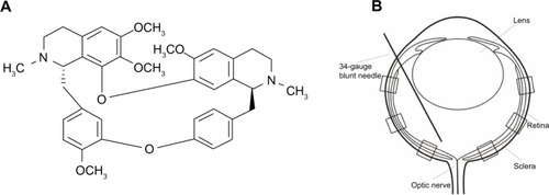
Figure 2 Effects of Tet on the survival of RGC-5 cells and SSP-induced neuron-like cells under serum deprivation conditions.
Abbreviations: ANOVA, analysis of variance; DMSO, dimethyl sulfoxide; FBS, fetal bovine serum; SD, standard deviation; SSP, staurosporine; Tet, tetrandrine.
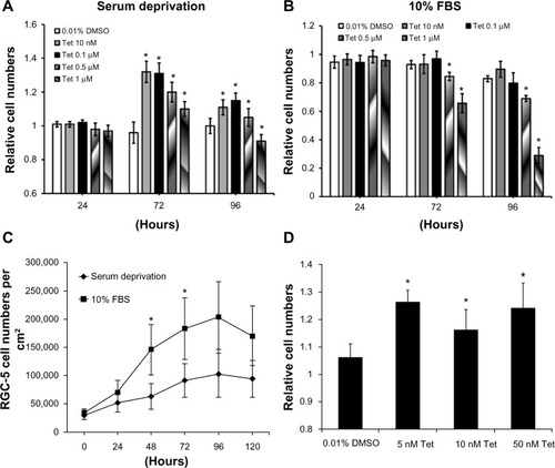
Figure 3 Tet protected RGC-5 cells from H2O2 injury and L-glutamate excitotoxicity.
Abbreviations: ANOVA, analysis of variance; L-glu, L-glutamate; SD, standard deviation; Tet, tetrandrine.
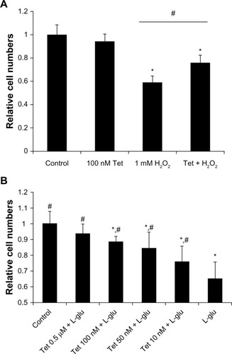
Figure 4 The identification, morphology, and survival curve of primary cultured RGCs.
Abbreviations: DAPI, 4, 6-diamidino-2-phenylindole; MAP2, microtubule associated protein 2; RGCs, retinal ganglion cells.
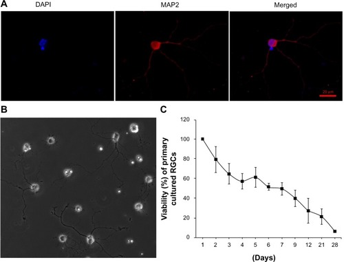
Figure 5 Time-dependent effect of Tet on the viability of primary cultured RGCs.
Abbreviations: ANOVA, analysis of variance; DMSO, dimethyl sulfoxide; RGCs, retinal ganglion cells; SD, standard deviation; Tet, tetrandrine.
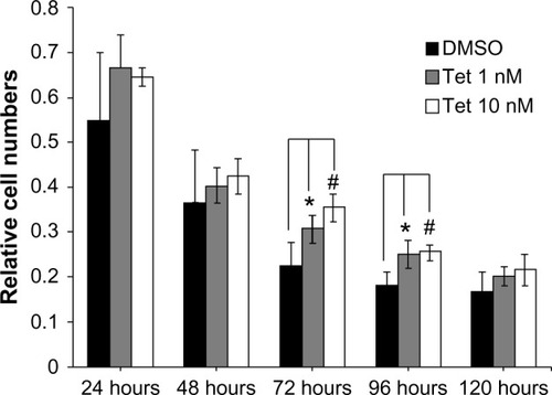
Figure 6 Tet protects RGCs from ischemic injury in vivo.
Abbreviations: CNTF, ciliary neurotrophic factor; GCL, ganglion cell layers; I/R, ischemia/reperfusion; PBS, phosphate-buffered saline; RGCs, retinal ganglion cells; Tet, tetrandrine.
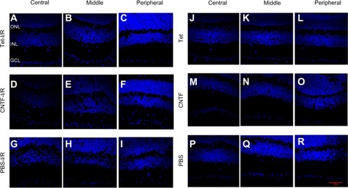
Figure 7 Statistical analysis of RGC survival following Tet application in vivo.
Abbreviations: ANOVA, analysis of variance; CNTF, ciliary neurotrophic factor; I/R, ischemia/reperfusion; PBS, phosphate-buffered saline; RGC, retinal ganglion cell; SD, standard deviation; Tet, tetrandrine.
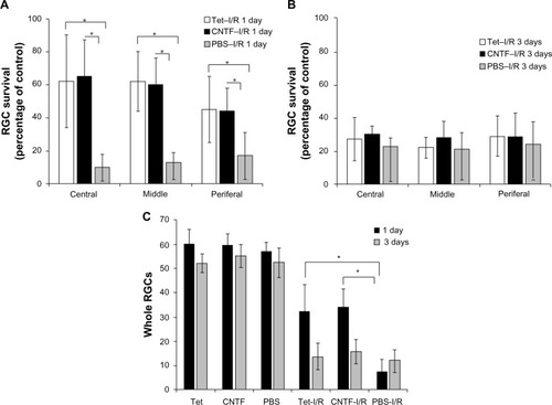
Figure 8 Effects of Tet and CNTF on primary cultured RGCs ΔΨm.
Abbreviations: ΔΨm, inner mitochondrial membrane potential; CNTF, ciliary neurotrophic factor; FCCP, carbonyl cyanide 4-trifluoromethoxyphenylhydrazone; FITC, fluorescein isothiocyanate; LL, lower left; LR, lower right; PBS, phosphate-buffered saline; PE, P-phycoerythrin; RGCs, retinal ganglion cells; Tet, tetrandrine; UL, upper left; UR, upper right.
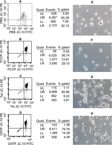
Figure 9 Tet and CNTF inhibited the expression of cleaved caspase-3 in the retinas 1 day after I/R insult.
Abbreviations: CNTF, ciliary neurotrophic factor; DAPI, 4, 6-diamidino-2-phenylindole; I/R, ischemia/reperfusion; PBS, phosphate-buffered saline; Tet, tetrandrine; GCL, ganglion cell layer; INL, inner nuclear layer; ONL, outer nuclear layer.
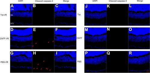
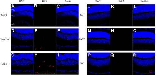
Figure 10 Tet and CNTF inhibited Bcl-2 expression 1 day after I/R insult.
Abbreviations: CNTF, ciliary neurotrophic factor; DAPI, 4′, 6-diamidino-2-phenylindole; I/R, ischemia/reperfusion; PBS, phosphate-buffered saline; Tet, tetrandrine.