Figures & data
Table 1 Dimensions and weight of fabricated small-size matrix intravaginal rings
Table 2 Dimensions of fabricated PU-93A and PU-60D res-intravaginal ring segments
Figure 1 Drug-free and hydroxychloroquine (HCQ)-loaded matrix and reservoir intravaginal rings (IVRs).
Notes: Schematic designs of (A) large- and small-matrix IVRs with HCQ (green) dispersed within the PU matrix and (B) small res-IVRs containing different quantities of reservoir segments with preformulated HCQ filled in (green). Photographs of hot-melt injection-molded (C) PU-93A matrix IVRs: a large, drug-free matrix IVR (left, 55×5.5 mm), a small drug-free IVR (middle, 25×5 mm), and a HCQ-loaded small IVR (right, 25×5 mm). (D) PU-60D res-IVR segments fabricated via hot-melt injection for in vitro release studies: a res-IVR segment (left), an end-capped res-IVR segment (middle), and an end-capped res-IVR containing HCQ-free K100M semisolid (right). (E) Full small-size PU-60D matrix IVR and res-IVRs: a small drug-free IVR (left, 25×5 mm), a small res-IVR containing one reservoir segment loaded with HCQ/K100M (1:1 wt ratio; middle) semisolid, and a small res-IVR containing three reservoir segments (right).
Abbreviation: res-IVRs, reservoir intravaginal rings; XD, cross-sectional diameter; OD, outer diameter; mm, millimeter; PU, polyether urethane.
Abbreviation: res-IVRs, reservoir intravaginal rings; XD, cross-sectional diameter; OD, outer diameter; mm, millimeter; PU, polyether urethane.
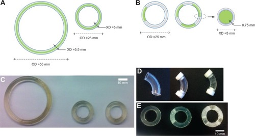
Figure 2 In vitro release of hydroxychloroquine (HCQ) from matrix intravaginal rings (IVRs) and res-IVRs.
Notes: (A) HCQ efflux from a 2% (w/w) noncoated PU-93A IVR and (B) 4% (w/w) noncoated, polyvinylpyrrolidone/poly(vinyl alcohol)-coated PU-93A IVRs. Data were scaled up to represent the release from a full-size small IVR. Inset graphs show daily release rates of different HCQ loadings from day 10 to day 18. (C) Cumulative release of HCQ from noncoated and polyvinylpyrrolidone/poly(vinyl alcohol)-coated PU-93A matrix full-size small IVRs. The total cumulative release (percentage) of HCQ from different IVRs is indicated in brackets. (D) Daily release of 4 mg HCQ total loading from either matrix or res-IVRs fabricated from (D) PU-93A or (E) PU-60D within initial 7 days. (F) Daily release and (G) cumulative release of HCQ from PU-60D res-IVR segments with either aqueous HCQ (purple) or HCQ/K100M (1:1 wt ratio) semisolid (red) containing 4 mg of total HCQ loaded. Linear cumulative HCQ release was calculated for both res-IVRs. Data is expressed as mean ± SD; n=6 for 2% and 4% (w/w) matrix IVRs, n=4 for polyvinylpyrrolidone/poly(vinyl alcohol)-coated 4% (w/w) matrix IVRs and n=3 for all the other IVRs.
Abbreviations: res-IVRs, reservoir intravaginal rings; PVP, polyvinylpyrrolidone; PVA, poly(vinyl alcohol).
Abbreviations: res-IVRs, reservoir intravaginal rings; PVP, polyvinylpyrrolidone; PVA, poly(vinyl alcohol).
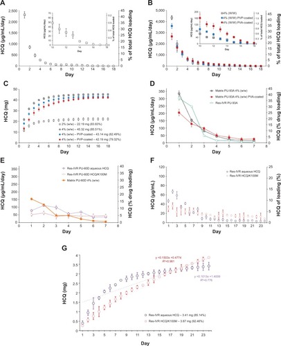
Figure 3 PU-93A matrix intravaginal ring accelerated stability test.
Notes: Hydroxychloroquine extracted from 2% (w/w), 4% (w/w), and polyvinylpyrrolidone, poly(vinyl alcohol)-coated matrix PU-93A intravaginal ring segments incubated for 30 days at (A) room temperature and (B) 40°C/75% relative humidity. Data expressed as mean ± SD, n=3.
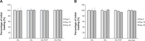
Figure 4 Swelling test of PU-93A matrix intravaginal ring segments.
Notes: Noncoated and coated 4% (w/w) hydroxychloroquine PU-93A intravaginal ring segments were incubated in sodium acetate buffer (pH 4) for 7 days at 37°C. Noncoated segments reached equilibrated state (no significant mass change) after 2 days, whereas PVP- or PVA-coated segments reached equilibrated state after 5 days of incubation. Data expressed as mean ± SD, n=3. *P<0.05 and **P<0.001 versus noncoated matrix segments.
Abbreviations: PVP, polyvinylpyrrolidone; PVA, poly(vinyl alcohol).
Abbreviations: PVP, polyvinylpyrrolidone; PVA, poly(vinyl alcohol).
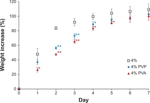
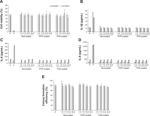
Figure 5 In vitro biocompatibility evaluations of hot-melt intravaginal ring segments.
Notes: (A) MTS assay was performed to determine cell viability. Cells cultured in drug-free medium were used as negative control; 1 M acrylamide prepared in culture medium was used to induce cell death as a positive control. (B) Interleukin (IL) 1β, (C) interleukin 6, and (D) interleukin 8 production were determined by enzyme-linked immunosorbent assay. Drug-free medium was used as a negative control, and 200 μg/mL of nonoxynol-9 or 50 μg/mL of lipopolysaccharide-treated cells were used as positive controls for interleukin 1β and interleukin 6/interleukin 8, respectively. (E) Colony formation assay was performed to determine the cell proliferation potential after cell incubation with elution medium for 12 days. Cells cultured in drug-free medium were used as a negative control. Data were normalized to the negative control and expressed as mean ± SD, n=3 except A (n=5). *P<0.05 versus negative control.
Abbreviations: N, negative control; P, positive control; PVP, polyvinylpyrrolidone; PVA, poly(vinyl alcohol).
Abbreviations: N, negative control; P, positive control; PVP, polyvinylpyrrolidone; PVA, poly(vinyl alcohol).