Figures & data
Table 1 Parameters for the electrospinning of drug–polymer solutions
Figure 1 Nanofiber morphology and diameter distribution of fabricated nanofibrous scaffolds.
Notes: Representative SEM images of SLS (A, B), FLS (D, E), and DLS (G–I). The surface morphologies were observed and photographed at 1,000× (A, D, G) and 10,000× (B, E). The fracture surface morphologies were observed and photographed at 1,000× (H) and 3,000× (I). Diameter distribution histogram of SLS (C) and FLS (F) nanofibrous scaffolds.
Abbreviations: DLS, double layer nanofibrous scaffolds; FLS, first layer of scaffolds; SEM, scanning electron microscopy; SLS, second layer of scaffolds.
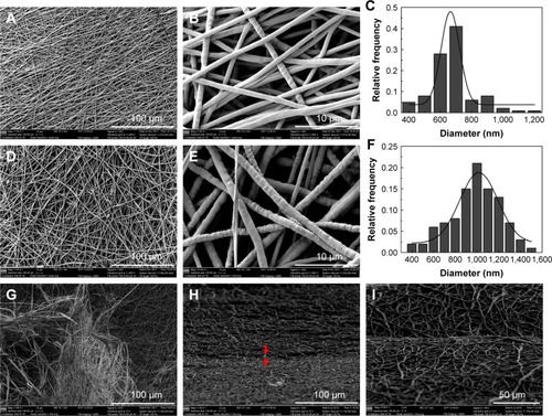
Table 2 Mechanical properties of scaffolds
Figure 2 Porosity and pore size distribution of the nanofibrous scaffolds.
Notes: Porosity percentage (A). Pore size distribution of SLS (B) and FLS (C). *The difference in mean is significant (P<0.05) with respect to chitosan. #The difference in mean is significant (P<0.05) with respect to PCL.
Abbreviations: DLS, double layer nanofibrous scaffolds; FLS, first layer of scaffolds; PCL, polycaprolactone; SLS, second layer of scaffolds.
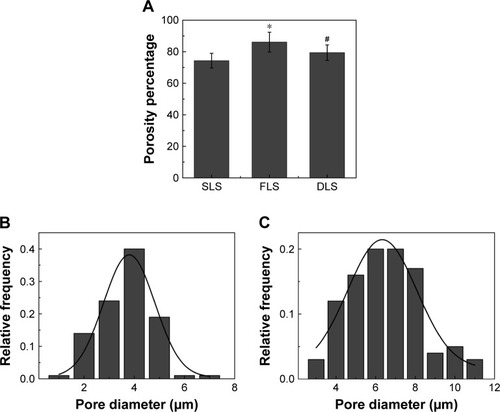
Figure 3 FTIR spectrum of LID (a), chitosan (b), SLS (c), mupirocin (d), PCL (e), FLS (f), and DLS (g).
Abbreviations: DLS, double layer nanofibrous scaffolds; FLS, first layer of scaffolds; FTIR, Fourier transform infrared spectra; LID, lidocaine hydrochloride; PCL, polycaprolactone; SLS, second layer of scaffolds.
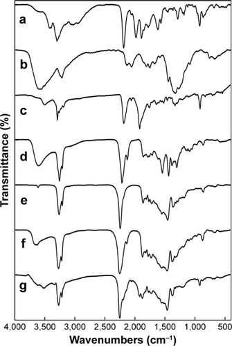
Figure 4 Thermal stability of chitosan and PCL nanofibrous scaffolds.
Notes: TGA graph (A) and DSC graph (B).
Abbreviations: DLS, double layer nanofibrous scaffolds; DSC, differential scanning calorimetry; FLS, first layer of scaffolds; LID, lidocaine hydrochloride; PCL, polycaprolactone; SLS, second layer of scaffolds; TGA, thermogravimetric analysis.
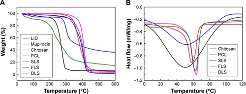
Figure 5 Water contact angles and swelling ratio of nanofibrous scaffolds.
Notes: Water contact angles of nanofibrous scaffolds (A). Swelling ratio of nanofibrous scaffolds (B). *The difference in mean is significant (P<0.05) with respect to SLS. #The difference in mean is significant (P<0.05) with respect to FLS.
Abbreviations: DLS, double layer nanofibrous scaffolds; FLS, first layer of scaffolds; SLS, second layer of scaffolds.
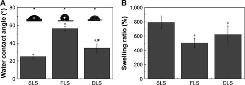
Figure 6 Comparative cytotoxicity of nanofibrous scaffolds.
Notes: Cell viability was determined by live/dead cell assay using calcein-AM (live) and propidium iodide (dead) (A–D). Representative images of control (A), SLS (B), FLS (C), and DLS (D). The viability of human dermal fibroblasts was evaluated using MTT assay (E). Scale bar denotes 1 µm.
Abbreviations: DLS, double layer nanofibrous scaffolds; FLS, first layer of scaffolds; SLS, second layer of scaffolds.
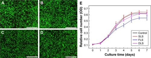
Figure 7 Release profiles of LID and mupirocin into PBS from DLS nanofibrous scaffolds.
Notes: LID eluted from DLS (A). Mupirocin eluted from DLS (B).
Abbreviations: DLS, double layer nanofibrous scaffolds; LID, lidocaine hydrochloride.
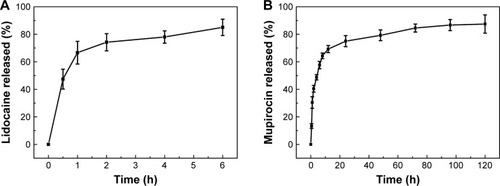
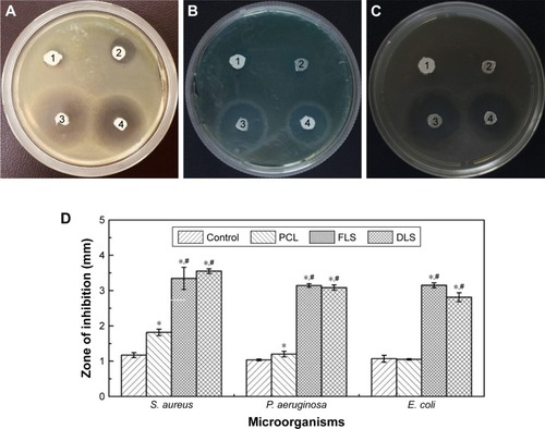
Figure 8 Evaluation of antibacterial activity of nanofibrous scaffolds.
Notes: (1) Filter paper control, (2) PCL, (3) FLS, (4) DLS against Staphylococcus aureus (A), Pseudomonas aeruginosa (B), and Escherichia coli (C) and evaluation of the inhibition zones for filter paper control, PCL, FLS, and DLS against S. aureus, P. aeruginosa, and E. coli (D). *The difference in mean is significant (P<0.05) with respect to control. #The difference in mean is significant (P<0.05) with respect to PCL.
Abbreviations: DLS, double layer nanofibrous scaffolds; FLS, first layer of scaffolds; PCL, polycaprolactone.