
Figures & data
Figure 1 Morphology and composition analysis of magnetic nonowires.
Notes: (A) SEM image of Ni NWs on top of a silicon wafer substrate. (B) TEM image of a single Ni NW. The inset shows the outer oxide layer that is 6–8 nm thick. (C) Point EDX spectra of a Ni NW core and (D) its surrounding layer. The insets show the corresponding STEM images. Red asterisks indicate the point from which the spectrum was measured. Scale bars: 40 nm.
Abbreviations: NW, nanowire; SEM, scanning electron microscopy; STEM, scanning transmission electron microscopy; TEM, transmission electron microscopy.
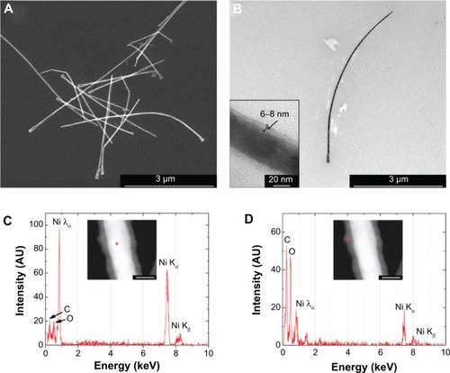
Figure 2 Magnetization loops of an array of Ni NWs embedded in the alumina membrane with magnetic field applied in the in-plane and out-of-plane directions.
Notes: Black: in-plane direction. Red: out-of-plane direction. Hc|| and and Ms||, refer to the coercive field and the saturation when the field is applied in the in-plane direction (black) of the nanowires.
Abbreviations: M, magnetization; NWs, nanowires.
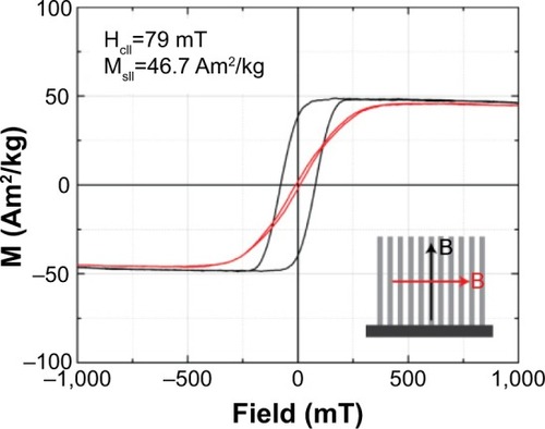
Figure 3 TEM images of colon cancer cells incubated for 1 hour with Ni NWs.
Notes: (A) Control cells (cells to which no NWs were added) with organelles such as M, GA, N, PM, and LD. (B) NWs in close proximity to the PM of colon cancer cells. Cells that internalized NWs as aggregates (C) and as single NWs (D). PM surrounding indivdual NWs are visible (red arrows). (E and F) Fully internalized NWs. A few NWs can be observed fully surrounded by a membrane vesicle (E) and a fully internalized large aggregate of NWs caused the reordering of organelles like the GA (red arrow) (F).
Abbreviations: GA, Golgi apparatus; LD, lipid droplets; M, mitochondria; N, nucleus; NW, nanowire; PM, plasma membrane; TEM, transmission electron microscopy.
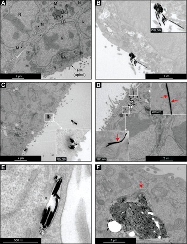
Figure 4 Viability of colon cancer (MTT assay) cells after 1 hour incubation with Ni NWs.
Notes: The NC sample (0 Ni μg/mL) corresponds to cells without NW addition. NW concentration values are expressed and Ni μg per mL of cell culture medium. Data represents means ± standard deviation, n=3, **P<0.01 versus NC.
Abbreviations: MTT, 3-(4,5-dimethylthiazol-2yl)-2,5-diphenyl tetrazolium bromide; NC, negative control; NW, nanowire.
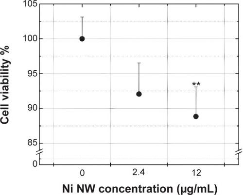
Figure 5 Viability of colon cancer cells (MTT assay) after exposure to AMFs of two different frequencies.
Notes: The negative control sample (0 minutes of AMF) corresponds to cells without AMF exposure. Data represent means ± standard deviation, n=3, *P<0.05.
Abbreviations: AMF, alternating magnetic field; MTT, 3-(4,5-dimethylthiazol-2yl)-2,5-diphenyl tetrazolium bromide.
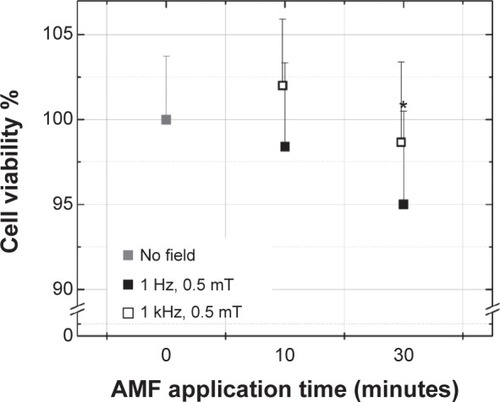
Figure 6 A simple model for estimating the force an AMF exerts on a NW that can be transmitted to a cell if the NW’s ends are attached to it.
Abbreviations: τm, magnetic torque; AMF, alternating magnetic field; Fm, magnetic force; M, midpoint; NW, nanowire.
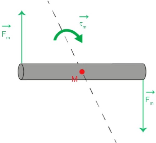
Figure 7 Cell viability of colon cancer (MTT assay) cells incubated with Ni NWs for 1 hour and then exposed to magnetic fields of different frequencies and amplitudes for 10 minutes.
Notes: In the NC cells (0 Ni μg/mL), no NWs were added (100% cell viability value). Data represent means ± standard deviation, n=3, *P<0.05; **P<0.01 versus NC.
Abbreviations: MTT, 3-(4,5-dimethylthiazol-2yl)-2,5-diphenyl tetrazolium bromide; NC, negative control; NW, nanowire.
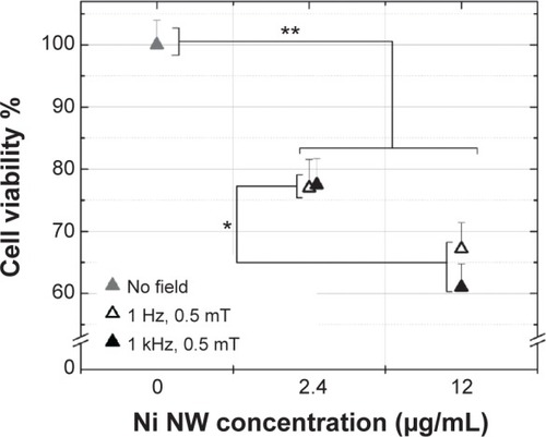
Figure 8 LDH leakage of colon cancer cells incubated with Ni NWs for 1 hour and then exposed to magnetic fields of different frequencies and amplitudes for 10 minutes.
Notes: There are three control samples in which no NWs or fields were added (gray triangles). A sample of lysed cells (§) was used as the PC, which corresponded to 100% leakage. No NWs were added to the NC (†) cells. Apoptosis was induced to cells (‡) by exposing them to ultraviolet radiation for 1 hour. Data represent means ± standard deviation, n=3, **P<0.01 versus NC. P<0.01 for PC versus all the other samples (data not shown).
Abbreviations: LDH, lactate dehydrogenase; NC, negative control; NW, nanowire; PC, positive control.
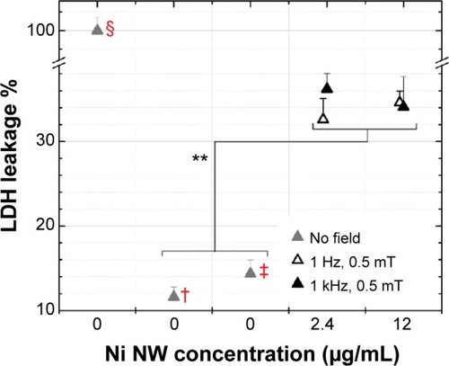
Figure 9 Schematic of the possible mechanism of cell death due to interaction of cancer cells with NWs and the subsequent application of a low-frequency, low-amplitude magnetic field.
Notes: The cytoskeleton fibers reorganize upon application of the magnetic field (evidence of F-actin reorganization due to magnetic field exposure is presented in Zablotskii et alCitation37).
Abbreviations: AMF, alternating magnetic field; NWs, nanowires.
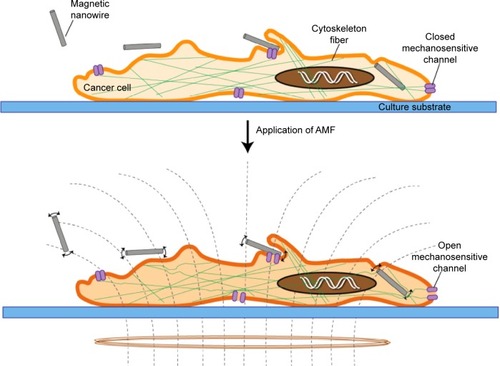
Figure S1 LDH leakage of colon cancer cells under different conditions.
Notes: A sample of lysed cells was used as the PC which corresponded to 100% leakage. No NWs were added to the NC cells. Apoptosis (APOP) was induced in cells by exposing them to ultraviolet radiation for 1 hour. Cells were incubated with NWs at two given concentrations for 1 hour (no AMF applied). Cells were exposed to AMF for 10 minutes (no NWs added). Data represent means ± standard deviation, n=3, *P<0.05 versus NC/APOP. P<0.01 for PC versus all the other samples (data not shown).
Abbreviations: AMF, alternating magnetic field; LDH, lactate dehydrogenase; NC, negative control; NWs, nanowires; PC, positive control.
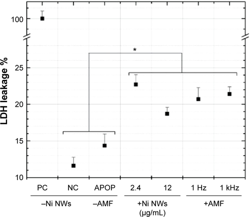
Table S1 Ni NW concentrations expressed in equivalent units