
Figures & data
Figure 1 SEM images of (A) CVC, (B) DCVC, (C and D) DCVC-Ag1, (E and F) DCVC-Ag2, and (G and H) DCVC-Ag3.
Abbreviations: CVC, central venous catheter; DCVC, central venous catheters coated with polydopamine films; SEM, scanning electron microscopy.
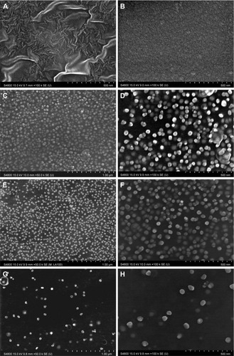
Table 1 Surface chemical composition and water contact angle of pristine CVC, DCVC, and DCVC-Ag samples (atomic percentage according to XPS analysis)
Figure 2 The survey XPS spectra of CVC, DCVC, and DCVC-Ag3.
Abbreviations: au, arbitrary unit; CVC, central venous catheter; DCVC, central ve nous catheters coated with polydopamine films; XPS, X-ray photoelectron spectroscopy.
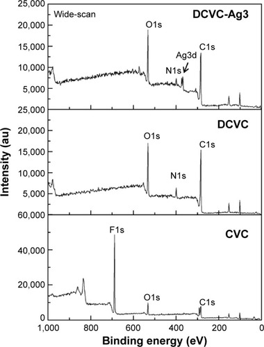
Figure 3 The high-resolution XPS spectra of DCVC-Ag1, DCVC-Ag2, and DCVC-Ag3.
Abbreviations: au, arbitrary unit; DCVC, central venous catheters coated with polydopamine films; XPS, X-ray photoelectron spectroscopy.
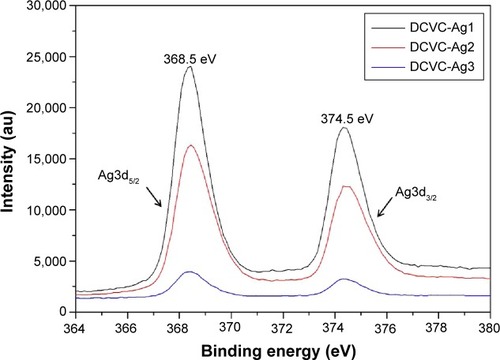
Figure 4 Raman spectra of various samples: (A) CVC, (B) DCVC, (C) DCVC-Ag3, (D) DCVC-Ag2, and (E) DCVC-Ag1.
Abbreviations: au, arbitrary unit; CVC, central venous catheter; DCVC, central venous catheters coated with polydopamine films.
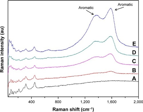
Figure 5 Photographs showing the zone of inhibition assay for DCVC-Ag samples.
Abbreviation: DCVC, central venous catheters coated with polydopamine films.
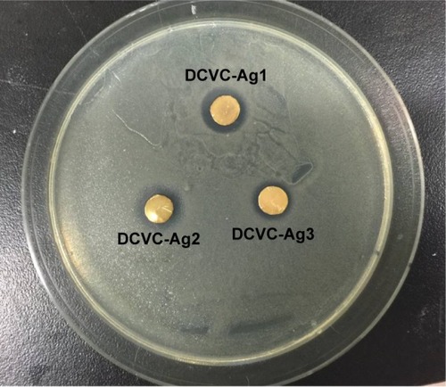
Figure 6 Fluorescent microscope images of Staphylococcus aureus stained on (A) DCVC, (B) DCVC-Ag1, (C) DCVC-Ag2, and (D) DCVC-Ag3 after 6 hours of culture.
Abbreviation: DCVC, central venous catheters coated with polydopamine films.
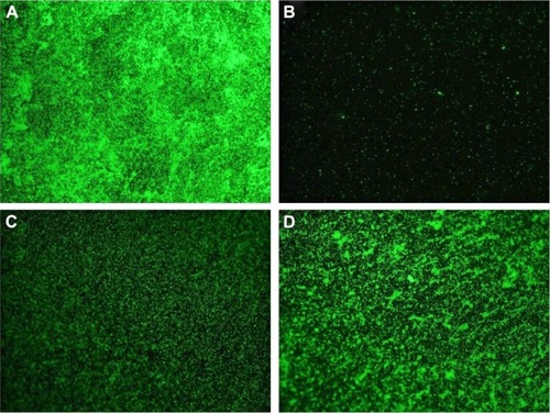
Figure 7 WST-1 assay for proliferation of MC3T3-E1 cell on different samples at various incubation periods.
Note: Error bars represent mean ± SD (n=5), *P<0.05, **P<0.01.
Abbreviations: CVC, central venous catheter; DCVC, central venous catheters coated with polydopamine films; SD, standard deviation; WST-1, water soluble tetrazolium salts.

Figure 8 Fluorescence microscopy images of Calcein-AM stained MC3T3-E1 cell attached on (A and B) DCVC, (C and D) DCVC-Ag1, (E and F) DCVC-Ag2, and (G and H) DCVC-Ag3 for 3 days.
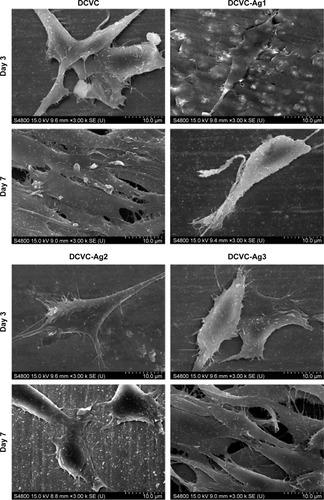
Figure 9 The SEM images of MC3T3-E1 cell attachment on DCVC, DCVC-Ag1, DCVC-Ag2, and DCVC-Ag3 samples for days 3 and 7.
Abbreviations: DCVC, central venous catheters coated with polydopamine films; SEM, scanning electron microscopy.
Abbreviation: DCVC, central venous catheters coated with polydopamine films.
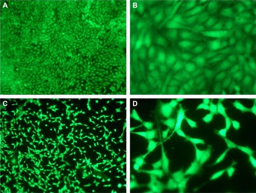
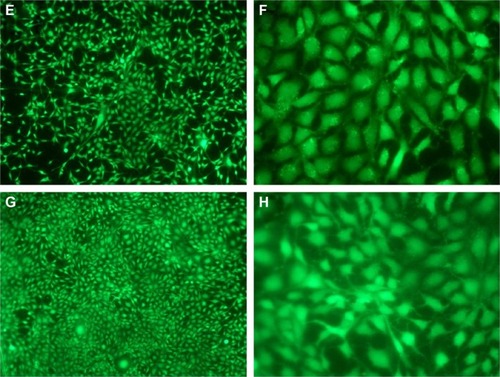
Figure 9 The SEM images of MC3T3-E1 cell attachment on DCVC, DCVC-Ag1, DCVC-Ag2, and DCVC-Ag3 samples for days 3 and 7.
Abbreviations: DCVC, central venous catheters coated with polydopamine films; SEM, scanning electron microscopy.