Figures & data
Table 1 Distribution of Rab18 status in gastric cancer according to clinicopathological characteristics
Figure 1 Expression of Rab18 in gastric cancers.
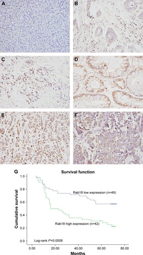
Figure 2 Expression of Rab18 in gastric cancer cell lines.
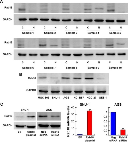
Figure 3 Rab18 regulates proliferation, colony formation, and cell cycle.
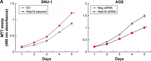
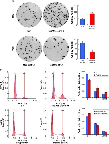
Figure 4 Rab18 regulates invasion, migration, and adhesion.
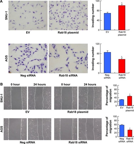
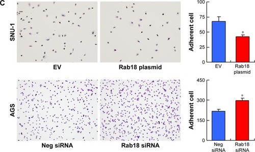
Figure 5 Rab18 regulates CDDP-induced apoptosis in gastric cancer cells.
Abbreviations: CDDP, cisplatin; PI, propidium iodide.
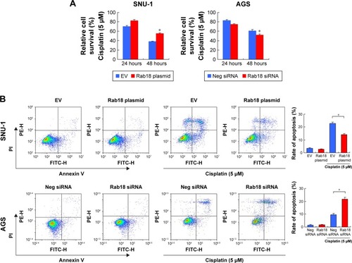
Figure 6 Rab18 regulates MMP and ROS production.
Abbreviations: CDDP, cisplatin; FITC, fluorescein isothiocyanate; MMP, mitochondrial membrane potential.
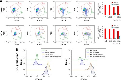
Figure 7 Rab18 regulates p-Rb, survivin, cytochrome c, cleaved PARP, and cleaved caspase 3.
Abbreviation: CDDP, cisplatin.
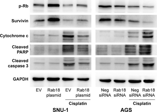
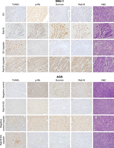
Figure 8 Rab18 promotes cancer cell growth in vivo.
Abbreviation: CDDP, cisplatin.
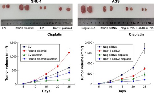
Figure S1 Validation of Rab18 antibody for Western blot and immunofluorescence.
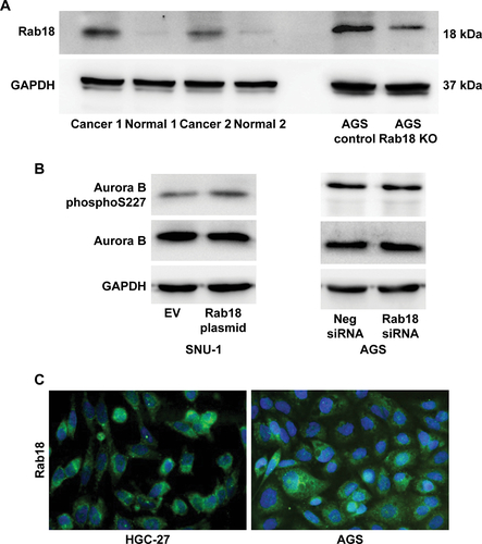
Figure S2 Immunohistochemistry for in vivo tumors formed by stably transfected cells.