Figures & data
Table 1 Immunohistochemical Stains in the Differential Diagnosis of Desmoplastic Melanoma
Table 2 (A and B) Clinical and Pathological Findings in Desmoplastic Melanoma
Figure 1 (A) Case 1, lesion presented as a reddish-brown nodule on the head and face, with uneven pigment and regression phenomenon. (B) Case 2, lesion presented as an isolated red papule on the left upper limb, without pigmentation, mimicking the “dermatofibroma”. (C) Case 3, lesion presented as a soybean-sized reddish nodule on the left corner of the eye with uneven pigment.
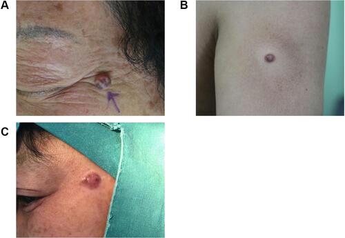
Figure 2 (A) Massive neoplasm cells with hyperchromatic nuclei, infiltrating through the tissue with associated thickening of interweaved collagen fibers (hematoxylin and eosin, original magnification ×100). (B) Neoplastic cells exhibited mild to moderate atypia. Cell morphology varied. They feathered either long or short spindles, accompanied with hyperchromatic nuclei, vesicular shape, increased nucleoplasm ratio, and sometimes mitotic figures (hematoxylin and eosin, original magnification ×400). (C) Melanocytes at the dermo-epidermal junction were arranged in a nesting or linear manner, similar to malignant freckles. Some of the melanocytes exhibited moderate atypia (hematoxylin and eosin, original magnification ×100). (D) Melanin deposition was prominent in the cytoplasm of neoplasm cells (hematoxylin and eosin, original magnification ×100). (E) Neurophilicity manifested by one or more well-defined spindle cell foci around the nerve or within the nerve sheath, squeezing the nerve sheath (hematoxylin and eosin, original magnification ×200). (F) Patchy lymphocytic infiltrates around and below the lesions, some of which formed lymphoid follicular-like structures (hematoxylin and eosin, original magnification ×100).
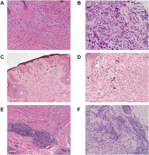
Figure 3 Immunophenotype of desmoplastic melanoma (IHC with SP method, × 100). (A) Diffuse, positive expression of S-100; (B) diffuse, positive expression of vimentin; (C) diffuse, positive expression of CD34; (D) positive expression of Melan-A.
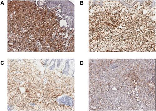
Figure 4 Immunophenotype (IHC with SP method, × 100). (A) Negative expression of MITF; (B) negative expression of HMB-45; (C) negative expression of SMA with surrounding vascular endothelium demonstrating positive internal control; (D) negative expression of P53.
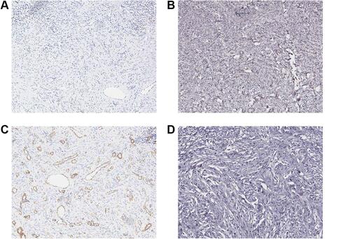
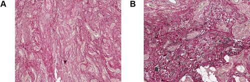
Figure 5 EVG staining (×100) showed that there were fewer elastic fibers in the desmoplastic melanoma lesion (A) than in the para-cancer lesion (B).