Figures & data
Table 1 Key Research Articles Contributing to the Field of Pulmonary Fibrosis
Figure 1 An illustration of AT2-AT1 cell transition during fibrotic lung injury. The figure represents the process of AT2-AT1 cell transition during fibrotic lung injury. AT2 cells are shown to proliferate after the lung injury, where a fraction of these cells differentiating into mature AT1 cells. Also included are the representative cell types involved in the repair process (top right). Created with BioRender.com.
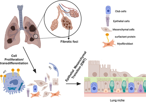
Figure 2 Lineage-tracing of cells during the repair phase of an injured lung by the Cre-ERT2 system. (a) Schematic representation of the Cre-ERT2 system. The presence of a stop codon flanked by loxP sites inhibits the expression of the reporter transgene (tdTomato). Upon tamoxifen treatment, the expressed Cre mediates site-specific recombination between loxP sites, which removes the intervening stop codon, and the reporter is expressed. (b) Lineage-tracing using Cre-ERT2 system enriches and identifies intermediate stages of transdifferentiated AT2 stem cells. Some of the identified intermediates are shown for representation. Similar enrichment can also be achieved using stem cell surface markers such as CD34, CD45, and Thy1. Created with BioRender.com.
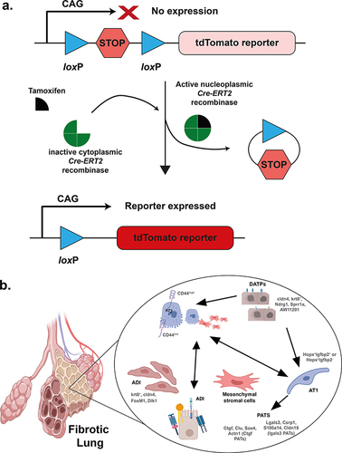
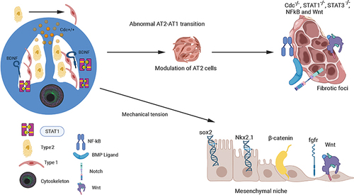
Figure 3 Mechanotransduction-Mediated Signaling in AT2-AT1 Transition. The figure illustrates the signaling factors and progenitor stem cells involved in the AT2-AT1 transition in fibrotic lungs, as well as the role of the mesenchymal niche microenvironment in this process. The complex mechanical signaling pathways that control this transition are depicted, with a focus on the involvement of the small Rho GTPase family member Cdc42 in regulating the STAT-3 and STAT-1 signaling pathways. The effects of mechanotransduction on Wnt, Sox2, EGF, β-catenin, and Nkx2 proteins in the lung mesenchyme, which can alter lung development and repair, have also been demonstrated (bottom right). Created with BioRender.com.