1,849
Views
76
CrossRef citations to date
0
Altmetric
Research Article

Propagation of target and organ at risk contours in radiotherapy of prostate cancer using deformable image registration

, , , &
Pages 1023-1032 | Received 20 May 2010, Accepted 21 Jun 2010, Published online: 13 Sep 2010

Figures & data

Table I. The criteria for each propagated contour in the clinical scoring, showing the number of slices that needed to be unapproved for each grade.

Table II. Patient average DSC including range in brackets for all registrations and contours.

Figure 1. DSC as a function of static image (CTnrX) to which the reference image (CTnr1) was registered for patient 3. The colors indicate organ contours; gray for bladder and black for rectum. Type of line indicate registration; dashed for rigid, dotted for algorithm A and solid for algorithm B.

Figure 1. DSC as a function of static image (CTnrX) to which the reference image (CTnr1) was registered for patient 3. The colors indicate organ contours; gray for bladder and black for rectum. Type of line indicate registration; dashed for rigid, dotted for algorithm A and solid for algorithm B.

Figure 2. DSC as a function of relative volume enclosed by the contour in the static and reference image including patient average DSC marked with solid lines for illustration. The colors indicate algorithm; black for algorithm A and gray for algorithm B. Different markers represent different patients as described in the legend.

Figure 2. DSC as a function of relative volume enclosed by the contour in the static and reference image including patient average DSC marked with solid lines for illustration. The colors indicate algorithm; black for algorithm A and gray for algorithm B. Different markers represent different patients as described in the legend.

Figure 3. Sensitivity as a function of PPV for all DIR propagated contours. The colors indicate algorithm; black for algorithm A and gray for algorithm B. Different markers represent different patients as described in the legend.

Figure 3. Sensitivity as a function of PPV for all DIR propagated contours. The colors indicate algorithm; black for algorithm A and gray for algorithm B. Different markers represent different patients as described in the legend.

Figure 4. Clinical scoring for all propagated contours. The colors indicate algorithm; black for algorithm A and gray for algorithm B. Different patterns in the bars represent different patients as described in the legend.

Figure 4. Clinical scoring for all propagated contours. The colors indicate algorithm; black for algorithm A and gray for algorithm B. Different patterns in the bars represent different patients as described in the legend.

Figure 5. Examples of DIR propagated contours seen on the static image for: CTV-p and rectum (to the left) and for CTV-ln, rectum and bladder (to the right). Colors of the contours are for rectum: darkgreen for the static image contour, green for algorithm A, light green for algorithm B, for CTV-p and CTV-ln: red contours for the static image, magenta for algorithm A and pink for algorithm B and for the bladder: orange contours for the static image, yellow for algorithm A and white for algorithm B.

Figure 5. Examples of DIR propagated contours seen on the static image for: CTV-p and rectum (to the left) and for CTV-ln, rectum and bladder (to the right). Colors of the contours are for rectum: darkgreen for the static image contour, green for algorithm A, light green for algorithm B, for CTV-p and CTV-ln: red contours for the static image, magenta for algorithm A and pink for algorithm B and for the bladder: orange contours for the static image, yellow for algorithm A and white for algorithm B.

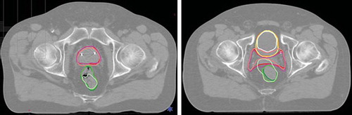
Table III. The relationship between the clinical scoring and DSC for all propagated contours with the two algorithms.

Reprints and Corporate Permissions

Please note: Selecting permissions does not provide access to the full text of the article, please see our help page How do I view content?

To request a reprint or corporate permissions for this article, please click on the relevant link below:

Academic Permissions

Please note: Selecting permissions does not provide access to the full text of the article, please see our help page How do I view content?

Obtain permissions instantly via Rightslink by clicking on the button below:

If you are unable to obtain permissions via Rightslink, please complete and submit this Permissions form. For more information, please visit our Permissions help page.