Figures & data
Figure 1. Endothelium-dependent relaxation responses to ACh in controls, ET-1-incubated, ET-1 and PEG-SOD plus apocynin-incubated, ET-1 and PJ34-incubated, and PJ34-incubated thoracic aorta rings. All values are expressed as mean ± standard error of the mean; *p < 0.05 as compared with controls; **p < 0.05 as compared with ET-1-incubated rings; n = 10 for all groups.
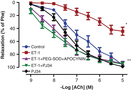
Figure 2. Effects of PEG-SOD plus apocynin, or PJ34 incubation on ET-1-induced vascular hyper-responsiveness to Phe in rat aortic rings. All values are expressed as mean ± standard error of the mean; *p < 0.05 as compared with controls; **p < 0.05 as compared with ET-1-incubated rings; n = 10 for all groups.
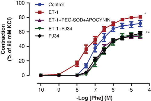
Figure 3. Endothelium-independent relaxation responses to SNP control, ET-1-incubated, ET-1 and PEG-SOD plus apocynin-incubated, ET-1 and PJ34-incubated, and PJ34-incubated thoracic aorta rings. All values are expressed as mean ± standard error of the mean; n = 10 for all groups.
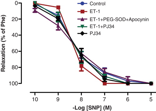
Figure 4. Representative photomicrographs of PARP-1 (a–f) immunohistochemistry in controls (a), ET-1-incubated (b), ET-1 and PEG-SOD plus apocynin-incubated (c), ET-1 and PJ34-incubated (d), and PJ34-incubated vessels (e). Strong immunostaining of PARP-1 was observed in ET-1-incubated endothelial cells (arrows). Expression of PARP-1 was minimal in both ET-1 and PEG-SOD plus apocynin-incubated, and ET-1 and PJ34-incubated vessel endothelium. Negative control sections with PARP-1 (f) primary antibodies. Note the absence of PARP-1 immunostainings. Original magnifications ×50.
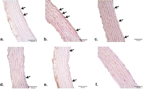
Figure 5. Representative photomicrographs of PAR (a–f) immunohistochemistry in controls (a), ET-1-incubated (b), ET-1 and PEG-SOD plus apocynin-incubated (c), ET-1 and PJ34-incubated (d), and PJ34-incubated vessels (e). Strong immunostaining of PAR was observed in ET-1-incubated endothelial cells (arrows). Expression of PAR was minimal in both ET-1 and PEG-SOD plus apocynin-incubated, and ET-1 and PJ34-incubated vessel endothelium. Negative control sections with PAR (f) primary antibodies. Note the absence of PAR immunostaining. Original magnifications ×50.
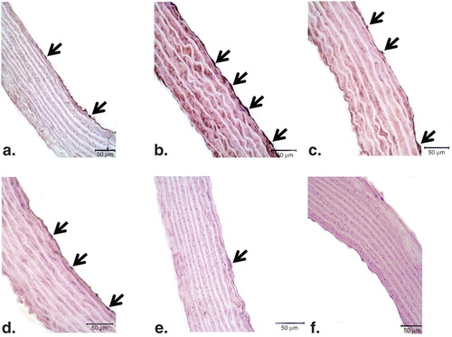
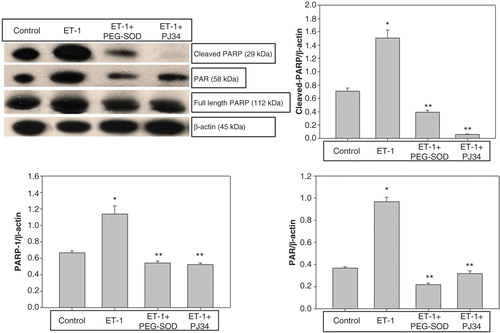
Figure 6. Evaluation of PARP-1 and PAR expression in thoracic aorta rings by Western blot analysis in controls, ET-1-incubated, ET-1 and PEG-SOD plus apocynin-incubated, and ET-1 and PJ34-incubated specimens. *p < 0.05 as compared with controls; **p < 0.05 as compared with ET-1-incubated rings.