Figures & data
Figure 1. HPLC fingerprint analysis of the standard SDG (A) and flax lignan concentrate (B). Peaks were detected at 282 nm.
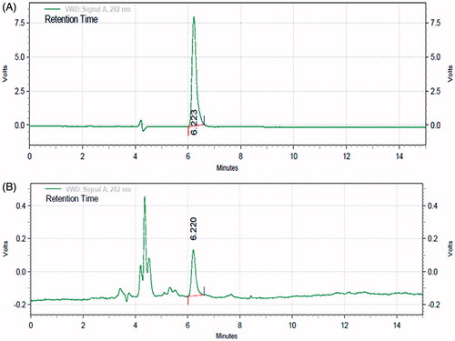
Figure 2. Effect of administration of captopril and FLC on various hemodynamic parameters viz. SBP (A), DBP (B), MABP (C), EDP (D), dP/dtmax (E), and dP/dtmin (F) in DOCA-salt hypertensive Wistar rats. Values are expressed as mean ± SEM (n = 6). Data were analyzed by one-way ANOVA followed by Dunnett's test. *p < 0.05, **p < 0.01, ***p < 0.001 as compared with UNTZD + DOCA control group and ###p < 0.001 compared to UNTZD control group.
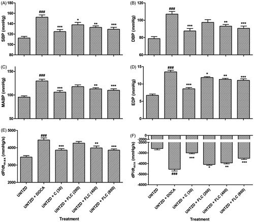
Table 1. Effects of captopril and FLC on organs weight and serum electrolytes in DOCA-salt induced hypertensive Wistar rats.
Table 2. Effect of captopril and FLC on serum cardiac, hepatic and renal markers in DOCA-salt induced hypertensive Wistar rats.
Table 3. Effect of captopril and FLC on lipid profile in DOCA-salt induced hypertensive Wistar rats.
Table 4. Effect of captopril and FLC on endogenous antioxidant enzymes in DOCA-salt induced hypertensive Wistar rats.
Figure 3. Effect of captopril and FLC on DOCA-salt induced alteration in kidney histology of rats. Photomicrograph of sections of kidney of (A) UNTZD control rats showed normal glomerulus cell and tubuli with the absence of any congestion, (B) UNTZD + DOCA-salt control group showed atrophy of tubular cell, necrosis and glomerulus congestion, (C) UNTZD + DOCA-salt + captopril (30 mg/kg) showed decrease in glomerulus congestion, atrophy and necrosis, (D) UNTZD + DOCA-salt + FLC (200 mg/kg) showed no significant protection against hypertension, (E) UNTZD + DOCA-salt + FLC (400 mg/kg) showed decrease in glomerulus atrophy, congestion and necrosis, and (F) UNTZD + DOCA-salt + FLC (800 mg/kg) showed significantly reduced glomerulus congestion and necrosis. Hematoxylin and eosin staining (at 40×).
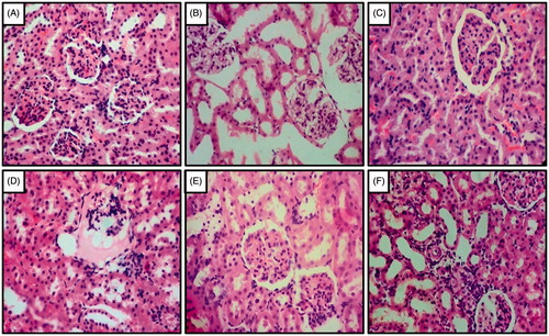
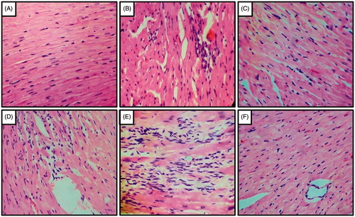
Figure 4. Effect of captopril and FLC on DOCA-salt induced alteration in heart histology of rats. Photomicrograph of sections of heart of (A) UNTZD rats showed normal cardiac muscle fibers, (B) UNTZD + DOCA-salt control showed severe myocardial degeneration, hypertrophy and infiltration of inflammatory cells, (C) UNTZD + DOCA-salt + captopril (30 mg/kg) showed minimal myocardial degeneration and infiltration of inflammatory cells, (D) UNTZD + DOCA-salt + FLC (200 mg/kg) showed no significant change in myocardial tissues, (E) UNTZD + DOCA-salt + FLC (400 mg/kg) showed decrease in myocardial degeneration and infiltration of inflammatory cells, and (F) UNTZD + DOCA-salt + FLC (800 mg/kg) showed significant reduction in myocardial degeneration and infiltration of inflammatory cells. Hematoxylin and eosin staining (at 40×).