Figures & data
Figure 1. Bar diagram representing the RS-induced changes in ulcer index of control, sham, and eugenol-treated [25 mg/kg (E-25), 50 mg/kg (E-50), and 100 mg/kg (E-100)] groups. Results are represented as mean ± SEM with n = 6 in each group. “n” is the number of rats used. *P < 0.05 compared to control, †P < 0.05 compared to sham, P < 0.05 compared to E-25, and P < 0.05 compared to E-50 (Student–Newmann–Keuls post hoc test).
![Figure 1. Bar diagram representing the RS-induced changes in ulcer index of control, sham, and eugenol-treated [25 mg/kg (E-25), 50 mg/kg (E-50), and 100 mg/kg (E-100)] groups. Results are represented as mean ± SEM with n = 6 in each group. “n” is the number of rats used. *P < 0.05 compared to control, †P < 0.05 compared to sham, P < 0.05 compared to E-25, and P < 0.05 compared to E-50 (Student–Newmann–Keuls post hoc test).](/cms/asset/cac06c44-bed7-44bc-a313-51d577a7d2f1/ists_a_521602_f0001_b.gif)
Figure 2. Bar diagram representing the RS-induced changes in plasma corticosterone (A) and NE levels (B) of control, sham, and eugenol-treated groups. Results are represented as mean ± SEM with n = 6 in each group. “n” is the number of rats used. *P < 0.05 compared to control, †P < 0.05 compared to sham, ‡P < 0.05 compared to E-25, ¶P < 0.05 compared to E-50, and P < 0.05 compared to E-100 (Student–Newmann–Keuls post hoc test).
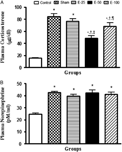
Figure 3. Bar diagram representing the RS-induced changes in the levels of NE (A), 5-HT (B), and 5-HIAA (C) of control, sham, and eugenol treated (50 mg/kg) groups in HIP, HYPO, PFC, and AMY regions of brain. Results are represented as mean ± SEM with n = 6 in each group. “n” is the number of rats used. *P < 0.05 compared to control and †P < 0.05 compared to sham group (Student–Newmann–Keuls post hoc test).
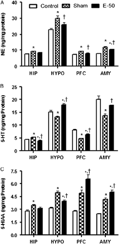
Figure 4. Bar diagram representing the RS-induced changes in the levels of DA (A), DOPAC (B), and HVA (C) of control, sham, and eugenol treated (50 mg/kg) groups in HIP, HYPO, PFC, and AMY regions of brain. Results are represented as mean ± SEM with n = 6 in each group. “n” is the number of rats used. *P < 0.05 compared to control and †P < 0.05 compared to sham group (Student–Newmann–Keuls post hoc test).
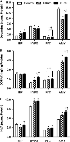
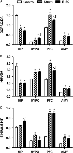
Figure 5. Bar diagram representing the RS-induced changes in the metabolite/monoamine ratios, DOPAC/DA (A), HVA/DA (B), and 5-HIAA/5-HT (C) of control, sham, and eugenol-treated (50 mg/kg) groups in HIP, HYPO, PFC, and AMY regions of brain. Results are represented as mean ± SEM with n = 6 in each group. “n” is the number of rats used. *P < 0.05 compared to control and †P < 0.05 compared to sham group (Student–Newmann–Keuls post hoc test).