Figures & data
Figure 1. TNFα, IL-1β, and TGFβ-2 increased FLS metabolic activity in vitro. The FLS metabolic activity was determined with Alamar blue after 14 d of culture. The FLS were untreated (w/o) or treated with 10 ng/mL tumour necrosis factor α (TNFα), interleukin (IL)-1β, oncostatin M (OSM), insulin growth factor-1 (IGF-1), transforming growth factor β-2 (TGFβ-2), or 1 ng/mL IL-6. Wells with no cells were included as negative control. Data shown is pooled data from three independent experiments with FLS from three different patients. Data are presented mean ± SEM. ****p < 0.0001.
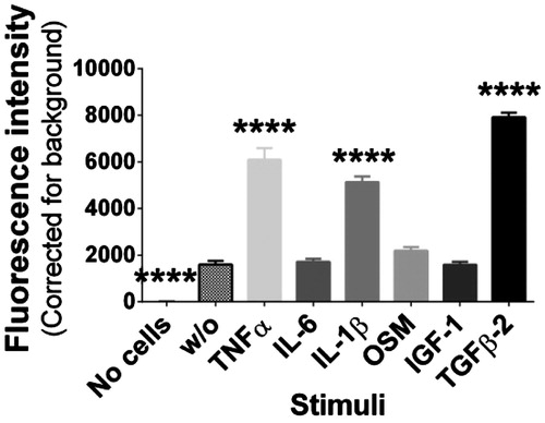
Figure 2. MMP-2, -3 or -9 were secreted by FLS, but not in the active form. Conditioned media from primary FLS untreated (w/o) or treated with 10 ng/mL TNFα, IL-1β, OSM, IGF-1, TGFβ-2, or 1 ng/mL IL-6 from days 0, 7, and 14 were used to detect the secretion of total MMP-3 with western blot (A) and total MMP-2 and -9 with gelatin zymography (B). (B) Control: bovine cartilage explants treated with 10 ng/ml oncostatin M and 20 ng/ml TNFα for 17 d used as ladder, as this is known to express both MMP-2 and MMP-9 (Sondergaard et al., Citation2006).
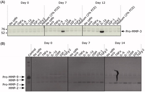
Figure 3. Preparation of synovial biopsies to synovial membrane explants. (A) When synovial biopsies are received from the hospital the fat is initially removed with a scalpel. Here are two biopsies from two different patients (A and B) showed before and after the fat was removed. Scale is 3 cm. (B) Small explants of 30 ± 2 mg are cut with the scalpel and the wet weight determined after the fat is removed. Scale is 1 cm. (C) Cut explants are transferred directly to a well containing culture media. Before start of the experiment the explants are washed 3 times in culture media.
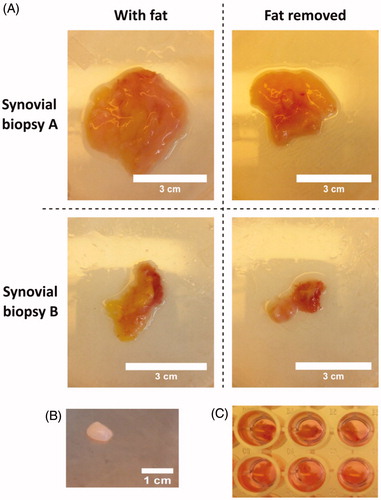
Figure 4. Hematoxylin and eosin staining of synovial membrane explants (SMEs). SMEs were fixated and embedded in paraffin, cut, and stained with hematoxylin and eosin after 14 d of culture. Here are two different explants from each treatment shown. The arrows indicate adipocytes found within the SMEs. The scale is 200 μM.
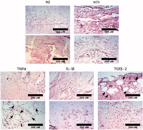
Figure 5. TNFα and IL-1β induce activation of MMP-3 and -9 in SMEs. Pooled conditioned medium from synovial membrane explants (SMEs) either metabolically inactivated (MI), untreated (w/o), or treated with 10 ng/ml TNFα, IL-1β, or TGFβ-2 were used to detect total MMP-3 with western blot (B + D) and total MMP-2 and -9 with gelatin zymography (A + C). The results shown are from two independent experiments with synovial tissue from four (A and D) and three (B and D) OA patients, respectively. (A + C) Control: bovine cartilage explants treated with 10 ng/ml oncostatin M and 20 ng/ml TNFα for 17 d used as ladder, as this is known to express both MMP-2 and MMP-9 (Sondergaard et al., Citation2006).
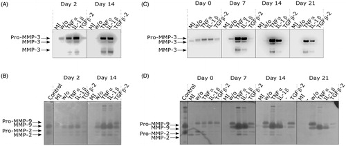
Figure 6. TNFα and IL-1β stimulated release C1M, C3M, and acMMP3 from the SMEs. Concentrations of C1M (A + D), C3M (B + E), and acMMP3 (C + F) were measured with ELISA in the conditioned medium from SMEs either metabolically inactivated (MI), untreated (w/o), or treated with 10 ng/ml TNFα, IL-1β, or TGFβ-2. The biomarker measurements are shown from two independent experiments with synovial tissue from four (A–C) and three (D–F) OA patients, respectively. Data are presented mean ± SEM. *p < 0.05, **p < 0.01, ***p < 0.001, ****p < 0.0001.
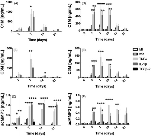
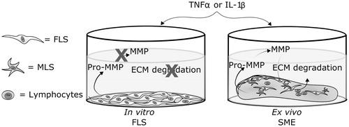
Figure 7. Synovial tissue generates active MMPs and degrades the ECM in response to TNFα or IL-1β ex vivo. In vitro primary fibroblast-like synoviocytes (FLS) cultured on type I and III collagen coating do not generate active metalloproteinases (MMPs), and, therefore, cannot generate extracellular matrix (ECM) degradations biomarkers like C1M, C3M, and acMMP3. Ex vivo cultured synovial membrane explants (SMEs) contain multiple cell types in their natural three-dimensional ECM, and generate active MMPs and release degradation biomarkers: C1M and C3M and the biomarker of activated MMP3, acMMP3, in a pro-inflammatory environment. MLS = macrophage-like synoviocytes.