Figures & data
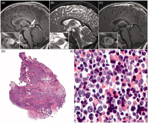
FIGURE 1. Sagittal T1-weighted magnetic resonance image (A) of the brain, showing a prominent pineal gland (arrow) with a multi-cystic anterior part and a solid posterior part. Sagittal T2-weighted magnetic resonance image (B), showing a fine nodular aspect of the cystic wall (arrowheads). The T1-weighted magnetic resonance image (C) shows prominent contrast enhancement of the solid part of the pineal gland combined with an increase in diameter compared with image (A). The histopathologic view (D) of the pineal tumor tissue stained with hematoxylin and eosin shows no necrosis or microvascular proliferation (original magnification × 2.5). At a higher magnification (E) moderately polymorphous cells with little cytoplasm and relatively large nuclei were visible; cells in a mitotic stage could easily be seen (original magnification × 40).