Figures & data
Table 1. Growth inhibitory effect of ebselen (compound 1) and its analogs on AH109 cells.
Figure 1. Effect of compounds 1, 2, and 3 on (A) yeast growth and (B–D) Pma1p activity. Statistical comparisons were against untreated control and found to be significantly different at †p < 0.05, ††p < 0.01, and †††p < 0.001.
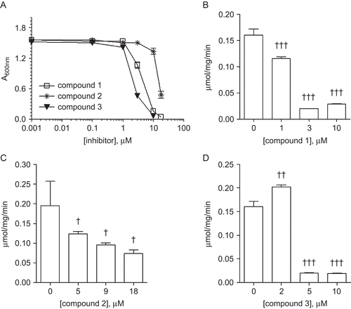
Table 2. Inhibition of the yeast plasma membrane H+-ATPase pump by ebselen (compound 1) and two of its analogs.
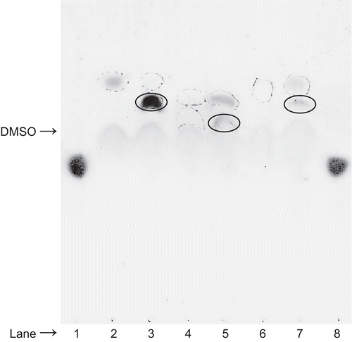
Figure 2. Thin layer chromatographic analysis of the products formed by reacting (30 mM) ebselen, (18 mM) compound 2, or (30 mM) compound 3 with (30 mM) l-cysteine. Lane 1, l-cysteine; lane 2, ebselen (dotted circle); lane 3, ebselen overspotted with l-cysteine; lane 4, compound 2 (dotted circle); lane 5, compound 2 overspotted with l-cysteine; lane 6, compound 3 (dotted circle); lane 7, compound 3 overspotted with l-cysteine; lane 8, l-cysteine. Note that lanes 3, 5, and 7 reveal that the l-cysteine spot has disappeared and that new spots (solid circles) with a slower migration rate than compounds 1, 2, and 3, respectively, are observed. l-cysteine was visualized with ninhydrin reagent followed by warming at 70°C in an oven.