827
Views
14
CrossRef citations to date
0
Altmetric
Research Article

Why acetylcholinesterase reactivators do not work in butyrylcholinesterase

, , , &
Pages 318-322 | Received 04 Mar 2009, Accepted 03 Jun 2009, Published online: 29 Oct 2009

Figures & data

Figure 1. AChE reactivators. Pralidoxime (2-PAM) as a representative of reactivators with one pyridinium ring and HI-6 as the most typical bisquaternary oxime.
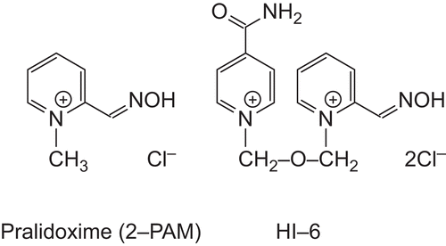
Figure 1.  AChE reactivators. Pralidoxime (2-PAM) as a representative of reactivators with one pyridinium ring and HI-6 as the most typical bisquaternary oxime.

Figure 2. Detail of 2GYU active site with the most important residues participating in reactivator binding displayed. The reactivator (HI-6) itself and the catalytic Ser203 are highlighted with bolder tubes.
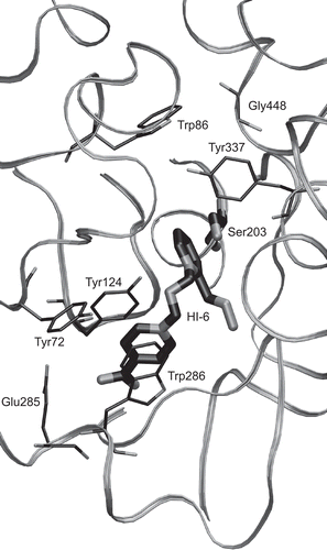
Figure 2.  Detail of 2GYU active site with the most important residues participating in reactivator binding displayed. The reactivator (HI-6) itself and the catalytic Ser203 are highlighted with bolder tubes.

Figure 3. Sequence alignment of BuChE (1P0I) and AChE (2GYU, chain A). The important residues influencing reactivator binding are marked by arrows. The alignment score is represented illustratively by symbols (star, colon, full stop, space) above the alignment rows. The far right column shows the number of the last residue in a row.
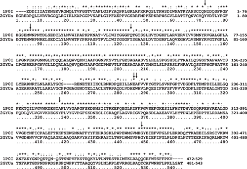
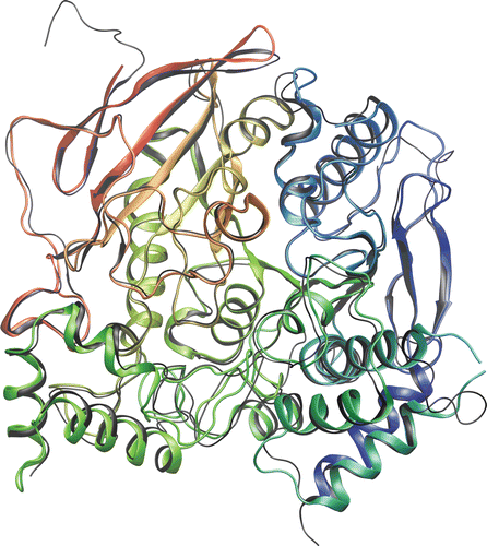
Figure 3.  Sequence alignment of BuChE (1P0I) and AChE (2GYU, chain A). The important residues influencing reactivator binding are marked by arrows. The alignment score is represented illustratively by symbols (star, colon, full stop, space) above the alignment rows. The far right column shows the number of the last residue in a row.
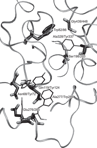
Figure 4. Aligned protein backbones of BuChE (1P0I) and AChE (2GYU) in rainbow colors and black, respectively.

Figure 4.  Aligned protein backbones of BuChE (1P0I) and AChE (2GYU) in rainbow colors and black, respectively.

Figure 5. 3-D alignment of BuChE (1P0I) and AChE (2GYU). Only the BuChE backbone is displayed and only residues influencing reactivator binding and catalytic Ser203 are shown. The relevant BuChE residues are highlighted by bolder tubes. Labels follow a pattern: BuChE residue/AChE residue.

Figure 5.  3-D alignment of BuChE (1P0I) and AChE (2GYU). Only the BuChE backbone is displayed and only residues influencing reactivator binding and catalytic Ser203 are shown. The relevant BuChE residues are highlighted by bolder tubes. Labels follow a pattern: BuChE residue/AChE residue.

Reprints and Corporate Permissions

Please note: Selecting permissions does not provide access to the full text of the article, please see our help page How do I view content?

To request a reprint or corporate permissions for this article, please click on the relevant link below:

Academic Permissions

Please note: Selecting permissions does not provide access to the full text of the article, please see our help page How do I view content?

Obtain permissions instantly via Rightslink by clicking on the button below:

If you are unable to obtain permissions via Rightslink, please complete and submit this Permissions form. For more information, please visit our Permissions help page.