Figures & data
Table 1. Physicochemical parameters and elemental details of the complexes.
Figure 1. Structure of the title complex [Cu(SPF)(A1)Cl].5H2O [Citation1].
![Figure 1. Structure of the title complex [Cu(SPF)(A1)Cl].5H2O [Citation1].](/cms/asset/fea1d112-3dae-48c6-b48c-5e58e9861e6d/ienz_a_487486_f0001_b.gif)
Table 2. Spectrophotometric titration data for the complexes.
Figure 2. Experimental cure from the spectrophotometric determination of metal content of 20.12 mg complex [Cu(SPF)(A1)Cl].5H2O dissolved in 20 ml of DD water by EDTA.
![Figure 2. Experimental cure from the spectrophotometric determination of metal content of 20.12 mg complex [Cu(SPF)(A1)Cl].5H2O dissolved in 20 ml of DD water by EDTA.](/cms/asset/78bfc75b-1b40-44e3-aa54-c75ed9209814/ienz_a_487486_f0002_b.gif)
Table 3. Characteristic absorptions bands of IR spectra of the complexes and drugs (cm−1).
Figure 3. The FAB-mass spectrum of [Cu(SPF)(A1)Cl].5H2O at an accelerating voltage of 10 kV.
![Figure 3. The FAB-mass spectrum of [Cu(SPF)(A1)Cl].5H2O at an accelerating voltage of 10 kV.](/cms/asset/4898c0a6-512b-4064-a63c-85f54ff7b8ae/ienz_a_487486_f0003_b.gif)
Figure 4. Proposed fragmentation pattern for [Cu(SPF)(A1)Cl].5H2O complex.
![Figure 4. Proposed fragmentation pattern for [Cu(SPF)(A1)Cl].5H2O complex.](/cms/asset/46ff3faf-9db3-4f72-bcb2-d03c9983a91b/ienz_a_487486_f0004_b.gif)
Table 4. MIC in terms of µM.
Table 5. The binding constants (Kb) and IC50 values of the complexes.
Figure 5. Electronic absorption spectra of [Cu(SPF)(A1)Cl].5H2O in phosphate buffer (Na2HPO4/NaH2PO4, pH 7.2) in the absence and presence of increasing amount of DNA. The [Cu] complex = 10 μM; [DNA] = 0–150 μM. The incubation period is 30 min at 37°C. Inset: Plot of [DNA]/(ϵa– ϵf) vs. [DNA]. Arrow shows the absorbance change upon increasing DNA concentrations.
![Figure 5. Electronic absorption spectra of [Cu(SPF)(A1)Cl].5H2O in phosphate buffer (Na2HPO4/NaH2PO4, pH 7.2) in the absence and presence of increasing amount of DNA. The [Cu] complex = 10 μM; [DNA] = 0–150 μM. The incubation period is 30 min at 37°C. Inset: Plot of [DNA]/(ϵa– ϵf) vs. [DNA]. Arrow shows the absorbance change upon increasing DNA concentrations.](/cms/asset/dca28004-5df4-42bc-970d-f7399cc258f8/ienz_a_487486_f0005_b.gif)
Figure 6. Effect on relative viscosity of DNA under the influence of increasing amount of complexes at 27°C ± 0.1°C in phosphate buffer (Na2HPO4 /NaH2PO4, pH 7.2).
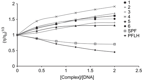
Table 6. Complex mediated DNA cleavage data by gel electrophoresis.
Figure 7. Photogenic view of cleavage of pUC19 DNA (300 µg/mL) with series of copper(II) complexes (200 µM) using 1% agarose gel containing 0.5 µg/mL ethidium bromide. All reactions were incubated in TE buffer (pH 8) in a final volume of 15 µL, for 24 h. at 37°C. Lane 1, DNA control; Lane 2, CuCl2·2H2O; Lane 3, Sparfloxacin; Lane 4, Pefloxacin; Lane 5, [Cu(SPF)(A1)Cl].5H2O; Lane 6, [Cu(SPF)(A2)Cl].5H2O; Lane 7, [Cu(SPF)(A3)Cl].5H2O; Lane 8, [Cu(PFL)(A1)Cl].5H2O; Lane 9, [Cu(PFL)(A2)Cl].5H2O; Lane 10, [Cu(PFL)(A3)Cl].5H2O.
![Figure 7. Photogenic view of cleavage of pUC19 DNA (300 µg/mL) with series of copper(II) complexes (200 µM) using 1% agarose gel containing 0.5 µg/mL ethidium bromide. All reactions were incubated in TE buffer (pH 8) in a final volume of 15 µL, for 24 h. at 37°C. Lane 1, DNA control; Lane 2, CuCl2·2H2O; Lane 3, Sparfloxacin; Lane 4, Pefloxacin; Lane 5, [Cu(SPF)(A1)Cl].5H2O; Lane 6, [Cu(SPF)(A2)Cl].5H2O; Lane 7, [Cu(SPF)(A3)Cl].5H2O; Lane 8, [Cu(PFL)(A1)Cl].5H2O; Lane 9, [Cu(PFL)(A2)Cl].5H2O; Lane 10, [Cu(PFL)(A3)Cl].5H2O.](/cms/asset/139bf8fc-3fd3-487c-88b8-3bda89577d47/ienz_a_487486_f0007_b.gif)
Figure 8. Absorbance values (Abs560) as a function of time (t) plotted for varying concentration of complex 1 from 0.25 µM to 3 µM for produced a good straight line are observed.
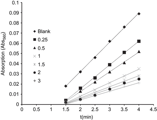
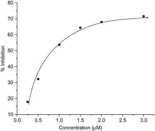
Figure 9. Plot of percentage of inhibiting NBT reduction with an increase in the concentration of complex 1.