1,595
Views
25
CrossRef citations to date
0
Altmetric
Research Article

Synthesis and biological screening of some pyridine derivatives as anti-malarial agents

, , , &
Pages 69-77 | Received 23 Jun 2010, Accepted 22 Mar 2011, Published online: 25 May 2011

Figures & data

Figure 1.  Chemical structures of pyridine derivatives having anti-malarial activity.

Figure 1.  Chemical structures of pyridine derivatives having anti-malarial activity.

Table 1.  Physical and analytical data of compounds 2ah, 4ae and 5.

Table 2.  Spectral data of compounds 2ah, 4ae and 5.

Scheme 1.  Synthesis of hydrazone derivatives.

Scheme 1.  Synthesis of hydrazone derivatives.

Scheme 2.  Synthesis of pyridone derivatives.

Scheme 2.  Synthesis of pyridone derivatives.

Table 3.  In vivo anti-malarial activity of compounds 2ah and 4ae.

Table 4.  In vitro anti-plasmodial activity against chloroquine-resistant (RKL9) strain of Plasmodium falciparum.

Figure 2.  3D view from a molecular modelling study, of the minimum-energy structure of the complex of WR99210 docked in DHFRE (PDB ID: 1J3I). Viewed using Molecular Operating Environment (MOE) module.
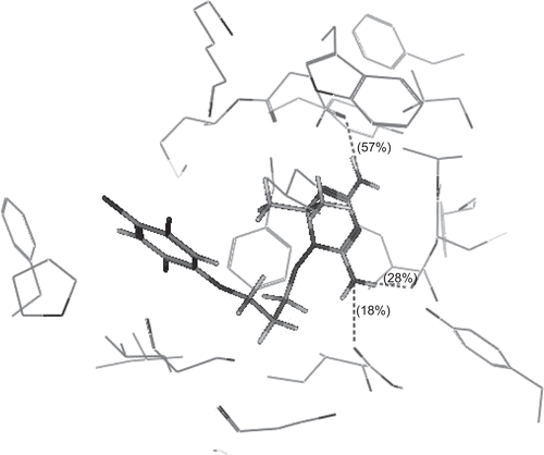
Figure 2.  3D view from a molecular modelling study, of the minimum-energy structure of the complex of WR99210 docked in DHFRE (PDB ID: 1J3I). Viewed using Molecular Operating Environment (MOE) module.

Table 5.  Calculated docking hydrogen bonding results for compounds WR99210, 2a, 2g and 2h.

Figure 3.  3D view from a molecular modelling study, of the minimum-energy structure of the complex of 2a docked in DHFRE (PDB ID: 1J3I). Viewed using Molecular Operating Environment (MOE) module.
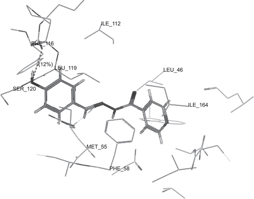
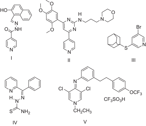
Figure 3.  3D view from a molecular modelling study, of the minimum-energy structure of the complex of 2a docked in DHFRE (PDB ID: 1J3I). Viewed using Molecular Operating Environment (MOE) module.
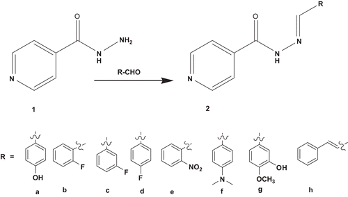
Figure 4.  3D view from a molecular modelling study, of the minimum-energy structure of the complex of 2g docked in DHFRE (PDB ID: 1J3I). Viewed using Molecular Operating Environment (MOE) module.
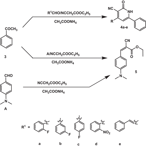
Figure 4.  3D view from a molecular modelling study, of the minimum-energy structure of the complex of 2g docked in DHFRE (PDB ID: 1J3I). Viewed using Molecular Operating Environment (MOE) module.
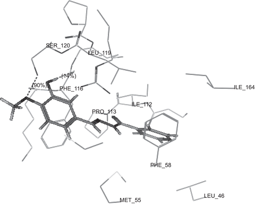
Figure 5.  3D view from a molecular modelling study, of the minimum-energy structure of the complex of 2h docked in DHFRE (PDB ID: 1J3I). Viewed using Molecular Operating Environment (MOE) module.
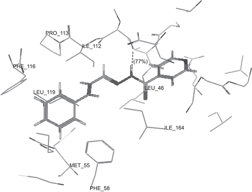
Figure 5.  3D view from a molecular modelling study, of the minimum-energy structure of the complex of 2h docked in DHFRE (PDB ID: 1J3I). Viewed using Molecular Operating Environment (MOE) module.

Reprints and Corporate Permissions

Please note: Selecting permissions does not provide access to the full text of the article, please see our help page How do I view content?

To request a reprint or corporate permissions for this article, please click on the relevant link below:

Academic Permissions

Please note: Selecting permissions does not provide access to the full text of the article, please see our help page How do I view content?

Obtain permissions instantly via Rightslink by clicking on the button below:

If you are unable to obtain permissions via Rightslink, please complete and submit this Permissions form. For more information, please visit our Permissions help page.