Figures & data
Figure 1. Carbazole derivatives (1a–g).
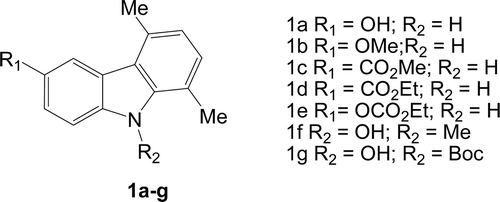
Figure 2. Effect of carbazole derivatives (1a–e) against MCF-7 and ISK cell proliferation. MCF-7 (A) and ISK (B) cells were treated for 96 or 24 h, respectively, after 24-h starvation, with the indicated concentrations of substances 1a, 1b, 1c, 1d and 1e. Cells proliferation was evaluated by MTT assay. Statistically significant differences are indicated. Histograms; mean of three independent experiments each performed with triplicate samples expressed as percent of basal; bars, SE (*P < 0.01 compared with basal).
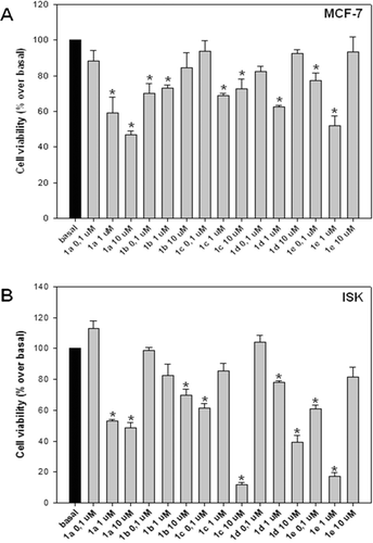
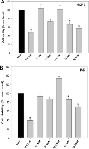
Figure 3. Effect of carbazole derivatives (1f–g) against MCF-7 and ISK cell proliferation. MCF-7 (A) and ISK (B) cells were treated for 96 or 24 h, respectively, after 24-h starvation, with the indicated concentrations of substances 1f and 1g. Cells proliferation was evaluated by MTT assay. Statistically significant differences are indicated. Histograms; mean of three independent experiments each performed with triplicate samples expressed as percent of basal; bars, SE (*P < 0.01 compared with basal).