760
Views
1
CrossRef citations to date
0
Altmetric
Research Article

Analysis of enzyme activity regulation by non-denaturing electrophoresis and application of this regulation for enzyme reactor production

&
Pages 894-899 | Received 06 Apr 2012, Accepted 10 May 2012, Published online: 18 Jul 2012

Figures & data

Table 1.  Quantitative data of LDH and esterase activities within non-denaturing IEF, non-denaturing stacking gel containing inhibitors (oxamate for LDH activity and acrinol for esterase activity) and non-denaturing separation gel after the separation of the cytosolic proteins in the mouse liver by non-denaturing IEF.

Figure 1.  LDH activities in the non-denaturing stacking gel containing 0.2% oxamate (a) and in the non-denaturing separation gel (b) after the separation of the cytosolic proteins in the mouse liver by non-denaturing IEF. LDH activity spots are indicated by arrows.
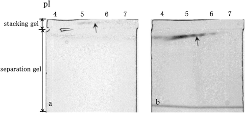
Figure 1.  LDH activities in the non-denaturing stacking gel containing 0.2% oxamate (a) and in the non-denaturing separation gel (b) after the separation of the cytosolic proteins in the mouse liver by non-denaturing IEF. LDH activity spots are indicated by arrows.

Figure 2.  Esterase activities in the non-denaturing stacking gel containing 0.1 M Tris–HCl (pH 6.8) (a), in the non-denaturing stacking gel containing 0.1 mM acrinol and 0.1 M acetate buffer (pH 4.8) (b) and in the non-denaturing separation gel (c) after the separation of cytosolic proteins in the mouse liver by non-denaturing IEF. Esterase activity spots are indicated by arrowheads.
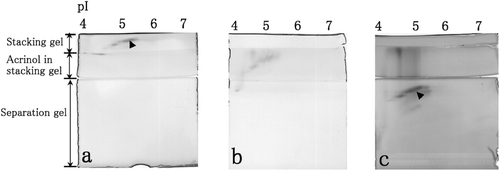
Figure 2.  Esterase activities in the non-denaturing stacking gel containing 0.1 M Tris–HCl (pH 6.8) (a), in the non-denaturing stacking gel containing 0.1 mM acrinol and 0.1 M acetate buffer (pH 4.8) (b) and in the non-denaturing separation gel (c) after the separation of cytosolic proteins in the mouse liver by non-denaturing IEF. Esterase activity spots are indicated by arrowheads.

Figure 3.  After separation by non-denaturing 2-DE and electroblotting onto membrane, LDH activities staining when the membrane was treated by 0.2% oxamate (a), and washed by Tris–HCl solution (b). After separation by non-denaturing 2-DE and electroblotting onto membrane, esterase activity staining when the membrane was treated by 0.1 mM acrinol (c), and washed by aspartic acid solution (d).
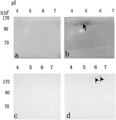
Figure 3.  After separation by non-denaturing 2-DE and electroblotting onto membrane, LDH activities staining when the membrane was treated by 0.2% oxamate (a), and washed by Tris–HCl solution (b). After separation by non-denaturing 2-DE and electroblotting onto membrane, esterase activity staining when the membrane was treated by 0.1 mM acrinol (c), and washed by aspartic acid solution (d).

Figure 4.  MALDI-TOF MS spectra of phosphatidylcholine and lysophosphatidylcholine when phosphatidylcholine was incubated with the esterase on membrane (arrow in 3d) treated with 0.1 mM acrinol (a), and washed by aspartic acid solution (b) after separation by non-denaturing 2-DE and electroblotting onto membrane. The relative amounts of lysophosphatidylcholine (m/z = 496.7) (c) when pure phosphatidylcholine was applied to esterase on membrane treated with 0.1 mM acrinol (acrinol in c), and washed by aspartic acid solution (acrinol release in c) after separation by non-denaturing 2-DE and electroblotting onto membrane. Each datum indicates mean standard error obtained from 15 individual measurements (number of measurements in c).
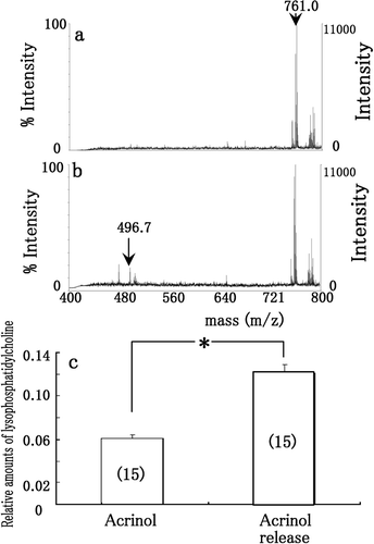
Figure 4.  MALDI-TOF MS spectra of phosphatidylcholine and lysophosphatidylcholine when phosphatidylcholine was incubated with the esterase on membrane (arrow in 3d) treated with 0.1 mM acrinol (a), and washed by aspartic acid solution (b) after separation by non-denaturing 2-DE and electroblotting onto membrane. The relative amounts of lysophosphatidylcholine (m/z = 496.7) (c) when pure phosphatidylcholine was applied to esterase on membrane treated with 0.1 mM acrinol (acrinol in c), and washed by aspartic acid solution (acrinol release in c) after separation by non-denaturing 2-DE and electroblotting onto membrane. Each datum indicates mean standard error obtained from 15 individual measurements (number of measurements in c).

Reprints and Corporate Permissions

Please note: Selecting permissions does not provide access to the full text of the article, please see our help page How do I view content?

To request a reprint or corporate permissions for this article, please click on the relevant link below:

Academic Permissions

Please note: Selecting permissions does not provide access to the full text of the article, please see our help page How do I view content?

Obtain permissions instantly via Rightslink by clicking on the button below:

If you are unable to obtain permissions via Rightslink, please complete and submit this Permissions form. For more information, please visit our Permissions help page.