773
Views
9
CrossRef citations to date
0
Altmetric
Research Article

(−)-Nyasol, isolated from Anemarrhena asphodeloides suppresses neuroinflammatory response through the inhibition of I-κBα degradation in LPS-stimulated BV-2 microglial cells

, , , , &
Pages 954-959 | Received 27 Mar 2012, Accepted 21 Mar 2012, Published online: 18 Jul 2012

Figures & data

Figure 1.  (A) Chemical structure of (−)-nyasol. (B) Effects of (−)-nyasol on lipopolysaccharide (LPS)-induced inflammatory cytokines in BV-2 microglial cells. The cells were stimulated with LPS in absence or presence of (−)-nyasol for 6 h. The levels of IL-1β and TNF-α mRNAs were determined by RT-PCR analysis. GAPDH was used as an internal control. The results shown are the representative of three independent experiments. (C) Effects of (−)-nyasol on LPS-induced nitrite production in BV-2 microglial cells. The amount of nitrite in culture medium was measured by using the Griess reagents, as described in Materials and methods. (D) Effects of (−)-nyasol on LPS-induced PGE2 production in BV-2 microglial cells. PGE2 concentrations were quantified in the culture supernatant by enzyme immunoassay (EIA). The values are expressed as the means ± SD of three individual experiments. *p < 0.01 and **p < 0.001 indicate significant differences from the LPS alone.
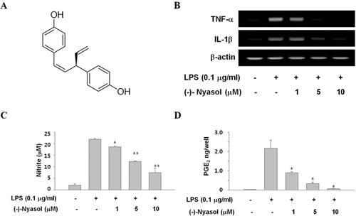
Figure 1.  (A) Chemical structure of (−)-nyasol. (B) Effects of (−)-nyasol on lipopolysaccharide (LPS)-induced inflammatory cytokines in BV-2 microglial cells. The cells were stimulated with LPS in absence or presence of (−)-nyasol for 6 h. The levels of IL-1β and TNF-α mRNAs were determined by RT-PCR analysis. GAPDH was used as an internal control. The results shown are the representative of three independent experiments. (C) Effects of (−)-nyasol on LPS-induced nitrite production in BV-2 microglial cells. The amount of nitrite in culture medium was measured by using the Griess reagents, as described in Materials and methods. (D) Effects of (−)-nyasol on LPS-induced PGE2 production in BV-2 microglial cells. PGE2 concentrations were quantified in the culture supernatant by enzyme immunoassay (EIA). The values are expressed as the means ± SD of three individual experiments. *p < 0.01 and **p < 0.001 indicate significant differences from the LPS alone.

Figure 2.  (A) The effects of (−)-nyasol on LPS-induced iNOS and COX-2 mRNA levels in BV-2 microglial cells. The mRNA expressions of iNOS and COX-2 were examined by RT-PCR. Cells were treated with lipopolysaccharide (LPS) (0.1 µg/mL) in the presence or absence of (−)-nyasol for 6 h. (B) The iNOS and COX-2 protein levels were determined by Western blot analysis. Cells were treated with LPS (0.1 µg/mL) in the presence or absence of (−)-nyasol for for 20 h. The results shown are the representative of three independent experiments.
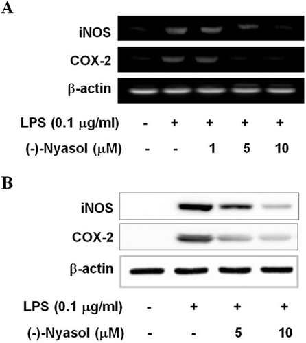
Figure 2.  (A) The effects of (−)-nyasol on LPS-induced iNOS and COX-2 mRNA levels in BV-2 microglial cells. The mRNA expressions of iNOS and COX-2 were examined by RT-PCR. Cells were treated with lipopolysaccharide (LPS) (0.1 µg/mL) in the presence or absence of (−)-nyasol for 6 h. (B) The iNOS and COX-2 protein levels were determined by Western blot analysis. Cells were treated with LPS (0.1 µg/mL) in the presence or absence of (−)-nyasol for for 20 h. The results shown are the representative of three independent experiments.

Figure 3.  Effect of (−)-nyasol on lipopolysaccharide (LPS)-induced I-κBα degradation and p65 translocation to the nucleus in BV-2 microglial cells. Cells were pretreated with (−)-nyasol for 30 min prior to stimulation of LPS. After treatment with LPS for an additional 15 min, cytosolic I-κBα and nuclear p65 were analyzed by Western blot. Images are the representative of three independent experiments that shows similar results.
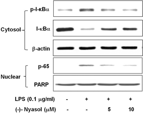
Figure 3.  Effect of (−)-nyasol on lipopolysaccharide (LPS)-induced I-κBα degradation and p65 translocation to the nucleus in BV-2 microglial cells. Cells were pretreated with (−)-nyasol for 30 min prior to stimulation of LPS. After treatment with LPS for an additional 15 min, cytosolic I-κBα and nuclear p65 were analyzed by Western blot. Images are the representative of three independent experiments that shows similar results.

Figure 4.  Effect of (−)-nyasol on lipopolysaccharide (LPS)-induced activation of mitogen-activated protein kinases (MAPKs) in BV-2 microglial cells. Cells were pretreated with (−)-nyasol for 30 min prior to stimulation of LPS. After treatment with LPS for an additional 15 min, proteins were extracted and analyzed for the levels of phosphorylated extracellular signal-regulated protein kinase (ERK), p38 and c-Jun N-terminal kinases (JNK) by Western blot. The results shown are the representative of three independent experiments.
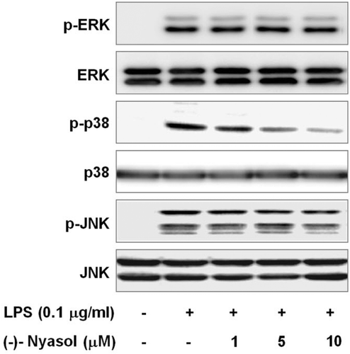
Figure 4.  Effect of (−)-nyasol on lipopolysaccharide (LPS)-induced activation of mitogen-activated protein kinases (MAPKs) in BV-2 microglial cells. Cells were pretreated with (−)-nyasol for 30 min prior to stimulation of LPS. After treatment with LPS for an additional 15 min, proteins were extracted and analyzed for the levels of phosphorylated extracellular signal-regulated protein kinase (ERK), p38 and c-Jun N-terminal kinases (JNK) by Western blot. The results shown are the representative of three independent experiments.

Reprints and Corporate Permissions

Please note: Selecting permissions does not provide access to the full text of the article, please see our help page How do I view content?

To request a reprint or corporate permissions for this article, please click on the relevant link below:

Academic Permissions

Please note: Selecting permissions does not provide access to the full text of the article, please see our help page How do I view content?

Obtain permissions instantly via Rightslink by clicking on the button below:

If you are unable to obtain permissions via Rightslink, please complete and submit this Permissions form. For more information, please visit our Permissions help page.