Figures & data
Table I. Comparison of several imaging modalities (Cheon and Lee Citation2008, Hachani et al. Citation2011, Massoud and Gambhir Citation2003).
Figure 1. An MRI (gradient echo, TR/TE = 100/4 ms, flip angle 30°, FOV = 5.5 × 2.5 cm, 256 × 256) of a mouse liver obtained at 9.4 T (A) before and (B) after injection of iron oxide (Nano-Ocean, Springdale, AR). The decrease of MR signal within the liver (yellow arrow) is visible (Blasiak et al. Citation2013).
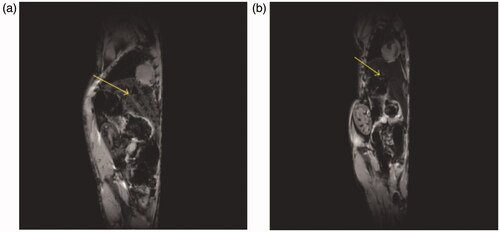
Figure 2. Effect of magnetic particle internalization in cells on relaxation times: (A) the protons in cells tagged by magnetic particles have a shorter
relaxation time than those in (B) untagged cells (Pankhurst et al. Citation2003, Yang et al. Citation2010).
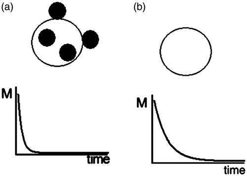
Table II. Comparison of some nanoparticles with high transverse relaxivity for T2 MRI (Benyettou et al. Citation2011, Hachani et al. Citation2011, Huang et al. Citation2011, Jung and Jacobs Citation1995, Ruiz et al. Citation2006).
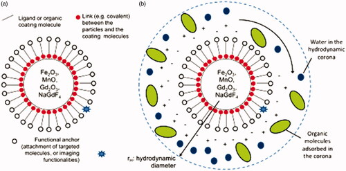
Figure 3. Schematic representations of (A) the structure of functional magnetic nanoparticles (MNPs) for MRI applications and (B) a colloidal nanoparticle suspended in biological media.