Figures & data
Figure 1. The mechanisms of acquired resistance of EGFR-TKIs. The secondary T790M mutation of EGFR leads to decrease the affinity to EGFR-TKIs. Irreversible TKIs bind with high affinity to receptors carrying the T790M mutation. MET or IGF activation induces activation of PI3K/Akt pathway independent of EGFR activation. In these cases MET-specific inhibitor or HGF-inhibitor, inhibition of parallel pathway is a feasible strategy.
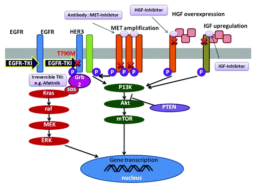
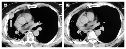
Figure 2. Tumor volume changes seen on CT (A) before the first erlotinib treatment; (B) 2 mo after the first erlotinib treatment.
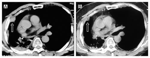
Figure 3. Tumor volume changes seen on CT (A) before the gemcitabine treatment; (B) 2 cycles after gemcitabine retreatment.
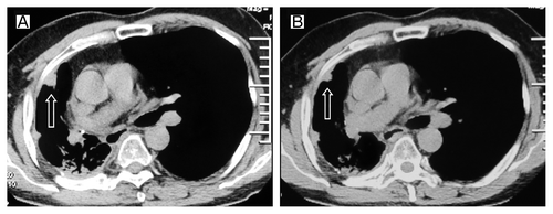
Figure 4. Tumor volume changes seen on CT (A) before the second erlotinib treatment; (B) 3 mo after the second erlotinib treatment.
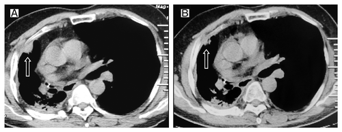
Figure 5. Tumor volume changes seen on CT (A) before the third erlotinib treatment; (B) 2 mo after the third time erlotinib treatment.