Figures & data
Table 1. Modules of aggressive behavior in male and female Drosophila
Figure 1. Different set-ups to study aggression in Drosophila. (A) Arena with a centrally placed food cup that can contain yeast paste or a virgin female, allowing the videotaping of aggressive behavior.Citation68 (B) Set-up allowing the simultaneous recording of 35 arenas, usually during a 15 min period.Citation72 (C) Scheme of a set-up compatible with CADABRA software allowing automated scoring of lunging, tussling, wing threats and chasing of two pairs of flies simultaneously. This set-up can be expanded to analyze eight pairs of flies simultaneously.Citation67 (D) Set-up allowing the instant analysis of the aggressive encounters between groups of eight flies over a 2 min period.Citation6,Citation12,Citation73
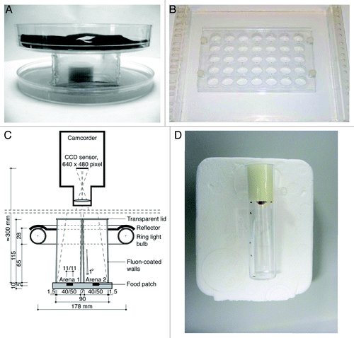
Figure 2. (A–C) Schematic representation of adult central brain cells reported to be serotonergic, dopaminergic or octopaminergic. Cholinergic cells were reported to be widespread throughout the entire brain making it difficult to present a schematic overview.Citation140-Citation143 PB, protocerebral bridge; FB, fan-shaped body; EB, ellipsoid body; NO, noduli; MB, mushroom bodies; P, mushroom body peduncle; CA, mushroom body calyx; AL, antennal lobe; SOG, subesophageal ganglion; LC, lobula complex; ME, medulla; LA, lamina. (A) Serotonergic cells. LLP1, posterior lateral protocerebrum; LP2a, between the medulla and the lateral protocerebrum; LP2b, between the medulla and the lateral protocerebrum; DP, dorsal protocerebrum; SP1, posterior to superior median protocerebrum; SP2, posterior median protocerebrum; IP, posterior inferior median protocerebrum; PLP, posterior lateral protocerebrum; SLP, superior lateral protocerebrum; AMP, anterior median protocerebrum; ALP, anterior lateral protocerebrum; AVP, anterior ventral protocerebrum; PAL, posterior to antennal lobe; DLS, dorsal lateral subesophageal ganglion; AS, anterior subesophageal ganglion; SE1, lateral subesophageal ganglion; SE2, anterior lateral subesophageal ganglion; SE3, most ventral subesophageal ganglion.Citation144-Citation146 (B) Dopaminergic cells. PAM, dorsomedial anterior protocerebral; PAL, dorsolateral anterior protocerebral; PPM, dorsomedial posterior protocerebral; PPL1, dorsolateral posterior protocerebral; PPL2, lateral posterior protocerebral; PPD, protocerebral posterial dorsal; T1, tritocerebrum; VUM, ventral unpaired medial neurons.Citation76,Citation147,Citation148 (C) Octopaminergic cells (nomenclature according to Sinakevitch and Strausfeld, 2006; nomenclature between brackets according to Monastirioti et al., 1995). Cluster G0a, Cluster G0b (LP, lateral protocerebrum cell), Cluster G1 (DMC, dorsal medial cluster), Cluster G2a (~DAC, dorsal anterior cluster?), Cluster G2b (~DAC, dorsal anterior cluster?), Cluster G3a (AL, antennal lobe cluster), Cluster G3b, Cluster G4a (DPC, dorsal posterior cluster), Cluster G4b (DPC, dorsal posterior cluster), Cluster G5a, Cluster G5b,c, Cluster G6, VUM, Ventral unpaired median neurons 1–3 (SM, subesophageal medial). (D-F) Diagrams representing the biosynthetic pathways of acetylcholine and the monoaminergic neurotransmitters: serotonin, dopamine and octopamine. (D) Serotonin synthesis. TPH, Tryptophan hydroxylase; DDC, DOPA decarboxylase. (E) Dopamine/octopamine synthesis. TH, Tyrosine hydroxylase; TDC, Tyrosine decarboxylase; DDC, DOPA decarboxylase; TβH, Dopamine β hydroxylase (F) Acetylcholine synthesis. ChAT, Choline acetyltransferase
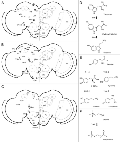
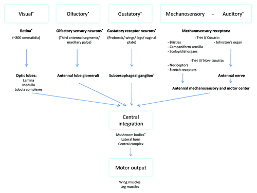
Figure 3. Scheme representing the flow of information provided by external stimuli to integration centers in the central brain. This central integration results in interpretation and integration of the different inputs and the generation of an appropriate response. The role of stimuli or neuronal structures marked with an asterisk (*) in aggression has been more closely analyzed. Visual information is received by the retinal ommatidia and travels through the different layers of the optic lobes, lamina, medulla and lobula complexes, to the central brain.Citation149 The described higher integration centers of this visual information include the mushroom bodies, the central complex and different neurons in the lateral protocerebrum.Citation97-Citation99,Citation150 Olfactory information is sensed by olfactory neurons expressing olfactory receptors located on the third antennal segments and the maxillary palps. Odorant cues travel via these neurons to the glomeruli in the antennal lobe from where projection neurons send this information to higher integration centers, including the mushroom bodies and the lateral horn.Citation93,Citation151-Citation155 Gustatory signals are sensed by gustatory receptors expressing gustatory receptors located on the proboscis, wings, legs and vaginal plate. These neurons all project to the SOG. The SOG has been shown to project toward multiple other brain regions including the antennal lobe, the lateral horn and the mushroom bodies.Citation96,Citation155,Citation156 Mechanosensory information is sensed by a variety of receptors, located all over the body, which can be subdivided into a ciliated and a non-ciliated group. Non-ciliated mechanosensory receptors include nociceptors and muscle and visceral stretch receptors. Ciliated mechanosensory receptors include: bristles responsible for touch perception, campaniform sensilla on wings and haltere providing info on flight parameters and chordotonal organs including scolopidial organs providing proprioceptive and gravireceptive information.Citation157,Citation158 The fly's largest chordotonal organ, Johnston’s organ, is located in the second antennal segment and represents the flies ear. Part of these neurons have been shown to innervate the antennal mechanosensory and motor center of the brain, a neuropil lateral to the SOG and the antennal lobes, further higher integration is mainly unknown.Citation100,Citation157,Citation158