Figures & data
Figure 1. Barrier sites in the CNS. The CNS contains three main barrier sites: (1) The blood-brain barrier which is formed by specialized brain capillary endothelial cells, (2) the barrier between the blood and the cerebrospinal fluid that exists at the choroid plexus epithelial cells and (3) the arachnoid epithelium presenting the middle layer of the meninges. While the endothelial cells of the BBB restrict the migration of potentially harmful blood-born agents to the central-nervous tissue, the choroid plexus epithelium and the arachnoid epithelium protect the cerebrospinal fluid. Tight junctions between endothelial and epithelial cells seal the intercellular spaces and minimize paracellular pathways.
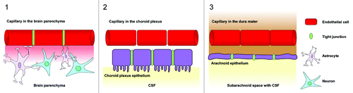
Figure 2. Components of the blood-brain barrier. The blood-brain barrier consists of specialized capillary endothelial cells that are lined by the basal lamina, astrocytic endfeet, pericytes and microglial cells. (A) Among several other transporters and receptors brain endothelial cells express excitatory amino acid transporters (EAAT1–3), glucose transporter 1 (GLUT1), L-system for large neutral amino acids (LAT1) and P-glycoprotein (Pgp). (B) Surrounding cells intensely interact with endothelial cells and release soluble agents in order to support the maintenance of BBB functions [5-HT (5-hydroxytryptamine [serotonin]), angiopoetin 1 (ANG1), basic fibroblast growth factor (bFGF), endothelin 1 (ET1), glial cell line-derived neurotrophic factor (GDNF), leukemia inhibitory factor (LIF), purinergic receptor (P2Y2), transforming growth factor-β, endothelium-specific receptor tyrosine kinase 2 (TIE2)] (from ref. Citation42, with permission).
![Figure 2. Components of the blood-brain barrier. The blood-brain barrier consists of specialized capillary endothelial cells that are lined by the basal lamina, astrocytic endfeet, pericytes and microglial cells. (A) Among several other transporters and receptors brain endothelial cells express excitatory amino acid transporters (EAAT1–3), glucose transporter 1 (GLUT1), L-system for large neutral amino acids (LAT1) and P-glycoprotein (Pgp). (B) Surrounding cells intensely interact with endothelial cells and release soluble agents in order to support the maintenance of BBB functions [5-HT (5-hydroxytryptamine [serotonin]), angiopoetin 1 (ANG1), basic fibroblast growth factor (bFGF), endothelin 1 (ET1), glial cell line-derived neurotrophic factor (GDNF), leukemia inhibitory factor (LIF), purinergic receptor (P2Y2), transforming growth factor-β, endothelium-specific receptor tyrosine kinase 2 (TIE2)] (from ref. Citation42, with permission).](/cms/asset/f2ae0426-9cc5-4211-800c-258276697c43/kvir_a_10919004_f0002.gif)
Figure 3. Assembly of endothelial tight junctions. Transmembranous molecules like claudins, occludin, junctional adhesion molecules (JAMs) and endothelial selective adhesion molecule (ESAM) are important tight junction components. On the cytoplasmic site these proteins are connected to adaptor and regulatory/signaling proteins [zonula occludens-1, -2 and -3 (ZO-1–3), cingulin, junction-associated coiled-coil protein (JACOP), the partitioning defective proteins 3 and 6 (PAR3/6), Ca2+-dependent serine protein kinase (CASK), tight junction-associated protein 7H6, Itch (E3 ubiquitin protein ligase), regulator of G-protein signaling 5 (RGS5), afadin (AF6), multi-PDZ-protein 1 (MUPP1), MAGI (membrane-associated guanylate kinase with inverted orientation of protein-protein interaction domains), ZO-1-associated nucleic acid-binding protein (ZONAB)], which link the membranous components to the actin/vinculin-based cytoskeleton. Vascular endothelial cadherin (VE-cadherin) and the platelet-endothelial cell adhesion molecule (PECAM) are components of endothelial adherens junctions and interact via homophilic bindings. Catenins, desmoplakin and p120 catenin (p120ctn) connect the adherence junction proteins with the cytoskeleton (modified from ref. Citation55, with permission).
![Figure 3. Assembly of endothelial tight junctions. Transmembranous molecules like claudins, occludin, junctional adhesion molecules (JAMs) and endothelial selective adhesion molecule (ESAM) are important tight junction components. On the cytoplasmic site these proteins are connected to adaptor and regulatory/signaling proteins [zonula occludens-1, -2 and -3 (ZO-1–3), cingulin, junction-associated coiled-coil protein (JACOP), the partitioning defective proteins 3 and 6 (PAR3/6), Ca2+-dependent serine protein kinase (CASK), tight junction-associated protein 7H6, Itch (E3 ubiquitin protein ligase), regulator of G-protein signaling 5 (RGS5), afadin (AF6), multi-PDZ-protein 1 (MUPP1), MAGI (membrane-associated guanylate kinase with inverted orientation of protein-protein interaction domains), ZO-1-associated nucleic acid-binding protein (ZONAB)], which link the membranous components to the actin/vinculin-based cytoskeleton. Vascular endothelial cadherin (VE-cadherin) and the platelet-endothelial cell adhesion molecule (PECAM) are components of endothelial adherens junctions and interact via homophilic bindings. Catenins, desmoplakin and p120 catenin (p120ctn) connect the adherence junction proteins with the cytoskeleton (modified from ref. Citation55, with permission).](/cms/asset/bed4efa0-b4ee-4b8a-9408-b773aaa0d8d4/kvir_a_10919004_f0003.gif)