Figures & data
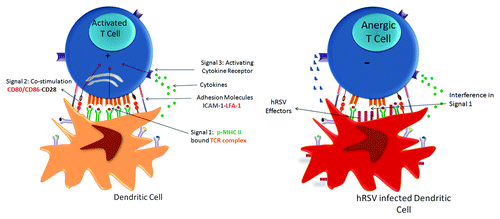
Figure 1. Schematic representation of the hRSV genome indicating the known function for each encoded protein. The figure shows the order of the 10 genes of hRSV and the known function of the 11 encoded proteins.
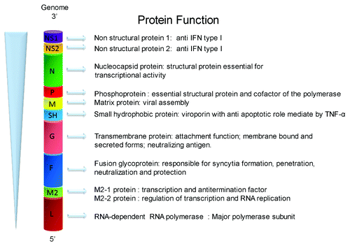
Table 1. HRSV interaction with host innate and adaptive immune response
Figure 2. HRSV infection blockade of IS assembly between infected DCs and T cells as a novel RSV virulence mechanism. A novel mechanism to avoid the host immune response elicit by hRSV is the impairment of immunological synapse (IS) assembly between hRSV-infected DCs and naïve T cells that impairs the T cells activation. During IS assembly DCs provide three different signals to promote the T-cell activation: Signal 1 (antigenic presentation in p-MHC), signal 2 (Co-stimulation) and signal 3 (cytokines) represented in the left part of cartoon. In hRSV infected DCs, as show the right part of cartoon, hRSV effectors are expressed in the surface membrane and interfering with the molecular interaction between the T-cell receptor (TCR) and antigenic pMHCs impairing the T-cell activation. This phenomenon is evidenced by a reduction in the Golgi apparatus polarization toward the DC and decreases in IL-2 secretion in T cells.