
Abstract
Background
Patients with head and neck squamous cell carcinoma of unknown primary (HNCUP) are often treated with extensive radiotherapy (RT). Frequently, the bilateral nodal clinical target volume (nCTV) and the volumes of suspected mucosal primary sites (mCTV) of the pharynx and larynx is irradiated. This treatment is effective but toxic. New data suggest that omission of the contralateral nCTV and mCTV, results in few recurrences. The present study explores photon versus proton therapy, in the primary and recurrent setting.
Material and methods
An analysis of twelve patients previously treated for HNCUP was performed. A fictitious recurrence was defined in patients treated for unilateral disease. Independently a volumetric arc photon plan and an intensity-modulated proton plan was made for all cases and scenarios.
Results
Compared to the standard bilateral treatment this study shows that limiting the target to unilateral nCTV leads to a significant decrease in dysphagia of 18% and 17% and xerostomia of 4.0% and 5% for photon and protons, respectively. Comparing photon RT directly to proton RT shows a small and often insignificant gain, using protons for both bilateral and unilateral targets. Focusing on re-irradiation, benefits from using protons in both the primary setting and at re-irradiation were limited. However, using protons for re-irradiation only leads to a decrease in the tissue volume receiving a specific dose outside the target overlapping region, e.g., V90Gymean was 31, 25, and 22 cm3 for photons-photons, photons-protons, and protons-protons, respectively. For V100Gy of the ipsilateral carotid artery, no differences were observed.
Conclusion
Omitting contralateral nCTV irradiation and mCTV irradiation will significantly reduce toxicity. The accumulated high dose volumes can be minimised using protons for re-irradiation. However, the use of protons for primary treatment provides limited benefit in most patients.
Background
Squamous cell carcinoma of the head and neck often gives rise to nodal metastasis in the neck, and this is the presenting symptom in many patients [Citation1,Citation2]. However, in some cases, the primary tumour is never located despite thorough investigations, including imaging, clinical examination in general anaesthesia and often tonsillectomy and base of tongue biopsies [Citation3]. The latter is increasingly being performed as the base of tongue mucosectomy, using transoral robotic surgery (TORS), further adding to the probability of identifying a primary tumour, decreasing the incidence of HNCUP [Citation4] and it might also change the character of HNCUP. When the primary tumour remains unidentified the patient is diagnosed with HNCUP. In Denmark the crude incidence rate of HNCUP is 0.7 cases per 100,000 people-year. Unfortunately, there is no high-level evidence to guide the treatment of HNCUP. No randomized clinical trial has ever been conducted to evaluate varying degrees of elective mucosal and neck node target volumes.
Surgery has been the treatment strategy for early nodal disease since 1998 in Denmark, and no adjuvant radiotherapy, were administered, provided the neck dissection was radical. The treatment of more extensive disease has been either primary or postoperative RT of bilateral neck, with elective irradiation of regions of the pharynx and larynx, considered likely origins of the metastasis [Citation5]. The chance of cure with this treatment is comparable to patients with the same nodal stage but with known primaries [Citation6]. Unfortunately, this is a very extensive treatment with severe side effects [Citation6–9].
Recent clinical retrospective studies [Citation10] have indicated that more focussed radiotherapy may be sufficient, especially for patients with unilateral neck node metastasis only. Unilateral RT only with limited or omitted mCTV is increasingly being utilized [Citation11]. The results are impressive, but concerns remain, regarding the risk of occurring primaries.
Proton radiotherapy for head and neck cancer is being investigated in several clinical trials, to minimize side effects (e.g., DAHANCA 35: NCT04607694 in Denmark, NCT01893307 in the US, and Torpedo in the UK). Dose planning studies have demonstrated that dose to uninvolved organs at risk (OAR) is reduced, due to the physical properties of the proton beam. This is thought to translate into a reduced risk of side effects, which can be estimated by using normal tissue complication (NTCP) models [Citation12]. We hypothesize that the use of intensity-modulated proton therapy (IMPT) can minimize dose to organs at risk and thereby treatment-related toxicity in patients with HNCUP. In the present study we compared the dose distribution of photon and proton radiotherapy for primary treatment of extensive and limited targets and for the treatment of occurring primaries. Furthermore, the concern regarding the risk of occurring primaries was addressed by comparing the volume of normal tissue receiving a high accumulated dose for both photon, proton, and mixed modality RT.
Material and methods
This is a dose planning study investigating the possible advantages of treating HNCUP patients with protons instead of photons both in the primary and the recurrent setting. The current study has been rated using the quality framework suggested by Hansen et al. [Citation13] (see supplementary material). Twelve cases treated for HNCUP at a single institution from 2014 to 2020 were selected. At the time of diagnosis, eight patients had N2a disease, two had 2b and 4 had N2c (TNM UICC 7th edition) and all were classified M0. Six patients were treated with primary (chemo)radiotherapy. Eight patients received neck dissection and postoperative chemo-radiotherapy, of which 3 were classified R0 and 5 R1 according to the residual tumour classification.
Nodal gross tumour volumes (GTV) and clinical target volumes (nCTV + mCTV) delineated at clinical treatment planning were used for this study. Normal tissue definitions were updated to present guidelines [Citation14]. For patients with bilateral disease photon and proton plans were generated with the original target volumes. Ten patients had unilateral neck disease at diagnosis. In these patients, the original GTV, nCTV, mCTV, were used to generate a unilateral treatment plan by deleting any contralateral nCTV and the mCTV. For the unilateral group, a fictitious, occurring primary was delineated. The recurrence volumes were located ipsilateral or in the midline and were similar in terms of location and size to relapses occurring in a cohort of 288 patients treated for HNCUP in the same period (unpublished data from DAHANCA). Photon and proton re-irradiation treatment plans were planned on the recurrence targets.
High-risk targets were treated to a prescribed dose of 66–68 Gy, intermediate risk, including postoperative target, to 60 Gy and elective volumes to 50 Gy. All with an integrated boost in 30–34 fractions. Re-irradiation targets were prescribed 60 Gy in 50 fractions, 10 weekly.
All plans were generated in the Eclipse treatment planning system v. 16.0 (Varian Medical Systems, Palo Alto, CA, USA). Photon plans were generated by an experienced photon planner, with more than 10 years clinical experience at a photon center and proton plans by an experienced proton planner with more than 4 years clinical experience at a proton therapy center. For the primary plans, both bilateral and unilateral targets, volumetric modulated arc therapy (VMAT) was used for photons, and 3–5 field IMPT for protons with either a 3 cm or 5 cm range shifter. The photon plans used 4 mm as PTV and PRV margins and the proton plan were robustly optimized and evaluated with 4 mm setup error and 3.5% range uncertainty. All targets were covered and the dose to organs at risk (OAR) was optimized according to the DAHANCA guidelines [Citation14]. Photon doses are calculated using AcurosXB v. 16.1 and proton doses using PCS – Proton Convolution Superposition v. 16.1. The study design is illustrated in .
Figure 1. Description of the strategy used for comparing photon RT to proton RT for HNCUP patients. The gross tumour volume (GTV) is shown in red and the elective target in orange. Selected OAR are the extended oral cavity (pink), the spinal cord (green) and the carotid arteries (brown). for bilateral disease, the treated targets were used, but OAR were adjusted to current guidelines. For cases with only unilateral nodal disease, the targets were adjusted by deleting mCTV and the contralateral nCTV and subsequently a fictitious, but probable, recurring primary were delineating and the carotid arteries were added as an OAR.
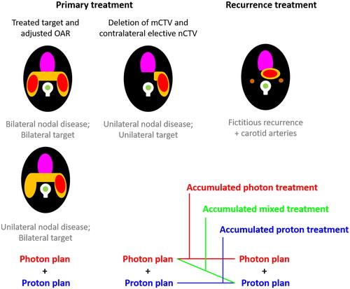
For the re-irradiation plans the PTV, PRV and setup error was decreased to 3 mm, and the carotid arteries were delineated as an additional OAR. Re-irradiation plans using photon therapy were optimized on the basis of the primary photon plan. Proton re-irradiation plans were optimized on the basis of the primary proton plan. In mixed modality treatment (photon + protons), the proton re-irradiation plans were accumulated, as is, with the primary photon plans, thus not reoptimized on top of the photon plan, as the optimal proton dose plan always was decided by the geometry of the re-irradiation CTV.
All accumulated plans respected the dose constraints for the spinal cord – the only critical OAR, without any EQD2 corrections or assumed recovery, and therefore only physical cumulated dose were used for simplicity.
Primary plans were compared by mean doses to OAR and ΔNTCP for grade 2 dysphagia and grade 3 xerostomia was calculated by a model taking dose to the pharyngeal constrictor muscles, oral cavity, and macroscopic salivary glands, into account [Citation15]. Re-irradiation plans were compared by evaluating the volume of the ipsilateral carotid artery receiving above 100 Gy (V100Gy) and the irradiated patient volume receiving 70 Gy–130 Gy, in 10 Gy increments.
Statistical significance was tested using the paired Wilcoxon signed-rank test. The statistical tests were performed in MATLAB and a p-value less than .05 was considered statistically significant.
The study was approved by the scientific ethics committee (case no. 1-10-72-208-22) and registered at the Central Jutland Region’s internal directory of research projects (serial no. 788459).
Results
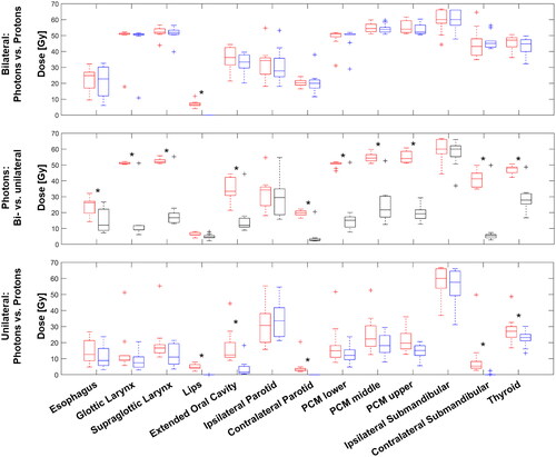
For bilateral HNCUP targets a comparison between photon and proton plans showed no significant differences in either NTCP for dysphagia or xerostomia or mean dose to OAR, except for the lips, where the median mean doses were decreased from 6.6 Gy to 0.0 Gy (see top).
Figure 2. Top: Boxplot comparisons of the mean doses to OAR for a subset of ten patients with simulated bilateral photon- (red) and proton treatment plans (blue). Middle: Boxplot comparison of the mean OAR doses for a subset of eight patients with either a bilateral- (red) or unilateral (black) target treated with photon RT. Bottom: Boxplot comparison of the mean OAR doses for a subset of ten patients with simulated unilateral photon- (red) and proton treatment plans (blue). statistically significant differences are marked with an asterisk.
Changing the treatment guidelines from bilateral- to unilateral treatment will certainly decrease the treated volume. High-risk targets (CTV1) are not changed, intermediate and low-risk targets (CTV2 and CTV3) are on average decreased by 21.9 cm3 [range: 0–92.5] and 189.4 cm3 [range: 134.4–258.8], respectively. This in turn leads to a significant decrease in mean NTCP for dysphagia grade 2 of 17.8% (SD: 5.9) and 17.2% (SD: 5.3) for photons and protons respectively. Similarly, the risk for xerostomia grade 3 was reduced with 4.0% (SD: 0.5) and 5.0% (SD: 0.5) for photons and protons respectively.
Mean doses to the OAR for photons, comparing bilateral and unilateral treatment are shown in (middle). All differences in mean doses are significant, except the dose to the lips and the two major ipsilateral salivary glands, which are partly included in the target.
Comparing photon treatment to proton treatment for unilateral nCTV and omission of mCTV showed a small, but statistically significant decrease in NTCP for dysphagia and xerostomia. Dysphagia was decreased from a mean of 2.1%–1.3% and xerostomia was decreased from 4.9% to 4.0%.
Also, a significant decrease in OAR mean doses were observed for the lips, the extended oral cavity, the contralateral salivary glands, and the thyroid gland. The largest sparing was observed for the extended oral cavity, going from 14.8 Gy in mean dose to 4.9 Gy and the contralateral submandibular gland, going from a mean dose of 6.1 Gy–0.3 Gy.
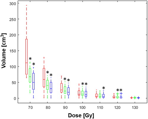
Primary and re-irradiation treatment plans were accumulated for the following treatment strategies; photons followed by photons (A), photons followed by protons (B), and protons followed by protons (C). The mean volume receiving 70 Gy (V70Gy) to 120 Gy, in increments of 10 Gy, is shown in . For doses below 120 Gy, the volume was highest for the photon only treatment (A), followed by the mixed modality treatment (B), and lowest for the proton only treatment (C), whereas the volume receiving 130 Gy is comparable for the three scenarios. The gain is highest for the lowest dose. For scenario B the gain for the four lowest doses are 51%, 39%, 22%, and 8%, for V70Gy, V80Gy, V90Gy, and V100Gy, respectively. For scenario C, they are slightly higher, being 64%, 49%, 31%, and 15%, for V70Gy, V80Gy, V90Gy, and V100Gy, respectively. For the volume receiving 100 Gy or more of the ipsilateral carotid artery, no significant differences were found, with a mean volume of 0.3 cm3 for A, B, and C.
Figure 3. Boxplot of the volumes receiving an accumulated dose between 70 Gy and 130 Gy. Photon + photon treatment is shown in red, photon + proton treatment in green, and proton + proton treatment in blue. Statistical significant differences to the photon + photon treatment are marked with asterisks.
Discussion
In this dose planning study, we found a large decrease in the risk of dysphagia and xerostomia, by a change in treatment strategy from bilateral nCTV plus mCTV to unilateral nCTV only. However, using protons in the primary setting seemed to have a limited added effect on risk of side effect.
Proton treatment is often used to decrease the risk of side effects. This can e.g., be estimated by a comparative treatment plan and a calculated NTCP reduction. In the Danish DAHANCA35 study, a threshold of 5% decrease in ΔNTCP for either dysphagia or xerostomia were chosen in order for patients to be enrolled into the study [Citation16]. Based on the study presented here, HNCUP patients, with either bilateral or unilateral disease, may not be good candidates for proton therapy in the primary setting, since ΔNTCP is below 4.8% for all cases. However, in case of emerging primaries or new primaries necessitating re-irradiation, proton therapy will probably decrease the high dose volume. No NTCP models are available for accumulated doses in re-irradiation [Citation17].
As the diagnostic strategy for patients with HNCUP is getting more efficient, patients who presents with unilateral nodal disease only, may be selected for only unilateral irradiation. This will substantially decrease the degree of grade 2 dysphagia and grade 3 xerostomia. However, this may come with a higher, but still low, risk of emerging primaries. In the present study, we have simulated the re-irradiation treatment of emerging primaries. It is possible to generate re-irradiation treatment plans, without compromising critical OAR after either a primary unilateral photon or proton plan. The volume receiving a high accumulated dose may be reduced by giving both the primary and re-irradiation treatment as IMPT. However, giving the primary treatment as VMAT-IMRT and the re-irradiation as IMPT results in almost the same reduction. Thus, in a Danish setting, HNCUP patients with new primaries or recurrences can be included in DAHANCA 37 (ClinicalTrials.gov Identifier: NCT03981068) where patients are offered proton radiotherapy for reirradiation of head and neck cancers.
Limitations of the present study are the small number of patients. Especially for the bilateral targets for primary radiotherapy as some patients may benefit from protons using a 5% risk reduction for dysphagia as a cut point as used in the DAHANCA 35 trial [Citation15]. The fictitious recurrences, used in the present study, are thought to represents a potential consequence of avoiding elective mucosal irradiation, and this may represent only a subset of potential recurrences.
In conclusion, in patients with a low risk of emerging primaries, limiting the target volumes will greatly reduce toxicity. Proton radiotherapy probably has a limited role in the primary treatment setting for HNCUP. However, re-irradiation for emerging primaries might be a suitable indication for IMPT.
Supplemental Material
Download MS Excel (82 KB)Disclosure statement
No potential conflict of interest was reported by the author(s).
References
- Overgaard J, Jovanovic A, Godballe C, et al. The Danish head and neck cancer database. Dove Medical Press Ltd; 2016. pp. 491–496. doi: 10.2147/CLEP.S103591.
- Johansen LV, Grau C, Overgaard J. Nodal control and surgical salvage after primary radiotherapy in 1782 patients with laryngeal and pharyngeal carcinoma. Acta Oncol. 2004;43(5):486–494. doi: 10.1080/02841860410034289.
- Grau C, Johansen LV, Jakobsen J, et al. Cervical lymph node metastases from unknown primary tumours: results from a national survey by the Danish society for head and neck oncology. Radiother Oncol. 2000;55(2):121–129. doi: 10.1016/s0167-8140(00)00172-9.
- Farooq S, Khandavilli S, Dretzke J, et al. Transoral tongue base mucosectomy for the identification of the primary site in the work-up of cancers of unknown origin: systematic review and meta-analysis. Oral oncology. 2019;91:97–106. doi: 10.1016/j.oraloncology.2019.02.018.
- Jakobsen J, Johansen LV, Grau C. National guidelines regarding carcinoma metastasis to the neck from unknown primary tumour. 2013. Available from: www.dahanca.dk
- Strojan P, Ferlito A, Langendijk JA, et al. Contemporary management of lymph node metastases from an unknown primary to the neck: II. A review of therapeutic options. Head Neck. 2013;35(2):286–293. doi: 10.1002/hed.21899.
- LaVigne AW, Margalit DN, Rawal B, et al. IMRT-based treatment of unknown primary malignancy of the head and neck: outcomes and improved toxicity with decreased mucosal dose and larynx sparing. Head Neck. 2019;41(4):959–966. doi: 10.1002/hed.25531.
- Richards TM, Bhide SA, Miah AB, et al. Total mucosal irradiation with intensity-modulated radiotherapy in patients with head and neck carcinoma of unknown primary: a pooled analysis of two prospective studies. Clin Oncol. 2016;28(9):e77–e84. doi: 10.1016/j.clon.2016.04.035.
- Sher DJ, Balboni TA, Haddad RI, et al. Efficacy and toxicity of chemoradiotherapy using intensity-modulated radiotherapy for unknown primary of head and neck. Int J Radiat Oncol Biol Phys. 2011;80(5):1405–1411. doi: 10.1016/j.ijrobp.2010.04.029.
- Ghatasheh H, Huang SH, Su J, et al. Evaluation of risk-tailored individualized selection of radiation therapy target volume for head and neck carcinoma of unknown primary. Radiother Oncol. 2022;175:56–64. doi: 10.1016/j.radonc.2022.07.016.
- Straetmans J, Stuut M, Wagemakers S, et al. Tumor control of cervical lymph node metastases of unknown primary origin: the impact of the radiotherapy target volume. Eur Arch Otorhinolaryngol. 2020;277(6):1753–1761. doi: 10.1007/s00405-020-05867-2.
- Tambas M, Steenbakkers R, van der Laan HP, et al. First experience with model-based selection of head and neck cancer patients for proton therapy. Radiother Oncol. 2020;151:206–213. doi: 10.1016/j.radonc.2020.07.056.
- Hansen CR, Crijns W, Hussein M, et al. Radiotherapy treatment plannINg study guidelines (RATING): a framework for setting up and reporting on scientific treatment planning studies. Radiother Oncol. 2020;153:67–78. doi: 10.1016/j.radonc.2020.09.033.
- National RKKP and DAHANCA Guidelines. Radiotherapy guidelines 2020 (National RKKP Guideline). 2020. https://www.dahanca.dk/
- Van den Bosch L, van der Schaaf A, van der Laan HP, et al. Comprehensive toxicity risk profiling in radiation therapy for head and neck cancer: a new concept for individually optimised treatment. Radiother Oncol. 2021;157:147–154. doi: 10.1016/j.radonc.2021.01.024.
- Hansen CR, Jensen K, Smulders B, et al. Evaluation of decentralised model-based selection of head and neck cancer patients for a proton treatment study. DAHANCA 35. Radiat Oncol. 2023:109812. doi: 10.1016/j.radonc.2023.109812.
- Embring A, Onjukka E, Mercke C, et al. Overlapping volumes in re-irradiation for head and neck cancer – an important factor for patient selection. Radiat Oncol. 2020;15(1):147. doi: 10.1186/s13014-020-01587-3.