
Abstract
To explore the potential use of CDK inhibitors in pancreatic ductal adenocarcinoma (PDAC) therapy, a series of novel 2-((4-sulfamoylphenyl)amino)-pyrrolo[2,3-d]pyrimidine derivatives was designed, synthesised, and investigated for inhibition on both CDK kinase activity and cellular proliferation of pancreatic cancer. Most of new sulphonamide-containing derivatives demonstrated strong inhibitory activity on CDK9 and obvious anti-proliferative activity in cell culture. Moreover, two new compounds suppressed cell proliferation of multiple human pancreatic cancer cell lines. The most potent compound 2g inhibited cancer cell proliferation by blocking Rb phosphorylation and induced apoptosis via downregulation of CDK9 downstream proteins Mcl-1 and c-Myc in MIA PaCa-2 cells. CDK9 knockdown experiment suggests its anti-proliferative activity is mainly mediated by CDK9. Additionally, 2g displayed moderate tumour inhibition effect in AsPC-1 derived xenograft mice model. Altogether, this study provided a new start for further optimisation to develop potential CDK inhibitor candidates for PDAC treatment by alone or combination use.
Introduction
Cancer is a major public health problem worldwide. Pancreatic ductal adenocarcinoma (PDAC) is one of the most lethal common malignances and ranks fourth in the United States and seventh worldwide among leading causes of cancer-related deathsCitation1. Gemcitabine with fluorouracil-based chemotherapies is the main treatment option for advanced PDACCitation2. New small molecule targeted therapy including of EGFR, tyrosine kinase, and PARP inhibitors, and immunotherapy such as PD-1 inhibitor might also be used for patients with PDACCitation3,Citation4. However, delayed diagnosis, deficiency of effective treatment, and chemoresistance led to a low survival rate of less than 10% for PDACCitation5,Citation6. Therefore, innovative approaches are urgently needed for PDAC patients.
Cyclin-dependent kinase (CDK) is a family of serine-threonine kinases, and consists of about 20 kinasesCitation7. Each CDK interacts with the specific cyclin to form a CDK-cyclin complex to produce enzyme activity. In addition to the cell-cycle CDKs (CDK1, 2, 4, 6, 7) that directly promote cell cycle progression, some transcriptional CDKs (CDK7, 8, 9) play an important role as regulator of transcription. Even though CDKs have been extensively studied in the past decades, their roles in the cell cycle and other related processes are not yet fully understood. Dysregulated CDKs have been linked to cancer initiation and progressionCitation8. Thus, it is not surprising that CDKs and their regulators are identified as drug targets in various human cancersCitation9. For instance, three CDK4/6 inhibitors (palbociclib, ribociclib, and abemaciclib) have been approved by the FDA for the treatment of advanced oestrogen receptor (ER)-positive breast cancerCitation10. Although few clinical evidence of CDK inhibitors was shown in PDAC, the emerging role of CDKs in the pathobiology of this dreadful cancer indicated the therapeutic potential to target CDKs as a novel treatment strategy for PDACCitation11–15. Recently, Sebti’s group also revealed a link between CDK hyperactivation and mutant Kras dependency through comparative phosphoproteomics and CRISPR/Cas9 gene editing, and consequently found that knocking out the CDK1, 2, 7, and 9 was as effective as knocking out Kras in mutant Kras-driven pancreatic cancerCitation11. Kras mutation is known a major driver and a desirable drug target in pancreatic cancers. Moreover, tumours that harbour Kras mutations are highly aggressive, invasive, metastatic, and associated with poor prognosisCitation16–18. A very high rate of activating mutations in Kras (>90%) exists in most human cancers of pancreas including the earliest stage of pancreatic intraepithelial neoplasia, common Kras mutations in pancreatic ductal adenocarcinoma are G12D, G12C, and G12VCitation19,Citation20. Besides of direct inhibit on KRAS protein, targeting RAS effectors or synthetic lethal partners appeared to be an alternative opportunity to treat Kras-driven cancersCitation21,Citation22. Therefore, CDK-targeting therapy could be exploited to efficaciously abrogate pancreatic cancer. There are a lot of studies necessary to further evaluate the promise of CDK-inhibitors in highly malignant PDAC. Here, we reported the design, synthesis, and structure-activity relationship (SAR) of a series of new 2-((4-sulfamoylphenyl)amino)-pyrrolo[2,3-d]pyrimidine derivatives as CDK inhibitors against PDAC (). Initial mechanism of action and in vivo efficacy study confirmed the potential of 2g as a lead compound to develop new approaches for treatment of PDAC in future study.
Figure 1. Discovery of 2-((4-sulfamoylphenyl)amino)-pyrrolo[2,3-d]pyrimidine derivatives as CDK inhibitors.
![Figure 1. Discovery of 2-((4-sulfamoylphenyl)amino)-pyrrolo[2,3-d]pyrimidine derivatives as CDK inhibitors.](/cms/asset/60a4466b-a052-40e1-9e01-267c973922d1/ienz_a_2169282_f0001_b.jpg)
Results and discussion
Chemistry
We previously reported a series of 6-anilinocarbonyl-substituted ribociclib derivatives with stronger anti-proliferative activity than ribociclib in pancreatic cancer cell culturesCitation23. These more potent derivatives contain the anilino group instead of dimethylamino group at C6-position of pyrrolo[2,3-d]pyrimidine in the chemical structure of ribociclib. Meanwhile, we were attracted by compound 1 (), which contains a distinct moiety of N-pyrimidine benzenesulfonamide in C2-substituent of pyrrolo[2,3-d]pyrimidine, in view of its higher selectivity to CDK9 than CDK4/6. 1 also displayed higher in vitro anti-proliferative activity than ribociclib in pancreatic cancer MIA PaCa-2 cell culture. To our knowledge, Wang et al reported anticancer activity of N-alkyl-benzenesulfonamide ribociclib derivatives as CDK9 inhibitor in non-small-cell lung cancerCitation24. This appeared to be a new chemical starting point to identify lead compound with novel mechanism of action and strong potency against PDAC cell proliferation. To understand the role of 4,6-dimethyl-pyrimidine in the benzenesulfonamide moiety of compound 1, we firstly designed and synthesised 2-((4-sulfamoylphenyl)amino)-pyrrolo[2,3-d]pyrimidine derivatives 2a–2j by bioisosteres replacement and fragment cleavage strategy. The synthetic route of 2a–2j was shown in Scheme 1. The classical Pd-catalyzed coupling was performed using commercial available 2-chloro-7-cyclopentyl-N,N-dimethyl-7H-pyrrolo[2,3-d]pyrimidine-6-carboxamide and aromatic amine as start materials at 110 °C under microwave irradiation to afford title compounds in the yield of 16%∼49%. In addition, several N-heterocycle benzenesulfonamide derivatives 5b–5e were designed and synthesised by the method shown in Scheme 2. Starting from 4-nitrobenzenesulfonyl chloride, traditional acylation and subsequent hydrogenation reduction of nitro group to amine were performed to give key 4-sulfonylaniline intermediates 4a–4d through intermediates 3a–3d. 4a–4d were used lately for coupling with 2-chloro-7-cyclopentyl-N,N-dimethyl-7H-pyrrolo[2,3-d]pyrimidine-6-carboxamide by same method as Scheme 1 to afford the corresponding intermediate 5a and final products 5c–5e. Removal of Boc protection from 5a in the presence of hydrochloride or trifluoroacetic acid gave title compound 5b.
Scheme 1. Synthesis of 2-((4-sulfamoylphenyl)amino) substituted derivatives. Reagents and conditions: (a) arylamine, Xantphos, Pd2 (db)3, Cs2CO3, DMF, 110 °C, microwave irradiation.
Scheme 2. Synthesis of 2-((4-sulfamoylphenyl)amino) substituted derivatives. Reagents and conditions: (a) K2CO3, DCM,RT; (b) Pd/C, Methanol; (c) 2-chloro-7-cyclopentyl-N,N-dimethyl-7H-pyrrolo[2,3-d]pyrimidine-6-carboxamide, Xantphos, Pd2 (db)3, Cs2CO3, DMF, 110 °C, microwave irradiation; (d) 2 M hydrochloride in Methanol.
![Scheme 2. Synthesis of 2-((4-sulfamoylphenyl)amino) substituted derivatives. Reagents and conditions: (a) K2CO3, DCM,RT; (b) Pd/C, Methanol; (c) 2-chloro-7-cyclopentyl-N,N-dimethyl-7H-pyrrolo[2,3-d]pyrimidine-6-carboxamide, Xantphos, Pd2 (db)3, Cs2CO3, DMF, 110 °C, microwave irradiation; (d) 2 M hydrochloride in Methanol.](/cms/asset/3ad1e174-2db1-4565-86b0-fd9beefbfc85/ienz_a_2169282_sch0002_b.jpg)
Alternatively, considering that replacing dimethylamino group of ribociclib with aniline increased anti-proliferative activity in previous study, we therefore designed and synthesised new derivatives 10a and 10b by introducing the aniline group into C6-substituent in 2-((4-sulfamoylphenyl)amino)-pyrrolo[2,3-d]pyrimidine. The synthetic route shown in Scheme 3 is similar to the previously reported methodCitation23. Briefly, the secondary amine intermediate 6 synthesised from 2-chloroacetyl chloride and specific amines by a one-pot two-step reaction was reacted with commercial available 4-chloro-2-thiomethylpyrimidine carboxaldehyde in the presence of N,N-diisopropyl ethylamine (DIPEA) to give intermediate 7, which further underwent the cyclisation reaction to provide pyrrolo[2,3-d]pyrimidine 8 in the presence of caesium carbonate by heating at 120 °C under microwave irradiation. The oxidation of 8 in the presence of m-CPBA in dichloromethane (DCM) afforded intermediate 9 in the yield of 57%. Finally, the aromatic nucleophilic substitution reaction of key intermediate 9 with N-(4-(N-(4,6-dimethyl-pyrimidin-2-yl)sulfamoyl)phenyl)formamide or N-(4-(N-(pyrimidin-2-yl)sulfamoyl)phenyl)formamide in the presence of Cs2CO3 gave title compounds 10a and 10b, respectively.
Scheme 3. Preparation of 2-((4-(N-(pyrimidin-2-yl)sulfamoyl)phenyl)amino)-7H-pyrrolo[2,3-d]pyrimidine derivatives. Reagents and conditions: (a) 2-methoxyphenylamine, DIPEA, acetonitrile, 0 °C; (b) cyclopentylamine, acetonitrile;(c) 4-chloro-2-(methylthio)pyrimidine-5-carbaldehyde, DIPEA, acetonitrile, MW, 80 °C; (d) Cs2CO3, MW, 120 °C, 30 min, acetonitrile; (e) m-CPBA, DCM; (f) N-(4-(N-(4,6-dimethylpyrimidin-2-yl)sulfamoyl)phenyl)formamide or N-(4-(N-(pyrimidin-2-yl)sulfamoyl)phenyl)formamide, Cs2CO3, DMSO, 85 °C
![Scheme 3. Preparation of 2-((4-(N-(pyrimidin-2-yl)sulfamoyl)phenyl)amino)-7H-pyrrolo[2,3-d]pyrimidine derivatives. Reagents and conditions: (a) 2-methoxyphenylamine, DIPEA, acetonitrile, 0 °C; (b) cyclopentylamine, acetonitrile;(c) 4-chloro-2-(methylthio)pyrimidine-5-carbaldehyde, DIPEA, acetonitrile, MW, 80 °C; (d) Cs2CO3, MW, 120 °C, 30 min, acetonitrile; (e) m-CPBA, DCM; (f) N-(4-(N-(4,6-dimethylpyrimidin-2-yl)sulfamoyl)phenyl)formamide or N-(4-(N-(pyrimidin-2-yl)sulfamoyl)phenyl)formamide, Cs2CO3, DMSO, 85 °C](/cms/asset/7858819e-7d32-4dc3-bf39-c61a36aff643/ienz_a_2169282_sch0003_c.jpg)
Biological evaluation
In vitro antiproliferative activity screen in MIA PaCa-2 cells
We screened these new derivatives with GCTB as positive drug in Kras mutant human pancreatic cancer MIA PaCa-2 cells. As shown in , in vitro anticancer activity of corresponding derivatives (2a and 2b) became weaker (IC50 > 10 µM) than compound 1 (IC50 = 4.98 µM) when the number of methyl substituent on the pyrimidine ring. The biological activity was further decreased after the pyrimidine was substituted with pyridine (2c, IC50 = 18.80 µM). It should be mentioned that poor solubility of compound 2b and 2c might exacerbate decreasing of in vitro efficacy. Replacement of dimethylpyrimidine with para-chlorophenyl group (2d) maintained similar activity (IC50 = 6.23 µM) to compound 1. This indicated that lipophilic terminal of C2-substituent is beneficial to anticancer activity when sulphonyl group was linked with aromatic ring. Interestingly, when we made a stepwise break down the sulphonamide moiety until sulphonamide was removed from compound 1, corresponding compounds 2e, 2f, and 2h displayed gradually decreased activity comparing to 1. Surprisingly, nearly ten-fold enhanced potency than 1 was observed for compound 2g (IC50 = 0.52 µM) which has a simple unsubstituted amino group of R2. However, further replacing the C2-phenylamino group of compounds 2e and 2g with pyridine-2-amino led to almost loss of activity of compounds 2i and 2j, respectively. In addition, comparable and even stronger anti-proliferative activity (IC50 = 7.8 0 ∼ 0.94 µM) than compound 1 was observed when R2 group is saturated heterocycle (5b–5e). Meanwhile, higher potency of compound 5b than 5c–5e suggests the role of free NH in the piperazine as hydrogen-bond donor to the biological activity. Alternatively, ten-fold decreased anticancer activity was observed for 6-anilinocarbonyl-substituted derivatives 10a and 10b comparing to 1 in this work.
Table 1. Anti-proliferative activity of 2-((4-sulfamoylphenyl)amino)-pyrrolo[2,3-d]pyrimidine derivatives in pancreatic cancer MIA PaCa-2 cell culture.
CDK protein kinase assay
In previous study, we found that CDK4 inhibitory activity of compound 1 is obviously weaker than highly selectively CDK4 inhibitor ribociclib, but compound 1 exhibited higher in vitro anti-proliferative activity compared to ribociclib. CDK kinase screening indicated that compound 1 possessed of the most potent inhibition on CDK9 than other CDK kinase. It led us to speculate that inhibition of CDK9 contributes much to the efficacy of compound 1 against PDAC cell growth. Thus, we firstly screened the CDK9 enzymatic activity inhibitory rate of new derivatives at concentration of 50 nM in a biochemical assay. As shown in , most of new sulphonamide derivatives possessed of significantly inhibitory activity (>80%) on CDK9. Moreover, these CDK9 inhibitors exhibited higher anti-proliferative activity in MIA PaCa-2 cells than non-CDK9 inhibitors, such as pyrrolo[2,3-d]pyrimidine derivatives with 2-aminopyridine side chain (2i and 2j) or 6-anilinocarbonyl side chain (10a and 10b). Low CDK9 inhibitory potency of compound 2i (27%) and 2j (25%) is consistent with the report that pyridin-2-amino moiety is key determinant of high selectivity to CDK4/6Citation25. In previous study CDK4/6 inhibition effect was less impaired when dimethylamino group of ribociclib was replaced with o-anisidine. However, such a change from 1 to 10a caused the loss of CDK9 inhibition activity. In general, terminal of C2-substituent of pyrrolo[2,3-d]pyrimidine skeleton reach out of cavity and expose to solvent region. Therefore, we assume that there is a size limitation for cavity outlet of CDK9. Steric hindrance of 3,5-dimethylpyrimidin-2-amino group stopped small molecule into deeper spacious cavity to accommodate planar anisidine fragment. Removal of two methyl substituents on pyrimidine from 10a could improve flexibility of small molecule within protein cavity. Thus, some CDK9 inhibitory activity was recovered in 10b (54%). These four inactive compounds on CDK9 also lack of the activity against PDAC cell growth. This result, at least to some extent, confirmed the anticancer activity of this class of 2-((4-sulfamoylphenyl)amino)-pyrrolo[2,3-d]pyrimidine derivatives is related to CDK9 inhibition activity. Admittedly, the anticancer activity of these compounds is not strictly dependent on CDK9 inhibition from the data in . Therefore, several active compounds (IC50 < 10 µM) were further investigated in multiple CDK (1, 2, 4, 7, 9) assays. As a result, all these compounds were identified as CDK9 inhibitors but not CDK7 inhibitors. Meanwhile, they presented the differential effect on CDK1, 2, and 4. Comparing to 1, compounds 5b, 5c, and 5e exhibited higher selectivity preference for both CDK4 and 9. However, the most potent compound 2g in PDAC cell culture behaves as a pan-CDK inhibitor which has strong inhibition on CDK1, 2, 4, and 9. Considering that ribociclib is a highly selective CDK4 inhibitor with low potency on CDK9, we estimated that inhibiting CDK9 enzyme activity played more important role to the anticancer performance of these derivatives in PDAC cells compared to other CDK subtypes.
Anti-proliferative activity screen in multiple PDAC cell lines
Thereafter, two potent anti-proliferative compounds 2g and 5b, which displayed different CDK inhibition profile, were investigated of anticancer activity in comparison with 1 in multiple human PDAC cell lines with different Kras genotypes. It could be seen that these compounds exerted higher sensitivity to BxPC-3 cells with wildtype Kras than Kras-mutant pancreatic cancer cell lines, which includes MIA PaCa-2, AsPC-1 and PANC-1. Moreover, two new derivatives 2g and 5b exhibited higher potency in some extend than compound 1 (). Surprisingly, all tested compounds showed significantly weaker potency in PANC-1 than other cell lines, and anti-proliferative activity of 1 was even slightly higher than both two new derivatives and gemcitabine in PANC-1 cell culture. To our knowledge, partial Kras G12D mutation was confirmed in PANC-1 while MIA PaCa-2 and AsPC-1 is fully Kras G12C and G12D mutation, respectively, by gene sequencing. We don’t know if Kras genetic status of cancer cells affects the biological performance of these CDK inhibitors in current study.
Table 2. Anti-proliferative activity of active 2-((4-sulfamoylphenyl)amino)-pyrrolo[2,3-d]pyrimidine derivatives in different PDAC cell lines.
2g induced cell cycle arrest and apoptosis in MIA PaCa-2 cells
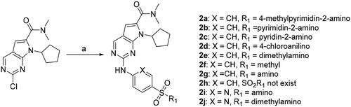
Based on above results, compound 2g was further examined for the initial mechanism of action in MIA PaCa-2 cells. In previous study compound 1 displayed a G2-arrested effect on cell cycle of MIA PaCa-2 cells.Citation23 Here, 2g induced cell cycle arrest at G1 phase at low concentration of 0.1 µM after 24 h treatment of MIA PaCa-2 cells, which might be owing to the inhibition of 2g on CDK4. As the concentration increased to 0.3 and 1.0 µM, a modest G2/M-arrested effect was observed (, Upper). This result is consistent with the expected effect of CDK inhibitor. In apoptosis study (, Lower), the number of late apoptotic cells increased in a dose-dependent manner after 24 h treatment with 2g. It suggests that this compound inhibits the growth of MIA PaCa-2 cells by inducing apoptosis. Meanwhile, down-regulation of phosphorylated Rb protein was observed after 2g treatment, this confirmed the effect of 2g on cell cycle induced by regulation of Rb protein. In addition, inhibition of CDK9 could decrease the level of RNA polymerase II C-terminal domain serine 2 phosphorylation, the pro-survival protein Myeloid Cell Leukaemia 1 (Mcl-1) and MYC oncoprotein, and induced rapid apoptosis in cancer cells.Citation26 In our study, same response in immunoblot assay was observed in MIA PaCa-2 cells after 6 h exposure to compound 2g (). These results suggest that 2g inhibited cancer cell proliferation by blocking Rb phosphorylation and induced apoptosis via downregulation of CDK9 downstream proteins. Compound 2g displayed slightly weaker and significantly stronger inhibitory activity on CDK4 and CDK9, respectively, compared to CDK4 inhibitor ribociclib (). However, compound 2g exhibited 20-fold higher in vitro anti-proliferative activity than the latter in MIA PaCa-2 cells (). Therefore, we thought that inhibition of CDK9 plays a key role in efficacy of compound 2g in PDAC cell culture. To investigate the effect of CDK9 expression levels on PDAC cellular susceptibility to our synthetic compound, we used a specific siRNA of CDK9 to alter the protein expression in MIA PaCa-2 cells (). An obviously lower cell viability was observed when the cells was transfected with siRNA (50 nM) for 48 h compared to siRNA-untreated cells. Moreover, the sensitivity of MIA PaCa-2 cells to compound 2g was significantly decreased when we treated cells with compound 2g after downregulation of CDK9 by siRNA (). It suggests that the anti-proliferative activity of compound 2g in MIA PaCa-2 cells relies on the inhibition of CDK9.
Figure 2. Compound 2g induced slightly cell cycle arrest and late apoptosis of MIA PaCa-2 cells. The cells were incubated without or with 2g at indicated concentrations for 24 h. The cell cycle distribution (Upper lane) or apoptosis (Lower lane) was detected using propidium iodide (PI) staining or both PI and annexin V-FITC staining, respectively, and analysed by flow cytometry assays. Graphs showing the results of a representative experiment, and the data was presented as mean ± standard deviation.
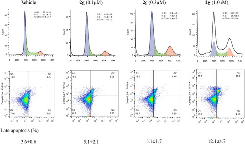
Figure 3. The anti-proliferative activity of new synthetic compounds is mainly mediated by CDK9. (A) 2g inhibited the phosphorylation of Rb and RNA polymerase II, induced apoptosis through down-regulation of Mcl-1 and c-Myc protein. MIA PaCa-2 cells were incubated with or without 2g at indicated concentration for 6 h. (B) CDK9 was dose-dependently knockdown by its specific siRNA after 48 h treatment in MIA PaCa-2 cells. (C) Downregulation of CDK9 using siCDK9 (50 nM) decreased the sensitivity of MIA PaCa-2 cell to compound 2g (0.2 µM). The cells were incubated with 2g for 72 h. NS no significance; ***p < 0.001; ****p < 0.0001. Whole-cell lysates were subjected to immunoblotting. A representative protein band of three independent experiments is shown. (D) Representative illustration of the binding of synthetic compound to CDK9/cyclin T. Small molecules 1 (left column), 2g (middle column), and 2i (right column) were shown in stick representation with carbons colored yellow.
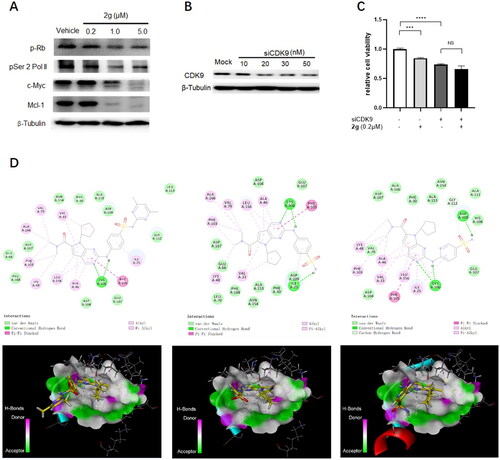
Table 3. IC50 (nM) values of anti-proliferative compounds against CDK activity.
Molecular docking study
To better understand the interaction mode of synthetic compounds with CDK9, molecular docking study was carried out using the X-ray cocrystal of CDK9 complexed with a 4‑(thiazol-5-yl)-2-(phenylamino)pyrimidine CDK inhibitor (PDB Code: 4BCF)Citation27. As shown in , the N3-pyrrolo[2,3-d]pyrimidine of compound 1 accepts a hydrogen bond from the peptide nitrogen of Cys106 in hinge region, while the C2-NH of the pyrrolo[2,3-d]pyrimidine fused ring donates a hydrogen bond to the carbonyl of Cys106. Meanwhile, pyrrolo[2,3-d]pyrimidine is sandwiched between Ala46 and Leu156 by hydrophobic interaction, and pyrimidine ring contacts the gatekeeper residue (Phe105) to form a long π–π interaction at the distance of 5.38 Å. In addition, multiple van der Waals interactions were formed between compound 1 and hydrophobic residues within the active site of CDK9 complex. Owing to the relative malleability of the CDK9 active site, compound 2g and other synthetic derivatives adopt a similar pose to compound 1 within the cavity of ATP binding site of CDK9 protein. In addition to above similar binding mode as compound 1, the hydrogen of sulphonamide group of 2g binds to Ile25 to form an additional hydrogen bond with CDK9. It was previously reported that replacing of aniline with 2-aminopyrimidine led to a clear preference to inhibition of CDK4/6 in the practical medicinal chemistry study. It was also observed from the comparison of CDK profiling between 2g and 2i in our research. However, we failed to explain the discrimination in CDK inhibitory activity through molecular docking analysis because both compounds exhibited similar interaction mode with CDK9/cyclinT complex (, upper lane). The only difference of 2i from 2g in molecular docking study is that a smaller dihedral orientation between pyrrolo[2,3-d]pyrimidine fused ring and 2-aniline ring of compound 2i was shown in 3 D diagram than that of the latter (, lower lane). Therefore, we supposed that it brought two unfavourable effects to compound 2i: one is that intramolecular exclusion due to excessive closing of 2-aminopyrimidine ring to 7-cyclopentyl ring; another is the space spatial location of N atom on the 2-aminopyrimidine ring is incompatible with the electrostatic environment of protein cavity in the practical experimental study. In summary, most of synthetic compounds kept the binding affinity to the active site of CDK9. The terminal of 2-aniline side chain of pyrrolo[2,3-d]pyrimidine scaffold extended to solvent exposure region. This explains why those active derivatives keep strong CDK9 inhibitory activity although they contain different substituents on sulphonyl group of 2-aniline moiety.
In vivo pharmacokinetics (PK) study
Although compounds 2g and 5b displayed different features in CDK selectivity, no significant difference of anticancer potency observed between them in multiple pancreatic cancer cell lines. So, oral absorption of both compounds was investigated in parallel by determining the drug concentration-time curve after single oral administration () to rats before the in vivo efficacy evaluation. Though solubility of 2g was found to be much poorer than 5b in the practical study, compound 2g was absorbed into the circulation more rapidly (Tmax = 0.5 h) and reached a slightly higher maximum plasma drug concentration (Cmax = 1161 ng/mL·h) than that of 5b (Tmax = 1.17 h, Cmax = 1080 ng/mL·h) after oral dosing to rats. Elimination half-life (T1/2) and mean retention time (MRT) of compound 2g was calculated as 7.54 h and 7.72 h, respectively, which is significantly longer than 5b. But we have to admit that rapid decrease of drug concentration to a low level within six hours occurred for both 2g and 5b. Finally, compound 2g displayed a higher systematic exposure (AUC0‑t = 4621 ng/mL·h) than 5b (AUC0‑t = 4427 ng/mL·h). Therefore, compound 2g was selected to undergo in vivo efficacy study due to its slightly better performance in both oral PK features and antiproliferative activity than compound 5b in this work.
Figure 4. Oral PK study of compounds 2g and 5 b after oral administration to rats (n = 3) at dose of 30 mg/kg.
In vivo efficacy study
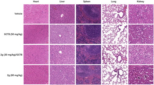
To test if above cellular action could be translated into anti-pancreatic cancer activity in vivo, we used AsPC-1 line, which forms tumour more rapidly than MIA PaCa-2 in mice, to generate CDX subcutaneous xenograft mice model for evaluation of in vivo efficacy of 2g. In addition of vehicle control, GCTB was used as a reference drug as it is the main chemotherapeutic agents used in clinic for pancreatic cancer. According to the PK data, we designed 30 mg/kg and 60 mg/kg oral dosages for 2g. Drug treatment duration was three weeks with consecutive five days treatment and two days off per week. In addition, combining low dosage 2g with GCTB was also included to explore the possibility of increasing GCTB susceptibility to pancreatic cancer in this work. As shown in , GCTB exhibited tumour inhibition effect with tumour growth inhibition (TGI) value of 36% compared to vehicle group while 2g at oral dose of 30 mg/kg only had a low TGI of 12% after 3-week administration. 2g at dose of 60 mg/kg led to reduction of tumour volume up to 46% compared to vehicle group. Combination of GCTB with 2g at dose of 30 mg/kg displayed higher TGI (43%) than either drug alone, but there is no significant difference between combination group and GCTB alone. Admittedly, an obvious decrease of body weight occurred in animals treated with high dose of 2g during the first ten days of administration, and body weight subsequently came back to the normal level. The maximal individual body weight loss was 23% on Day 21 when 2g was co-administered with GCTB. In addition, in vivo toxicity is a major concern due to the inhibitory activity of 2g on multiple CDK kinase. Encouragingly, no obvious pathological change was observed in main organs of mice after 3-week treatment with either 2g or GCTB ().
Figure 5. In vivo efficacy evaluation in AsPC-1 cell-derived xenograft mice model (n = 6).
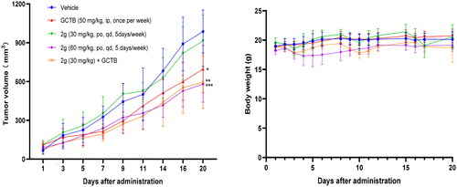
Figure 6. Representative H&E-stained tissue section (scale bar, 50 µM for all) from CDX mice treated with vehicle, GCTB or 2g alone, or combination of GCTB and 2g for 3 weeks.
To better understand in vivo efficacy of 2g, we determined drug concentrations in both tumour tissues and plasma samples at 1 h and 4 h after 30 mg/kg single oral dosing to tumour-bearing mice. As shown in , drug concentration of 2g in both plasma and tumour tissue is higher than 2-fold of cellular IC50 value. However, drug concentration significantly dropped down to an insufficient level for efficacy in either plasma and tumour tissue at 4 h after administration. This suggests that 2g was easily omogenizat and eliminated, the resulted short target occupation might be the reason for its moderate in vivo efficacy.
Table 4. Drug concentrations in tumour tissue and plasma sample after 30 mg/kg single dosing of 2g to tumour-bearing mice (n = 3).
Conclusions
A series of new 2-((4-sulfamoylphenyl)amino)-pyrrolo[2,3-d]pyrimidine derivatives was designed, omogenizat, and investigated for the inhibition effect on both human pancreatic cancer cell proliferation and CDK enzymatic activity. All anti-proliferative compounds in pancreatic MIA PaCa-2 cells displayed strong inhibition on CDK9 but no inhibition on CDK7. In addition, CDK4 activity might play a role in pancreatic cancer cell growth from the performance of active compounds in both in vitro anticancer activity and CDK inhibition pattern. Anti-proliferation MTT screening identified two highly active compounds 2g and 5b which kept a sensitivity at low micromolar level to both Kras -mutant and © PDAC cell lines. Especially, compound 2g mediated cancer cell proliferation blockage and induced apoptosis in pancreatic cancer MIA PaCa-2 cells, and rapid down-regulation of Mcl-1 and c-Myc oncoprotein were observed after 2g treatment. Moreover, CDK9 knockdown experiment suggests that the anti-proliferative activity of compound 2g in MIA PaCa-2 cells is mainly mediated by the inhibition of CDK9. Initial molecular docking study revealed the principle binding mode of 2-((4-sulfamoylphenyl)amino)-pyrrolo[2,3-d]pyrimidine derivatives to CDK9/cyclin T complex. Detailed analysis might provide valuable cues for future design of new chemotype of selective CDK9 inhibitor. Oral PK and in vivo efficacy study further confirmed 2g could inhibit tumour growth by oral administration for 3 weeks in AsPC-1 cell-derived xenograft mice model. Considering of function compensation of CDK7 and 9 in cancer cell proliferation, continuous effort to improve in vivo exposure of 2-((4-sulfamoylphenyl)amino)-pyrrolo[2,3-d]pyrimidine derivatives and screening effective anti-proliferative partners is worthy to provide promising regimens for PDAC treatment. Admittedly, CDK inhibition might not the only mechanism responsive for the anticancer activity of these 2-((4-sulfamoylphenyl)amino)-pyrrolo[2,3-d]pyrimidine derivatives. Benzenesulfonamide moiety appeared in the chemical structure of a variety of bioactive substances. For example, some benzenesulfonamide derivatives have been investigated and developed for the treatment of hypoxic tumours as carbonic anhydrase inhibitorsCitation28. Thus, deep study on anticancer mechanism of 2-((4-sulfamoylphenyl)amino)-pyrrolo[2,3-d]pyrimidine is necessary for further chemical omogenizatiation. Together, current study provided a new start to develop promising CDK inhibitor candidates for PDAC treatment by alone or combination use.
Experimental methods
General information for chemical experiments
All commercially available chemicals and solvents were used without further purification. The reaction was monitored by Merck TLC silica gel 60 F254 aluminium sheets or LC-MS instrument. Column chromatography was performed on RediSep silica gel column using the Teledyne ISCO CombiflashRf + system (Teledyne, Lincoln, USA) for purification. Samples purity were determined on a Shimadzu 2020 UFLC-MS instrument (Kyoto, Japan) which was equipped with a Shim-pack VP-ODS column (2.0 mm × 150 mm, 5 µm) and DAD detector, an electrospray omogenizaation source (ESI) and a single-quadrupole mass analyser. The elution phase was composed of acetonitrile and 0.1% formic acid aqueous solution in a gradient mode at a flow rate of 0.4 ml/min. The 1H spectrum were recorded using a WNMR-I 500 MHz spectrometer (Wuhan Zhongke-Niujin, China) or an INOVA 400 MHz spectrometer (Varian). HR-MS was collected with triple TOF 5600 + LC/MS/MS system (CADM-YQ-086) with an ESI mass selective detector (SCIEX Manufacturing, Singapore). All title compounds are >95% pure by UFLC analysis.
Chemical synthesis
Preparation of 7-cyclopentyl-N,N-dimethyl-2-((4-(N-(4-methylpyrimidin-2-yl)sulfamoyl)phenyl)amino)-7H-pyrrolo[2,3-d]pyrimidine-6-carboxamide (2a)
To a solution of 2-chloro-7-cyclopentyl-N,N-dimethyl-7H-pyrrolo[2,3-d]pyrimidine-6-carboxamid-e (200 mg, 0.683 mmol) in DMF (10 ml) were added 4-amino-N-(4-methylpyrimidin-2-yl) benzenesulfonamide (163.94 mg, 1.35 mmol), XantPhos (107 mg, 0.186 mmol), Pd2(dba3) (113 mg, 0.124 mmol) and Cs2CO3 (606 mg, 186 mmol) at room temperature. The reaction was stirred at 100 °C until 13a was completely conversed into aim product by TLC monitoring. The mixture was filtered, concentrated and the residue was extracted with dichloromethane and distilled water. The water phase was reextracted with DCM for two times. The combined organic phase was washed with saline, dried with anhydrous magenism sulphate and filtered for purification by column chromatography eluted with dichloromethane/methamol (93/7) to afford the faint yellow solid 2a with yield of 16.5%. 1H NMR (500 MHz, DMSO-d6) δ 10.04 (s, 1H), 8.81 (d, J = 1.6 Hz, 1H), 8.32 (d, J = 4.8 Hz, 1H), 7.97 (t, J = 10.5 Hz, 2H), 7.89 (d, J = 8.6 Hz, 2H), –.95–6.86 (m, 1H), –.65–6.60 (m, 1H), 5.75 (d, J = 1.5 Hz, 1H), 4.75 (q, J = 9.0 Hz, 1H), 3.06 (d, J = 8.7 Hz, 6H), 2.89 (d, J = 1.7 Hz, 1H), 2.73 (s, 1H), 2.44 (s, 2H), 2.32 (s, 3H), 1.98 (d, J = 17.8 Hz, 4H), 1.69 (s, 2H). HRMS(ESI+) m/z calcd C25H29N8O3S for [M + H] 521.2083, found 521.2069.
7-Cyclopentyl-N,N-dimethyl-2-((4-(N-(pyrimidin-2-yl)sulfamoyl)phenyl)amino)-7H-pyrrolo[2,3-d]pyrimidine-6-carboxamide (2b)
It was omogenizatsised as yellow solid with yield of 31.8% from 2-chloro-7-cyclopenty-N,N-dimethyl-7H-pyrrolo[2,3-d]pyrimidine-6-carboxamide and 4-amino-N-(pyrimidin-2-yl)benzenesulfonamide by similar method as 2a. 1H NMR (400 MHz, TFA-d) δ 8.99 (d, J = 5.5 Hz, 2H), 8.88 (s, 1H), –.33–8.26 (m, 2H), –.05–7.97 (m, 2H), 7.56 (t, J = 5.5 Hz, 1H), 6.97 (s, 1H), 4.75 (p, J = 8.9 Hz, 1H), –.41–3.23 (m, 6H), 2.50 (qd, J = 10.9, 8.7, 4.7 Hz, 2H), –.36–2.16 (m, 2H), –.14–2.00 (m, 2H), 1.84 (dq, J = 11.5, 7.1, 6.1 Hz, 2H). HRMS(ESI+) m/z calcd C24H27N8O3S for [M + H] 507.1927, found 507.1920.
7-Cyclopentyl-N,N-dimethyl-2-((4omogeniridin-2-yl)sulfamoyl)phenyl)amino)-7H-pyrrolo[2,3-d]pyrimidine-6-carboxamide (2c)
It was omogenizatsised as yellow solid with yield of 24% from 2-chloro-7-cyclopenty-N,N-dimeth-yl-7H-pyrrolo[2,3-d]pyrimidine-6-carboxamide and 4-aminomogeniridin-2-yl)benzenesulfonamide by similar method as 2a. 1H NMR (500 MHz, DMSO-d6) δ 10.01 (s, 1H), 8.83 (s, 1H), 8.09 (s, 1H), 7.98 (d, J = 8.5 Hz, 2H), 7.82 (d, J = 8.5 Hz, 2H), 7.72 (t, J = 7.6 Hz, 1H), 7.17 (d, J = 8.5 Hz, 1H), 6.92 (s, 1H), 6.65 (s, 1H), 4.78 (p, J = 8.8 Hz, 1H), 3.08 (s, 6H), 2.45 (s, 2H), –.08–1.91 (m, 4H), 1.70 (s, 2H). HRMS(ESI+) m/z calcd C25H28N7O3S for [M + H] 506.1974, found 506.1960.
2-((4-(N-(4-chlorophenyl)sulfamoyl)phenyl)amino)-7-cyclopentyl-N,N-dimethyl-7H-pyrrolo[2,3-d]pyrimidine-6-carboxamide (2d)
It was omogenizatsised as white solid with yield of 28.3% from 2-chloro-7-cyclopenty-N,N-dimethyl-7H-pyrrolo[2,3-d]pyrimidine-6-carboxamide and 4-amino-N-(4-chlorophenyl)benzenesulfonamide by similar method as 2a. 1H NMR (500 MHz, DMSO-d6) δ 10.14 (dd, J = 97.2, 5.9 Hz, 2H), 8.81 (d, J = 6.5 Hz, 1H), 7.93 (q, J = 5.7, 3.7 Hz, 2H), –.74–7.52 (m, 2H), 7.28 (d, J = 7.1 Hz, 2H), –.19–7.03 (m, 2H), 6.63 (d, J = 6.3 Hz, 1H), –.82–4.65 (m, 1H), 3.05 (s, 6H)2.38 (d, J = 14.1 Hz, 2H), 1.94 (d, J = 43.3 Hz, 4H), 1.65 (s, 2H). HRMS(ESI+) m/z calcd C26H28ClN6O3S for [M + H] 539.1632, found 539.1634.
7-Cyclopentyl-2-((4-(N,N-dimethylsulfamoyl)phenyl)amino)-N,N-dimethyl-7H-pyrrolo[2,3-d]pyrimidine-6-carboxamide (2e)
It was omogenizatsised as white solid with yield of 49.5% from 2-chloro-7-cyclopenty-N,N-dimethyl-7H-pyrrolo[2,3-d]pyrimidine-6-carboxamide and 4-amino-N,N-dimethylbenzenesulfonamide by similar method as 2a. 1H NMR (500 MHz, Chloroform-d) δ 8.68 (s, 1H), 7.93 (s, 1H), 7.89 (d, J = 8.3 Hz, 2H), 7.77 − 7.70 (m, 2H), 6.48 (s, 1H), 4.80 (p, J = 9.0 Hz, 1H), 3.16 (s, 6H), 2.72 (s, 6H), –.59–2.51 (m, 2H), –.14–2.03 (m, 4H), –.78–1.69 (m, 2H). HRMS(ESI+) m/z calcd C22H29N6O3S for [M + H] 457.2022, found 457.2025.
7-Cyclopentyl-N,N-dimethyl-2-((4-(methylsulfonyl)phenyl)amino)-7H-pyrrolo[2,3-d]pyrimidine-6-carboxamide (2f)
It was omogenizatsised as white solid with yield of 31.9% from 2-chloro-7-cyclopenty-N,N-dimethyl-7H-pyrrolo[2,3-d]pyrimidine-6-carboxamide and 4-(methylsulfonyl)aniline by similar method as 2a. 1H NMR (500 MHz, Chloroform-d) δ 8.69 (s, 1H), –.95–7.84 (m, 5H), 6.47 (s, 1H), –.87–4.74 (m, 1H), 3.16 (s, 6H), 3.06 (s, 3H), –.64–2.51 (m, 2H), 2.08 (dd, J = 17.5, 7.6 Hz, 4H), –.79–1.70 (m, 2H). HRMS(ESI+) m/z calcd C21H26N5O3S for [M + H] 428.1751, found 428.1761.
7-Cyclopentyl-N,N-dimethyl-2-((4-sulfamoylphenyl)amino)-7H-pyrrolo[2,3-d]pyrimidine-6-carboxamide (2 g)
It was omogenizatsised as white solid with yield of 22.9% from 2-chloro-7-cyclopenty-N,N-dimethyl-7H-pyrrolo[2,3-d]pyrimidine-6-carboxamide and 4-aminobenzenesulfonamide by similar method as 2a. 1H NMR (600 MHz, DMSO-d6) δ 9.96 (s, 1H), 8.82 (s, 1H), 8.00 (d, J = 8.9 Hz, 2H), 7.73 (d, J = 8.9 Hz, 2H), 7.15 (s, 2H), 6.63 (s, 1H), 4.76 (p, J = 8.8 Hz, 1H), 3.06 (s, 6H), 2.46 (dd, J = 14.7, 6.0 Hz, 2H), –.08–1.94 (m, 4H), –.74–1.64 (m, 2H). HRMS(ESI+) m/z calcd C20H25N6O3S for [M + H] 429.1709, found 429.1692.
7-Cyclopentyl-N,N-dimethyl-2-((5-sulfamoylpyridin-2-yl)amino)-7H-pyrrolo[2,3-d]pyrimidine-6-carboxamide (2i)
It was omogenizatsised as white solid with yield of 29.8% from 2-chloro-7-cyclopenty-N,N-dimethyl-7H-pyrrolo[2,3-d]pyrimidine-6-carboxamide and 6-aminopyridine-3-sulphonamide by similar method as 2a. 1H NMR (500 MHz, DMSO-d6) δ 10.44 (s, 1H), 8.91 (s, 1H), 8.68 (d, J = 2.5 Hz, 1H), 8.49 (d, J = 9.0 Hz, 1H), 8.12 (dd, J = 9.0, 2.6 Hz, 1H), 7.41 (s, 2H), 6.69 (s, 1H), 4.78 (p, J = 8.8 Hz, 1H), 3.07 (d, J = 8.2 Hz, 6H), –.48–2.39 (m, 2H), –.07–1.95 (m, 4H), 1.70 (dd, J = 11.5, 5.4 Hz, 2H).HRMS(ESI+) m/z calcd C19H24N7O3S for [M + H] 430.1661, found 430.1661.
7-Cyclopentyl-2-((5-(N,N-dimethylsulfaomogeniridin-2-yl)amino)-N,N-dimethyl-7H-pyrrolo[2,3-d]pyrimidine-6-carboxamide (2j)
It was omogenizatsised as white solid with yield of 31.6% from 2-chloro-7-cyclopenty-N,N-dimethyl-7H-pyrrolo[2,3-d]pyrimidine-6-carboxamide and 6-amino-N,N-dimethylpyridine-3-sulphonamide by similar method as 2a. 1H NMR (500 MHz, DMSO-d6) δ 10.51 (s, 1H), 8.93 (s, 1H), 8.62 (d, J = 2.5 Hz, 1H), 8.51 (d, J = 9.0 Hz, 1H), 8.09 (dd, J = 9.0, 2.5 Hz, 1H), 6.71 (s, 1H), 4.82 (p, J = 8.9 Hz, 1H), 3.09 (s, 6H), 2.68 (s, 6H), 2.42 (dd, J = 14.8, 6.7 Hz, 2H), 2.03 (q, J = 9.9, 8.7 Hz, 4H), –.73–1.64 (m, 2H). HRMS(ESI+) m/z calcd C21H28N7O3S for [M + H] 458.1974, found 458.1958.
Preparation of tert-Butyl 4-((4-nitrophenyl)sulphonyl)piperazine-1-carboxylate (3a)
To a solution of tert-butyl piperazine-1-carboxylate (252 mg, calculated as 1.35 mmol) and K2CO3 (373 mg, 2.7 mmol) in acetonitrile (10 ml) p-nitrobenzenesulfonyl chloride (300 mg, 1.35 mmol) was added slowly, and the reaction was started at room temperature. The reaction was completed as shown in TLC plate. The mixture was filtered, concentrated and the residue was extracted with dichloromethane and distilled water. The organic phase was washed with saline, concentrated and dried to afford 3a as yellow solid by yield of 73%. 1H NMR (400 MHz, DMSO-d6) δ 8.43–8.36(m, 2H), 8.00–7.91 (m, 2H), 3.35 (t, = 5.1 Hz, 4H), 2.90 (t, J = 5.1 Hz, 4H), 1.29 (s, 9H).
1-Methyl-4-((4-nitrophenyl)sulphonyl)piperazine (3b)
It was omogenizatsised as faint red solid with yield of 83.6% from p-nitrobenzenesulfonyl chloride and 1-methylpiperazine by similar method as 3a. 1H NMR (400 MHz, DMSO-d6) δ 8.47–8.42(m, 2H), 8.03–7.98 (m, 2H), 2.97 (t, J = 5.0 Hz, 4H), 2.37 (t, J = 4.9 Hz, 4H), 2.15 (s, 3H).
4-Methyl-1-((4-nitrophenyl)sulphonyl)piperidine (3c)
It was omogenizatsised as white-like solid solid with yield of 87% from p-nitrobenzenesulfonyl chloride and 4-methylpiperidine by similar method as 3a. 1H NMR(500 MHz,Chloroform-d) δ 8.40(d, J = 8.4 Hz,2H),7.97(d, J = 8.3 Hz,2H), 3.82 (d, J = 11.0 Hz, 2H),2.35(t,J = 11.3 Hz,2H),1.72(d, J = 10.4 Hz, 2H),1.41–1.31 (m, 2H), 1 0.29 (dd, J = 11.3, 3.9 Hz, 1H), 0.95 (d, J = 5.3 Hz, 3H).
4-((4-Nitrophenyl)sulphonyl)morpholine (3d)
It was omogenizatsised as white-like solid solid with yield of 83% from p-nitrobenzenesulfonyl chloride and morpholine by similar method as 3a. 1H NMR (500 MHz, Chloroform-d) δ 8.49–8.40(m,2H), 7.99 (dd, J = 8.9, 2.1 Hz, 2H), 3.80 (t, J = 4.7 Hz, 4H), 3.10 (dd, J = 5.8,3.5 Hz, 4H).
Preparation of tert-Butyl 4-((4-aminophenyl)sulphonyl)piperazine-1-carboxylate (4a)
To a solution of 3a (500 mg, 1.75 mmol) in DCM (20 ml) Pd/C (50 mg) was added. The reaction was stirred for 3 h at 30 psi. The reaction was completed as shown in TLC plate. The mixture was filtered and the filtrate was concentrated and dried to afford 4a as white solid by yield of 96.1%. 1H NMR (400 MHz, Chloroform-d) δ 7.59–7.53(m, 2H), 6.77–6.70 (m, 2H), 4.20 (s, 2H), 3.57–3.51(m, 4H), 2.97 (t, J = 5.0 Hz, 4H), 1.45 (s, 10H).
4-((4-Methylpiperazin-1-yl)sulphonyl)aniline (4b)
It was omogenizatsised as white solid solid with yield of 41.7% from 3b and Pd/C by similar method as 4a. 1H NMR (400 MHz, DMSO-d6) δ 7.37–7.31 (m, 2H), 6.67–6.61 (m, 2H), 6.07 (s, 2H), 2.79 (s, 4H), 2.35 (s, 4H), 2.14 (s, 3H).
4-((4-Methylpiperidin-1-yl)sulphonyl)aniline (4c)
It was omogenizatsised as white solid solid with yield of 97.4% from 3c and Pd/C by similar method as 4a. 1H NMR (400 MHz, Chloroform-d) δ 7.56 (d, J = 8.3 Hz, 2H), 6.73 (d, J = 8.3 Hz, 2H), 4.15 (s, 2H), 3.73 (d, J = 10.9 Hz, 2H), 2.33–2.11 (m, 2H), 1.69 (d, J = 9.5 Hz, 2H), 1.33 (d, J = 10.8 Hz, 3H), 0.94 (d, J = 4.4 Hz, 3H).
4-(Morpholinosulfonyl)aniline (4d)
It was omogenizatsised as white solid solid with yield of 91.9% from 3d and Pd/C by similar method as 4a. 1H NMR (400 MHz, Chloroform-d) δ 7.58–7.50 (m, 2H), 6.74–6.64(m, 2H), 4.15 (s, 2H), 3.14 (t, J = 4.7 Hz, 4H), 2.96 (t, J = 4.7 Hz, 4H).
Tert-Butyl 4-((4-((7-cyclopentyl-6-(dimethylcarbamoyl)-7H-pyrrolo[2,3-d]pyrimidin-2-yl)amino)phenyl)sulphonyl)piperazine-1-carboxylate (5a)
It was omogenizatsised from 2-chloro-7-cyclopenty-N,N-dimethyl-7H-pyrrolo[2,3-d]pyrimidine-6-carboxamide and 4a by similar method to 2a as white-like solid (yield: 71%). 1H NMR (500 MHz, DMSO-d6) δ 10.12 (s, 1H), 8.85 (s, 1H), 8.07 (d, J = 8.5 Hz, 2H), 7.65 (d, J = 8.4 Hz, 2H), 6.66 (s, 1H), 4.80 (s, 1H), 3.42 (s, 4H), 3.08 (s, 6H), 2.85 (s, 4H), 2.44 (s, 2H), 2.00 (d, J = 9.4 Hz, 4H), 1.69 (s, 2H), 1.35 (s, 9H).
Preparation of 7-Cyclopentyl-N,N-dimethyl-2-((4-(piperazin-1-ylsulfonyl)phenyl)amino)-7H-pyrrolo[2,3-d]pyrimidine-6-carboxamide trifluoroacetate (5b)
To a solution of 5a (1.53 g) in the dichloromethane (15 ml) trifluoroacetic acid (2.0 ml) was added. The reaction was heated at 55 °C and stirred until 5a was absence by LC-MS. The mixture was concentrated to remove the solvent and resuspended in the ether to afford white-like powder 5b in the yield of 90%. 1H NMR (500 MHz, DMSO-d6) δ 10.29 (s, 1H), 9.13 (s, 1H), 8.89 (s, 1H), 8.13 (d, J = 8.7 Hz, 2H), 7.71 (d, J = 8.7 Hz, 2H), 6.70 (s, 1H), 4.81 (dd, J = 17.4, 8.8 Hz, 1H), 3.18 (d, J = 14.7 Hz, 8H), 3.06 (d, J = 24.0 Hz, 6H), 2.42 (d, J = 29.8 Hz, 2H), –.10–1.94 (m, 4H), 1.71 (d, J = 5.0 Hz, 2H). HRMS(ESI+) m/z calcd C24H32N7O3S for [M + H] 498.2287, found 498.2274.
7-Cyclopentyl-N,N-dimethyl-2-((4-((4-methylpiperazin-1-yl)sulphonyl)phenyl)amino)-7H-pyrrolo[2,3-d]pyrimidine-6-carboxamide (5c)
It was omogenizatsised as white solid with yield of 33% from 2-chloro-7-cyclopenty-N,N-dimethyl-7H-pyrrolo[2,3-d]pyrimidine-6-carboxamide and 4b by similar method as 2a. 1H NMR (600 MHz, DMSO-d6) δ 10.08 (s, 1H), 8.84 (s, 1H), 8.06 (t, J = 5.7 Hz, 2H), 7.63 (d, J = 8.9 Hz, 2H), 6.64 (s, 1H), 4.78 (p, J = 8.9 Hz, 1H), 3.06 (s, 6H), 2.88 (s, 4H), 2.44 (dd, J = 19.9, 8.1 Hz, 2H), 2.38 (d, J = 17.1 Hz, 4H), 2.14 (s, 3H), –.05–1.92 (m, 4H), –.72–1.63 (m, 2H). HRMS(ESI+) m/z calcd C25H34N7O3S for [M + H] 512.2444, found 512.2436.
7-Cyclopentyl-N,N-dimethyl-2-((4-((4-methylpiperidin-1-yl)sulphonyl)phenyl)amino)-7H-pyrrolo[2,3-d]pyrimidine-6-carboxamide (5c)
It was omogenizatsised as white solid with yield of 61% from 2-chloro-7-cyclopenty-N,N-dimethyl-7H-pyrrolo[2,3-d]pyrimidine-6-carboxamide and 4c by similar method as 2a. 1H NMR (500 MHz, DMSO-d6) δ 10.07 (s, 1H), 8.83 (d, J = 1.9 Hz, 1H), 8.03 (d, J = 8.5 Hz, 2H), 7.62 (d, J = 8.5 Hz, 2H), 6.64 (d, J = 1.8 Hz, 1H), 4.78 (p, J = 8.8 Hz, 1H), 3.58 (d, J = 11.3 Hz, 2H), 3.06 (d, J = 10.1 Hz, 6H), 2.42 (t, J = 9.9 Hz, 2H), 2.17 (t, J = 11.9 Hz, 2H), 1.98 (dt, J = 15.8, 9.1 Hz, 4H), 1.65 (t, J = 11.8 Hz, 4H), 1.28 (s, 1H), –.19–1.05 (m, 2H), 0.84 (d, J = 6.3 Hz, 3H). HRMS(ESI+) m/z calcd C26H35N6O3S for [M + H] 511.2491, found 511.2468.
7-Cyclopentyl-N,N-dimethyl-2-((4-(morpholinosulfonyl)phenyl)amino)-7H-pyrrolo[2,3-d]pyrimidine-6-carboxamide (5d)
It was omogenizatsised as white solid with yield of 31.9% from 2-chloro-7-cyclopenty-N,N-dimethyl-7H-pyrrolo[2,3-d]pyrimidine-6-carboxamide and 4d by similar method as 2a. 1H NMR (500 MHz, DMSO-d6) δ 10.12 (s, 1H), 8.84 (d, J = 1.9 Hz, 1H), 8.07 (d, J = 8.5 Hz, 2H), 7.63 (d, J = 8.5 Hz, 2H), 6.64 (d, J = 2.0 Hz, 1H), 4.78 (p, J = 8.9 Hz, 1H), 3.63 (d, J = 9.3 Hz, 4H), –.12–2.99 (m, 6H), 2.85 (t, J = 4.7 Hz, 4H), 2.43 (q, J = 9.9, 9.1 Hz, 2H), 2.00 (dq, J = 24.6, 8.2, 7.7 Hz, 4H), 1.67 (q, J = 6.7, 6.0 Hz, 2H). HRMS(ESI+) m/z calcd C24H31N6O4S for [M + H] 499.2127, found 499.2113.
Preparation of 2-(cyclopentylamino)-N-(2-methoxyphenyl)acetamide (6)
As previously described,Citation23 to a solution of 2-methoxyphenylamine in acetonitrile, diisopropylethylamine (1.0 equiv) and 2-chloroacetyl chloride (1.0 equiv) were slowly added under ice-salt bath. After 0.5 h, cyclopentylamine (4.0equiv) was added into above mixture and the reaction was stirred at room temperature overnight. The resultant reaction was filtered, and concentrated under vacuo to remove the solvent and excessive cyclopentylamine. The residue was extracted with ethyl acetate and water, organic phase was washed with saline, dried with anhydrous sodium sulphate and concentrated under vacuo to afford crude 6 as a dark yellow oil (56%). 1H NMR (500 MHz, DMSO-d6) δ 10.01 (s,1H), 8.76 (s, 1H), 8.29 (d, J = 4.1 Hz, 1H), 8.09 (d, J = 8.0 Hz, 1H), 7.36 (dt, J = 27.1, 13.5 Hz, 1H), 3.41–3.38 (s,3H), –.15–2.97 (m, 1H), –.81–1.59 (m, 4H), –.58–1.26 (m, 4H).
One-pot method for synthesis of 7-cyclopentyl-N-(2-methoxyphenyl)-2-(methylthio)-7H-pyrrolo[2,3-d]pyrimidine-6-carboxamide (8)
To solution of 6 (1.53 g, 6.0 mmol) and DIPEA (1.04 g, 8.36 mmol) in acetonitrile (20 ml), commercially available 4-chloro-2-(methylthio)pyrimidine-5-carbaldehyde (1.30 g, 6.9 mmol) was added slowly at room temperature. The reaction was stirred for 3 h at 50 °C to afford intermediate 7. Caesium carbonate (5.6 g, 17.28 mmol) was added into above reaction and stirred under MW irradiation for 0.5 h at 120 °C. LC-MS showed that the reaction was completed. The resulted mixture was filtered, evaporated and extracted with ethyl acetate and water. The organic phase was dried and evaporated to remove the solvent. The residue was recrystallized with methanol to afforded 8 as white solid (1.9 g) in the yield of 68%. 1H NMR (500 MHz, DMSO-d6) δ 9.68 (s, 1H), 8.99 (s, 1H), 7.80 (d, J = 7.7 Hz, 1H), 7.23 (t, J = 7.4 Hz, 2H), 7.13 (d, J = 8.1 Hz, 1H), 7.00 (t, J = 7.6 Hz, 1H), 5.52 (p, J = 8.6 Hz, 1H), 3.87 (s, 3H), 2.60 (s, 3H), –.50–2.38 (m, 2H), 2.04 (dd, J = 12.8, 6.7 Hz, 4H), 1.67 (d, J = 5.4 Hz, 2H).
7-Cyclopentyl-N-(2-methoxyphenyl)-2-(methylsulfonyl)-7H-pyrrolo[2,3-d]pyrimidine-6-carboxamide (9)
To a solution of 8 (1 equiv) in DCM (20 ml), m-CPBA (3 equiv) was slowly added and the reaction was heated under reflux for 2 – 3 h, then cooled to room temperature and evaporated under vacuo. The residue was extracted with ethyl acetate and saturated aqueous sodium bicarbonate, and washed with brine. The organic phase was dried with sodium sulphate and evaporated to remove the solvent. The residue was purified by flash column chromatography to give the desired product as light pink solid with yield of 57%. 1H NMR (500 MHz, Chloroform-d) δ 9.22 (s, 1H), 8.62 (s, 1H), 8.50 (d, J = 7.7 Hz, 1H), 7.21 (t, J = 7.8 Hz, 1H), –.13–7.07 (m, 2H), 7.02 (d, J = 8.1 Hz, 1H), 5.65 (dd, J = 17.3, 8.6 Hz, 1H), 4.00 (s, 3H), 3.46 (s, 3H), 2.51 (d, J = 7.8 Hz, 2H), –.31–2.11 (m, 4H), 1.79 (d, J = 4.8 Hz, 2H).
7-Cyclopentyl-2-((4-(N-(4,6-dimethylpyrimidin-2-yl)sulfamoyl)phenyl)amino)-N-(2-methoxyphenyl)-7H-pyrrolo[2,3-d]pyrimidine-6-carboxamide (10a)
To a solution of 9 (150 mg) in DMF (5 ml), N-(4-(N-(4,6-dimethylpyrimidin-2-yl)sulfamoyl)phenyl)formamide (110 mg) and caesium carbonate (235 mg) were added. The reaction was heated at 85 °C for 24 h. Then the mixture was extracted with ethyl acetate and the distilled water, the organic phase was washed with brine, concentrated under vacuo, and suspended in the methanol. The product 10a was obtained after filtration as white-like solid. (96 mg, 43%). 1H NMR (400 MHz, DMSO-d6) δ 10.13 (s, 1H), 9.54 (s, 1H), 8.93 (s, 1H), 8.01 (d, J = 8.8 Hz, 2H), 7.92 (d, J = 8.8 Hz, 2H), 7.79 (dd, J = 7.9, 1.6 Hz, 1H), –.26–7.16 (m, 2H), 7.11 (dd, J = 8.4, 1.4 Hz, 1H), 6.98 (td, J = 7.6, 1.4 Hz, 1H), 6.78 (s, 1H), 5.51 (p, J = 9.2 Hz, 1H), 3.85 (s, 3H), 2.55 (s, 3H), 2.26 (s, 7H), 2.01 (s, 5H), 1.71 (s, 2H). HRMS(ESI+) m/z calcd C31H33N8O4S for [M + H] 613.2349, found 613.2349.
7-Cyclopentyl-N-(2-methoxyphenyl)-2-((4-(N-(pyrimidin-2-yl)sulfamoyl)phenyl)amino)-7H-pyrrolo[2,3-d]pyrimidine-6-carboxamide (10b)
It was prepared by the reaction of 9 with N-(4-(N-(pyrimidin-2-yl)sulfamoyl)phenyl)formamide using the same method as 10a in the yield of 38% as a white-like solid. 1H NMR (400 MHz, TFA-d1) δ 9.02 (t, J = 7.9 Hz, 2H), 8.96 (s, 1H), 8.34 (d, J = 8.4 Hz, 2H), 8.05 (t, J = 7.8 Hz, 3H), 7.61 (t, J = 5.3 Hz, 1H), –.42–7.34 (m, 2H), 7.13 (t, J = 8.2 Hz, 2H), 5.40 (p, J = 8.9 Hz, 1H), 4.02 (s, 3H), 2.58 (d, J = 7.9 Hz, 2H), 2.33 (d, J = 20.3 Hz, 2H), 2.11 (s, 2H), 1.87 (d, J = 5.1 Hz, 2H). HRMS(ESI+) m/z calcd C29H29N8O4S for [M + H] 585.2032, found 585.2016.
Biology
Cell culture
The human pancreatic carcinoma MIA PaCa-2 was purchased from China Centre for Type Culture Collection, AsPC-1 and BxPC-3 cell Lines were purchased from Cell Centre of the Institute of Basic Medical Sciences, Chinese Academy of Medical Sciences and Peking Union Medical College. PANC-1 was kindly provided by Nankai Hospital (Tianjin, China). These cancer cell lines were cultured according to the manufacturer’s instruction.
Proliferation assays
Cells were planted in 96-well plates at a density of 3000–4000 cells per well and incubated at 37 °C in a humidified CO2 incubator for 24 h. Then 3-fold diluted concentrations of tested compound were added respectively which was dissolved by DMSO in the ratio of 1:1000. After indicated incubation period, MTT was added to each well and incubated for 4 h at 37 °C. Then, the supernatant was discarded gently, and the precipitate was dissolved in 100 µL of DMSO in each well. The absorbance (A) at 570 nm was detected with a microplate reader (Multiskan MK3; Thermo Fisher Scientific, MA, USA). Vehicle-treated cells were served as the control for the calculation of relative cell viability (%) of treated group at single concentration. IC50 value was calculated directly from the inhibition curve of serial dilution of test compound and was expressed as mean ± SD using the SPSS statistical software (SPSS17.0 Inc., Chicago, IL, USA).
Kinase assays
CDK kinase activity (IC50 values and inhibitory rate at single concentration) was measured using the kinase profiling services (Shanghai ChemPartner Co., Ltd). Compounds were sent to ChemPartner as a solution in DMSO at concentration of 10 mM.
Immunoblots
Lysates were prepared from cells treated for 24 h. Harvested cells were lysed in 20 mmol/L Tris pH 7.7, 100 mmol/L NaCl, 20 mmol/L β-glycerophosphate, 5 mmol/L MgCl2, 1 mmol/L NaF, 0.1% Triton X-100, 5% glycerol plus freshly added 1 mmol/L DTT, 0.1 µmol/L okadaic acid, 1 mmol/L sodium vanadate, and cOmplete Mini Protease Inhibitor Tablets (Roche, #11836170001). For SDS-PAGE, equal amounts of protein/lane were loaded. After gel transfer onto PDV membrane, immunoblots were performed using the following antibodies: CDK9 (Cell Signalling Technology #C12FT; 1:1,000), RNA polymerase II (Abcam #ab5131; 1:1000), MCL-1 (Abcam #Y37; 1:10omogeniospho-Rb (Cell Signalling Technology #d2b12; 1:1,000), c-Myc (Abcam #YB69; 1:1000). Secondary antibodies were from Cell Signalling Technology (#7076 or #7074). ECL detection was performed.
Cell cycle and apoptosis analysis
MIA PaCa-2 cells were incubated with test compound at given concentration for indicated time in 6 well plates. The cells were collected and washed with cold PBS for two times. For cell cycle analysis, cells were fixed with 1 ml of 70% ethanol at −20 °C overnight then re-suspended with 400 µL binding buffer at density of 106 cells/mL, with PI staining for 30 min at room temperature. DNA contents were analysed by a BD Accuri C6 flow cytometry (BD Biosciences, Mountain View, CA, USA). For cell apoptosis analysis, after centrifugation and removal of the supernatants, cells were resuspended in 500 µL of 1 ×binding buffer which was then added to 5 µL of annexin V-FITC and incubated at room temperature for 10 min. After adding 10 µL of PI the cells were incubated at room temperature for another 10 min, and their fluorescence intensity evaluated by a BD Accuri C6 (BD Biosciences, Mountain View, CA, USA).
Transfection with specific siRNA
Specific siRNAs against CDK9 (Catelog ID: SIGS0004694-1) were purchased from RiboBio (Guangdong, China) together with a scrambled siRNA. For siRNA transfection, cells were transfected with a pooled sample of 3 CDK9-specific siRNA oligos at specific concentrations using riboFECT CP Transfection Kit (Catelog ID: C10511-05, RiboBio, Guangdong, China) according to the manufacturer’s instructions. A scrambled siRNA was applied as a negative control. Approximately 48 h after transfection, the cells were analysed for protein expression by Western blot. The most effective siRNA was used for RNA interference experiment. Statistical analysis was performed using GraphPad Prism (Ver 8.0.2).
Molecule docking
The X-ray crystal structure of CDK9 (4BCF) was downloaded from the Protein Data Bank (https://www.rcsb.org). The protein was prepared according to standard operations. The co-crystallized water molecules were deleted, and the “Clean Protein” and “Prepare Protein” tools of Discovery Studio were used to resolve potential problems in protein structures, such as the model missing loop regions, delete alternative conformations, add hydrogen atoms, and generate the protonation state at pH 7.0. The active site was identified according to small molecule ligand and previous reports, and the docking radius of the binding site sphere was set to 9 Å. The compounds were “Full Minimization.” Discovery Studio 4.0 was used to carry out the CDOCKER method to perform docking-based virtual screening.
Animal studies
Compound formulations
Synthetic compound was administered as a suspension or solution in the saline with 5% DMSO and 5% Solutol. GCTB was dissolved in saline. All of the experiments were approved by the Institutional Animal Care and Use Committee of Institute of Medicinal Biotechnology. All experiments were carried out under ethics committee–approved protocols and in compliance with the guidelines for the care of laboratory animals of Institute of Medicinal Biotechnology, CAMS.
Oral PK study
SD rats were purchased from Beijing Vital River Laboratory Animal Technology Co., Ltd and randomly divided into three groups (n = 3, male). The animals were housed under standard conditions and fastened for 12 h before the treatment. They had free access to water and consumed a standard laboratory diet from 4 h after administration. Blood was taken from the orbital venous plexus at 0, 0.083, 0.25, 0.5, 1, 2, 4, 6, 8, and 24 h after intragastric administration of the test compound (30 mg/kg). The blood samples were collected into heparin anticoagulated EP tubes and centrifuged at 8000 r/min for 5 min at 2−8 °C; the plasma was separated and stored at −70 °C for use. Plasma was centrifuged at 4000 rpm for 10 min at 2−8 °C and stored at −70 °C until analysis. Plasma samples were analysed by LC-MS/MS (AB Sciex API4000 QTRAP) equipped with an ESI ion resource and Analyst 1.5.1 software. The noncompartmental model analysis was used for calculating pharmacokinetic parameters with the WinNolin professional v6.3 program (Pharsight, USA).
Mouse xenograft studies
AsPC-1 (5 × 10⁷) cells were injected subcutaneously into 5-week-old female BALB/C nude mice (Beijing Huafukang Bioscience CO. Inc, Beijing, China). After two weeks, mice were omogenizamised and treated with vehicle, gemcitabine (GCTB), or test compound. Blank solvent and test compound was administered by oral gavage (10 ml/kg) with once daily of five days and followed by 2 days off in each week, GCTB was given once a week by 50 mg/kg intraperitoneal injection. Mice were inspected daily for clinical signs, both tumour diameters and body weight were determined 3 times weekly. Tumour volumes were calculated according to the formula “tumor volume (V) = length × width2/2”. Median TGI values were calculated as follows: TGI = 100 × [1-(Vtreated group at day20-Vtreated group at day1)/(Vvehicle group at day20-Vvehicle group at day1)]. Data were plotted as average with SD in the graphical representation of tumour growth. The p values obtained from the two-way ANOVA analysis. Meanwhile, main organs of mice were collected, formalin-fixed and paraffin-embedded for pathological analysis by H&E dyeing. For drug concentration determination, blood and tumour tissues were collected at indicated time after single administration. Standard curve of test compound was established in the acquisition mode of SIM using a SHIMADZU LC-MS instrument. Blood kept in anticoagulant tube coated with heparin sodium was centrifuged to provide the supernatant, which was followed by added with 3-fold acetonitrile to precipitate plasma protein. After centrifugation at 10 × 103 for 5 min, the supernatant was injected directedly into LC-MS instrument for quantification of drug concentration. As for tumour tissues, omogenizationation (Precellys Evolution) was done after weighed accurately and added with 3-fold saline. After centrifugation under same condition as plasma samples, supernatant was separated and injected into LC-MS for quantification.
Disclosure statement
No potential conflict of interest was reported by the authors.
Additional information
Funding
References
- Khalaf N, El-Serag HB, Abrams HR, Thrift AP. Burden of pancreatic cancer: from epidemiology to practice. Clin Gastroenterol Hepatol. 2021;19(5):876–884.
- Hajatdoost L, Sedaghat K, Walker EJ, Thomas J, Kosari S. Chemotherapy in pancreatic cancer: a systematic review. Medicina. 2018;54(3):48.
- Malone ER, Oliva M, Sabatini PJB, Stockley TL, Siu LL. Molecular profiling for precision cancer therapies. Genome Med. 2020;12(1):8.
- Lakkakula BVKS, Farran B, Lakkakula S, Peela S, Yarla NS, Bramhachari PV, Kamal MA, Saddala MS, Nagaraju GP. Small molecule tyrosine kinase inhibitors and pancreatic cancer-Trials and troubles. Semin Cancer Biol. 2019;56:149–167.
- Siegel RL, Miller KD, Fuchs HE, Jemal A. Cancer Statistics. CA Cancer J Clin. 2021;71(1):7–33.
- Spadi R, Brusa F, Ponzetti A, Chiappino I, Birocco N, Ciuffreda L, Satolli MA. Current therapeutic strategies for advanced pancreatic cancer: a review for clinicians. World J Clin Oncol. 2016;7(1):27–43.
- Malumbres M. Cyclin-dependent kinases. Genome Biol. 2014;15(6):122.
- Malumbres M, Barbacid M. To cycle or not to cycle: a critical decision in cancer. Nat Rev Cancer. 2001;1(3):222–231.
- Panagiotou E, Gomatou G, Trontzas IP, Syrigos N, Kotteas E. Cyclin-dependent kinase (CDK) inhibitors in solid tumors: a review of clinical trials. Clin Transl Oncol. 2022;24(2):161–192.
- Spring LM, Wander SA, Andre F, Moy B, Turner NC, Bardia A. Cyclin-dependent kinase 4 and 6 inhibitors for hormone receptor-positive breast cancer: past, present, and future. Lancet. 2020;395(10226):817–827.
- Kazi A, Chen L, Xiang S, Vangipurapu R, Yang H, Beato F, Fang B, Williams TM, Husain K, Underwood P, et al. Global phosphoproteomics reveal CDK suppression as a vulnerability to KRas addiction in pancreatic cancer. Clin Cancer Res. 2021;27(14):4012–4024.
- Ma Y, Sender S, Sekora A, et al. Inhibitory response to CK II inhibitor silmitasertib and cdks inhibitor dinaciclib is related to genetic differences in pancreatic ductal adenocarcinoma cell lines. Int J Mol Sci. 2022;23(8):4409.
- Garcia-Reyes B, Kretz AL, Ruff JP, et al. The emerging role of Cyclin-Dependent Kinases (CDKs) in pancreatic ductal adenocarcinoma. Int J Mol Sci. 2018;19(10):3219.
- Pecoraro C, Parrino B, Cascioferro S, et al. A new oxadiazole-based topsentin derivative modulates cyclin-dependent kinase 1 expression and exerts cytotoxic effects on pancreatic cancer cells. Molecules. 2021;27(1):19.
- Wijnen R, Pecoraro C, Carbone D, et al. Cyclin Dependent Kinase-1 (CDK-1) inhibition as a novel therapeutic strategy against Pancreatic Ductal Adenocarcinoma (PDAC). Cancers. 2021;13(17):4389.
- Lièvre A, Bachet J-B, Le Corre D, Boige V, Landi B, Emile J-F, Côté J-F, Tomasic G, Penna C, Ducreux M, et al. KRAS mutation status is predictive of response to cetuximab therapy in colorectal cancer. Cancer Res. 2006;66(8):3992–3995.
- Gautschi O, Huegli B, Ziegler A, Gugger M, Heighway J, Ratschiller D, Mack PC, Gumerlock PH, Kung HJ, Stahel RA, et al. Origin and prognostic value of circulating KRAS mutations in lung cancer patients. Cancer Lett. 2007;254(2):265–273.
- Slebos RJ, Kibbelaar RE, Dalesio O, Kooistra A, Stam J, Meijer CJ, Wagenaar SS, Vanderschueren RG, van Zandwijk N, Mooi WJ, et al. K-ras oncogene activation as a prognostic marker in adenocarcinoma of the lung. N Engl J Med. 1990;323(9):561–565.
- Almoguera C, Shibata D, Forrester K, Martin J, Arnheim N, Perucho M. Most human carcinomas of the exocrine pancreas contain mutant c-K-ras genes. Cell. 1988;53(4):549–554.
- Kanda M, Matthaei H, Wu J, Hong S-M, Yu J, Borges M, Hruban RH, Maitra A, Kinzler K, Vogelstein B, et al. Presence of somatic mutations in most early-stage pancreatic intraepithelial neoplasia. Gastroenterology. 2012;142(4):730–733 e739.
- Downward J. RAS synthetic lethal screens revisited: still seeking the elusive prize? Clin Cancer Res. 2015;21(8):1802–1809.
- Kaelin WG. Jr. The concept of synthetic lethality in the context of anticancer therapy. Nat Rev Cancer. 2005;5(9):689–698.
- Shi X, Quan Y, Wang Y, Wang Y, Li Y. Design, synthesis, and biological evaluation of 2,6,7-substituted pyrrolo[2,3-d]pyrimidines as cyclin dependent kinase inhibitor in pancreatic cancer cells. Bioorg Med Chem Lett. 2021;33:127725.
- Wang X, Yu C, Wang C, Ma Y, Wang T, Li Y, Huang Z, Zhou M, Sun P, Zheng J, et al. Novel cyclin-dependent kinase 9 (CDK9) inhibitor with suppression of cancer stemness activity against non-small-cell lung cancer. Eur J Med Chem. 2019;181:111535.
- Toogood PL, Harvey PJ, Repine JT, Sheehan DJ, VanderWel SN, Zhou H, Keller PR, McNamara DJ, Sherry D, Zhu T, et al. Discovery of a potent and selective inhibitor of cyclin-dependent kinase 4/6. J Med Chem. 2005;48(7):2388–2406.
- Frame S, Saladino C, MacKay C, Atrash B, Sheldrake P, McDonald E, Clarke PA, Workman P, Blake D, Zheleva D, et al. Fadraciclib (CYC065), a novel CDK inhibitor, targets key pro-survival and oncogenic pathways in cancer. PLoS One. 2020;15(7):e0234103.
- Hole AJ, Baumli S, Shao H, Shi S, Huang S, Pepper C, Fischer PM, Wang S, Endicott JA, Noble ME, et al. Comparative structural and functional studies of 4-(thiazol-5-yl)-2-(phenylamino)pyrimidine-5-carbonitrile CDK9 inhibitors suggest the basis for isotype selectivity. J Med Chem. 2013;56(3):660–670.
- Supuran CT. Carbonic anhydrase inhibitors: an update on experimental agents for the treatment and imaging of hypoxic tumors. Expert Opin Investig Drugs. 2021;30(12):1197–1208.