Abstract
Background Laboratory tests and early clinical studies have shown that highly cross-linked polyethylene (PE) markedly improves wear resistance compared to conventional PE. We evaluated this type of PE in two randomized clinical studies using radiostereometric analysis (RSA). The 2- and 3-year follow-up of these studies have already been reported. We found a lower penetration rate for the highly cross-linked PE than for conventional PE. We now report the outcome after 5 years.
Patients and methods 60 patients (61 hips) with a median age of 55 years were randomized to receive either highly cross-linked PE (Durasul) or conventional cemented all-polyethylene sockets of the same design. 55 patients (56 hips) were followed for 5 years. In the second study, 32 patients (64 hips) with a median age of 48 years and with bilateral primary or secondary osteoarthritis of the hip had hybrid total hip arthroplasty with liners made of highly cross-linked PE (Longevity) on one side and conventional PE on the other. 23 patients in this study have passed the 5-year follow-up.
Results Bedding-in and creep for the Durasul all-PE was reached by 2 years, and by 1 year for the Longevity liners. In both control groups with conventional PE, this was reached by 6 months. The steady-state wear rate was close to zero in the two study groups and 0.06 mm/ year for the conventional PE in the two control groups.
Interpretation We found that use of highly crosslinked PE instead of conventional PE reduced the wear by more than 95%, which supports continued use of this type of PE in young, active patients.
Cell-mediated inflammatory responses to polyethylene wear debris, resulting in periprosthetic osteolysis and loosening, are thought to be the most important contributory factor in failure of a joint implant (Harris Citation2001, Dumbleton et al. Citation2002). Many studies have confirmed a relationship between wear and osteolysis (Han et al. Citation1999, Dumbleton et al. Citation2002, Orishimo et al. Citation2003, Wilkinson et al. Citation2005). If the polyethylene wear rate is less than 0.05 mm per year, this complication is rarely observed (Dumbleton et al. Citation2002). Methods to improve the wear performance of ultra-high-molecular-weight polyethylene (UHMWPE)—such as high-pressure crystallization and fober reinforcements—have not been successful in the past, mainly because little information on the mechanism(s) of wear of UHMWPE was available (Jasty et al. Citation1997, Sychterz et al. Citation2000). Implant retrieval studies have indicated that during multidirectional motion in vivo, the surface orientation of the PE ruptures and particles of various sizes are released into the joint (Jasty et al. Citation1997). One method to reduce abrasion-related wear is to increase the cross-link density of the PE (McKellop et al. Citation1999, Muratoglu et al. Citation2001). Highly cross-linked PE is made by irradiating UHMWPE either with an electron beam or with gamma rays and eliminating the free radicals produced during irradiation by heating. In vitro simulator wear studies of such materials have predicted that abrasive wear may be reduced by up to 100% as compared to conventional PE (McKellop et al.Citation1999, Muratoglu et al. Citation2001). Recent early clinical reports have shown wear reduction of between 50% and 80% (Digas et al. Citation2003, Citation2004, Heisel et al. Citation2004, Manning et al. Citation2005, Röhrl et al. Citation2005).
In 1998 and 2000, we initiated two prospective randomized studies comparing electron beam highly cross-linked and melted PE with conventional gamma-irradiated PE in cemented and uncemented socket designs using radiostereometric analysis. We have already reported the 2- and 3-year results (Digas et al. Citation2003, Citation2004). In this extended follow-up, we investigated whether the initial findings of lower femoral head penetration in the highly cross-linked PE were still valid after 5 years.
Material and methods
“Cemented” study
60 patients (61 hips) with a median age of 55 (35–70) years and median weight of 82 (47–120) kg participated in the study. The patients were randomized using closed envelopes and they received either highly cross-linked or conventional all-PE cups (Sulene (Centerpulse); Zimmer GmbH, Winterthur, Switzerland). 1 patient who was operated on bilaterally received different PE on each side. We report demographics and RSA data for 55 patients (56 hips) who have passed the 5-year control.
All patients received all-PE cups of the same design (Weber (Centerpulse); Zimmer). The sockets were machined from either highly cross-linked PE (Durasul; Zimmer Inc., Warsaw, IN) (Muratoglu et al. Citation2001, Digas et al. Citation2003) or conventional UHMWPE (Sulene; Zimmer). The conventional UHMWPE sockets were machined from compres-sion-molded sheets (GUR 1050). The highly crosslinked cups were sterilized using ethylene oxide gas and the controls were sterilized by gamma irradiation (25–40 kGy) in an inert gas atmosphere (nitrogen).
All patients received Spectron stems (Smith and Nephew, Memphis, TN) with 12/14 global taper (Digas et al. Citation2003). 28-mm femoral heads made of Co-Cr alloy were used in all hips. The hips were operated using a modified Hardinge approach. The components were fixed with Palacos with gentamicin cement (Schering Plough, Heraeus Kultzer, Wehrheim, Germany). A third-generation cementing technique was used.
The manufacturer inserted 10 tantalum markers (diameter 1.0 mm) into the periphery and the inferior part of the dome of the cup. 6–9 additional tantalum markers (diameter 0.80 mm) were inserted into the acetabulum during the surgical procedure.
Hybrid study
32 patients with bilateral primary or secondary osteoarthritis of the hip (11 men and 21 women; 64 hips) and with a median age and weight of 48 (29–70) years and 75 (51–97) kg, respectively, participated. All patients received bilateral hybrid total hip arthroplasties (THAs). The most painful side was operated first and was randomized (using closed envelopes) to either a highly cross-linked PE liner or a conventional PE liner (Zimmer, Warsaw, IN). A different PE type was always used on the contralateral side. We present the demographic and radiostereometric analysis (RSA) data only for those 23 patients (46 hips) who passed the 5-year control at the time of writing.
All patients received Trilogy cups with 3 cluster holes and liners of the same design (Zimmer) (Digas et al. Citation2004). The liners were machined from either highly cross-linked PE (Longevity, Zimmer) or conventional UHMWPE (Zimmer). In both groups, compression-molded UHMWPE with resin type GUR 1050 was used. The highly cross-linked liners in the study group had first been irradiated with an electron beam (100 kGy) and then melted to eliminate the free radicals. The Longevity liners had been sterilized using gas plasma and the control liners were sterilized with gamma irradiation (25–37 kGy) in an inert gas atmosphere (nitrogen).
Surgical approach, stem type, bone cement, and cementing technique were the same as used in the “cemented” study described previously. 8–10 tantalum markets (diameter 1.0 mm) were inserted into the periphery of the liner and 6–9 tantalum markers (diameter 0.80 mm) were inserted into the acetabulum during the surgical procedure.
In both studies, the patients were mobilized on the day after the operation and they were allowed as much weight bearing as tolerated. Both studies were conducted according to the Helsinki Declaration and were approved by the local ethics committee.
Radiostereometry (RSA)
RSA examinations were done with the patient supine, within 7 days of the operation and after 3 months, 6 months, 1, 2, 3, and 5 years. Radiostereometric examinations were also done with the patient standing—starting at 3 months and continuing thereafter at the same time intervals as for the supine position. A uniplanar technique with the calibration cage positioned under the examination table was used (Kärrholm et al. Citation1997).
Translation of the center of the head using the PE markers as a fixed reference segment represented the penetration of the femoral head. Migration of the cup was measured as rotations about the three cardinal axes and translations of the center of the cup. To maintain the precision of the measurements, the RSA was only done if at least 3 well-defined markers could be identified. These markers should have an acceptable configuration (a condition number of < 130) (Söderkvist and Wedin Citation1993) and stability (a mean error of rigid body fitting of < 0.3) (Selvik Citation1989). The precision of the measurements was evaluated in each study. The precision (99% confidence limits) for the cemented cups varied from 0.08 to 0.22 mm concerning cup migration and femoral head penetration, and less than 0.8° for cup rotation. The corresponding values in the “uncemented” study were 0.07–0.32 mm and less than 0.6° (Digas et al. Citation2003).
RSA for “cemented” study
In 1 patient (1 hip, highly cross-linked group) the postoperative supine examination and the standing examination at 5 years were missing. Furthermore, in 1 patient (1 hip, control group) the migration of the cup could not be evaluated because of an insufficient number of tantalum markers in the acetabulum. In 6 patients (6 hips, 5 in the highly cross-linked group and 1 in the control group), no data were available—either at the 3month or the 5-year control—and 3 patients (3 hips, 1 in the highly cross-linked group and 2 in the control group) were excluded because a sufficient number of tantalum markers could not be seen at the 3-month or 5-year follow-up (standing examinations). In total, 1 observation was missing in the supine examinations and 10 were missing in the standing examinations in the studies of wear, and 1 observation was missing in the studies of migration.
RSA for “hybrid” study
In 19 of 23 patients with 5-year follow-up, both hips could be evaluated when the patients was examined in supine position. RSA data are shown for these patients. In the remaining 4 patients, 1 or both hips could not be studied because a sufficient number of tantalum markers could not be visualized at the immediate postoperative examinations or the 5-year followup examinations. In 1 patient, instability of the left joint was observed at the postoperative examination. Wear rate and migration was measured in this patient, starting at three months for both hips. 12 patients could be evaluated bilaterally at 5 years in the standing position. In 2 patients, no data were available at 3 months or at 5 years. In the remaining 9 patients, 1 or both hips could not be evaluated at 3 months or at 5 years. All 23 patients attended the clinical follow-up at 5 years. Sex, age, diagnoses, weight, offset, cup size, and inclination of the cup were similar in the two groups in both studies (p < 0.05)
Clinical evaluation
55 patients attended the clinical follow up at 5 years (56 hips). The Harris hip score was recorded preoperatively and at 1, 2, 3, and 5 years postoperatively. The operating surgeons and doctors from our joint replacement unit evaluated the patients at each clinical follow-up.
Statistics
Mann-Whitney test was used to evaluate any difference between the groups at a given time interval in both studies. Wilcoxon's matched pairs signed ranks test was used to evaluate changes between observations over time. Probability values less than 0.05 were considered a statistically significant difference.
Results
“Cemented” study
At 5 years, the proximal and total (3-dimen-sional) head penetration was lower in the study group regardless of positioning of the patient. At 6 months, the proximal head penetration was similar in the two groups when the patient was in the supine position (highly cross-linked, 0.08 mm; control, 0.09 mm; p = 0.6). Between 6 months and 2 years, the proximal wear reached 0.15 and 0.18 mm in the group that received highly cross-linked PE and the control group, respectively (p = 0.2). During the remaining follow-up period (2–5 years), the proximal head penetration was unchanged in the study group while it increased linearly in the control group (highly cross-linked, 0.15 mm; control, 0.36 mm; p < 0.001) ( and ). 24 of 28 patients in the study group and 5 of 27 in the control group had a proximal penetration rate (including the bedding-in) of less than 0.05 mm/ year throughout the follow-up period. The total (3dimensional) head penetration in supine position showed the same pattern as the proximal head penetration in the same position. At 2 years, the total penetration was almost identical in the two groups (highly cross-linked, 0.21 mm; control, 0.22 mm; p = 0.4). At the end of follow up, the total head penetration was minimally changed in the study group (0.23 mm) while it had increased to 0.41 mm in the control group (p < 0.001) ().
Figure 1. Graph showing the degree of penetration of the femoral head (proximally) into the cemented acetabular component, in 28 hips with highly cross-linked PE (Durasul) and in 27 hips with conventional (control) PE. The examination was done with the patient in the supine position. Mean ± SE.
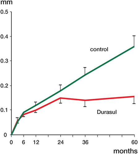
Table 1. Supine examination (at 0–5 years) for the “cemented” study
The mean amount of proximal head penetration from 3 months to 2 years, measured in standing position, was 0.08 mm in the study group and 0.14 mm in the control group (p = 0.05). During the remaining follow-up period (2–5 years), the penetration rate increased in the control group but remained almost unchanged in the study group (highly cross-linked PE, p = 0.6; control, p < 0.001; Wilcoxon signed rank test). At 5 years, the mean proximal head penetration was 3 times higher in the control group (highly cross-linked PE, 0.10 mm; control, 0.29 mm; p < 0.001) ( and ). The mean total or 3-dimensional penetration at the end of follow-up (for the standing position) reached 0.20 mm and 0.32 mm in the study group and control group, respectively (p = 0.001) ().
Figure 2. Graph showing the degree of penetration of the femoral head (proximally) into the cemented acetabular component, in 22 hips with highly cross-linked PE (Durasul) and in 24 hips with conventional (control) PE. This examination was done with the patient in the standing position. Mean ± SE.
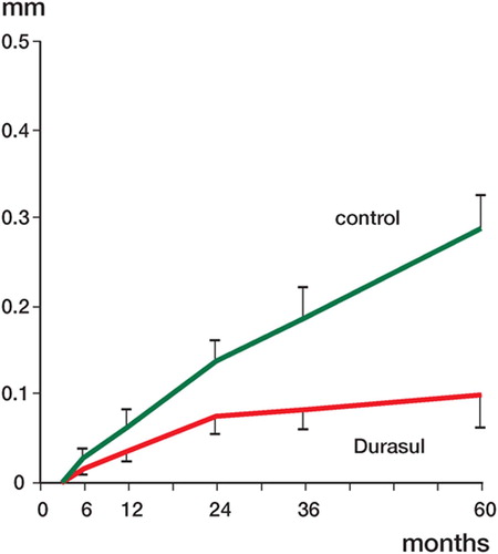
Table 2. Standing examination (at 3 months to 5 years) for the “cemented” study
The mean mediolateral and AP head penetration at 5 years was 0.05 mm or less in both groups of patients and for both positions (p ≥ 0.2) ( and ). 1 patient—a 42-year-old woman with highly cross-linked PE in the “cemented” study—was revised 5 months after the 5-year control because of stem loosening. This patient complained of pain. The RSA data at 5 years revealed stem subsidence of 6.6 mm, retroversion of 7.7o, and varus rotation of 5.2o. The cup had migrated 0.43 mm laterally and 0.34 mm proximally. Radiolucent lines were observed in regions 1 and 3. During surgery, the Weber cup was found to be well fixed whereas the stem was loose. Because of suspicion of a low-virulence infection, both components were revised—to an uncemented Trilogy cup and an Epoch stem. Multiple bacteriological specimens taken perioperatively did not yield positive cultures.
Hybrid study
The highly cross-linked PE liner showed lower proximal and total (3-dimensional) head penetration at 5 years, both in the supine and standing positions. At 1 year, the mean proximal head penetration in supine position was not significantly different in the highly cross-linked PE group and the control group (0.08 and 0.12 mm, respectively; p = 0.07). At 5 years, the mean proximal head penetration reached 0.34 mm in the control group but remained unchanged (0.08 mm) in the highly crosslinked PE group (p < 0.001) ( and ).
Figure 3. Graph showing the degree of penetration of the femoral head (proximally) into the uncemented acetabular component, in 19 patients with bilateral total hip arthroplasty. The examination was done with the patient in the supine position. Mean ± SE.
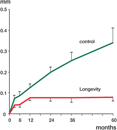
Table 3. Supine examination (at 0–5 years) for the hybrid study
On the side with highly cross-linked PE, 12 of 19 cups showed a degree of proximal penetration at 5 years that was less than 0.1 mm, whereas only 1 of the sockets on the control side showed the same low degree of penetration. The highest proximal penetration rate recorded was 0.05 mm/year in highly cross-linked group and 0.23 mm/year in the control group. The mean total (3-dimensional) head penetration was similar for both groups at the 1 year follow-up (highly cross-linked PE, 0.24 mm; control, 0.25 mm). At 5 years, the mean total (3-dimensional) head penetration was only slightly changed in the highly cross-linked PE group (0.20 mm), but had increased to 0.41 mm in the control group (p < 0.001) ().
At 5 years, the penetration could be studied bilaterally in standing position in only 12 of 23 patients. The mean proximal penetration at the end of follow-up for the highly cross-linked PE group was identical to that in the supine position (0.08 mm), while in the control group the mean proximal penetration had reached 0.28 mm (p = 0.001) (). The mean total (3-dimensional) penetration at five years was 0.31 mm and 0.37 mm in the highly crosslinked PE and control groups, respectively (p = 0.2) ().
Table 4. Standing examination (at 3 months to 5 years) for the hybrid study
Penetration in the mediolateral and AP direction throughout the follow-up period was less than 0.1 mm in both groups and for both positions, without any statistically significant difference (p ≥ 0.2) ( and ).
The mean proximal-distal migration for uncemented cups was similar for the two sides (highly cross-linked, 0.04mm; control, 0.10 mm; p = 0.09). In the study of cemented cups, the two groups showed almost equal proximal migration (highly cross-linked, 0.15 mm; control, 0.13 mm; p = 0.7). The mean mediolateral and anterior-posterior migration was less than 0.21 mm in both studies, regardless of the type of polyethylene (p ≥ 0.09). Small rotations of the liner and all-polyethylene sockets were observed (absolute mean values of ≤ 0.30°; p ≥ 0.2).
The hip and pain scores and the changes in these scores between the preoperative evaluation and the 5-year follow-up were similar between the two groups in both studies (p ≥ 0.3).
Discussion
In order to minimize the effect of confounding factors, we designed two randomized clinical stud-ies—each with a uniform socket design, femoral head diameter, head material, and type of stem. Sex, age, weight, and diagnosis were not significantly different in the study involving cemented cups. In the study involving uncemented cups, patients with bilateral hip disease were operated on simultaneously to minimize any influence of confounding factors. The two types of highly cross-linked PE studied showed significantly lower wear at 5 years of follow-up as compared to control hips.
The reduction in wear previously observed in clinical studies is lower than what was predicted in some laboratory studies (Martell et al. Citation2003, Hopper et al. Citation2003, Heisel et al. Citation2004). During the postoperative months, a large proportion of the total femoral head penetration is attributed to the bedding-in process. The total magnitude of penetration during the early follow-up overstates true material loss, whereas the steady-state penetration after the early bedding-in period almost exclusively indicates material loss or true wear.
According to our study, the bedding-in and creep for the Longevity liners was 0.08 mm. After the steady state is reached at 1 year, the wear of the polyethylene liner is undetectable using RSA. The same situation was observed in the Durasul study, but the steady state was not reached until after the 2-year follow-up. The steady state for conventional PE appears to have been reached at 6 months, and when the penetration had reached 0.09 mm in both studies. In the “cemented” study, steady state seemed to occur when the “creep” amounted to 0.15 mm—as previously observed by Dorr et al. (Citation2005) in a study of 31 patients with liners made of Durasul. In their study, steady state was also found after 2 years. Manning et al. (Citation2005) used Martell's method to study the penetration of 28-mm metal femoral heads into Durasul and Longevity liners. In that study, the femoral head penetration was statistically unchanged after 2 years of follow-up for both types of highly cross-linked PE. Thus, the run-ning-in period for Durasul PE in all 3 studies was 2 years. The different findings concerning Longevity polyethylene between the study by Manning et al. (Citation2005) and ours are difficult to explain, but may be related to the use of different methodologies. Nonetheless, these findings suggest that highly cross-linked PE creeps over a longer period of time than conventional polyethylene, perhaps because of different modulus of elasticity. This slower rate of early deformation did not appear to have any adverse effects. The only patient who was revised in our two studies had a cemented Weber cup of Durasul PE. At 5 years, the proximal femoral head penetration was 0.03 mm and the total (3-dimen-sional) penetration was 0.28 mm. The reason for the revision was stem loosening that was probably not related to PE wear, but rather for mechanical reasons such as debonding, poor cementing technique, and/or inferior bone quality.
The overall proximal penetration rates for Durasul and conventional PE cups in the “cemented” study were 0.03 and 0.072 mm/year. Excluding the bed-ding-in and creep—probably present for two years in the Durasul group and 6 months in the control group—the penetration rate could be estimated at 0.001 and 0.06 mm/year, respectively, for the two types of PE. Thus, there was 98% reduction of the wear rate for highly cross-linked PE compared to conventional PE. The corresponding penetration rate for the highly cross-linked and conventional PE liners in the hybrid study were 0.016 and 0.068 mm/ year. After the steady state had been reached (estimated to be one year for highly cross-linked PE and 6 months for conventional PE), no further penetration rate was detected on the side with highly crosslinked PE (mean 0.079 mm at 1 year and 0.078 mm at 5 years) while it was 0.057 mm/year on average on the side with conventional PE. Assuming that there is almost no wear for Longevity liners after the 1-year control (not measurable with our method), the reduction in wear compared to conventional PE would be between 99% and 100%.
Medium-term clinical results for different types of highly cross-linked PE have been published by several institutions. At 5-year follow-up, Dorr et al. (Citation2005) reported a mean linear wear rate of 0.029 mm/year for Durasul PE. In a study by Eng et al. (Citation2006), Marathon PE liners were followed for 6 years. The mean wear rate for these liners was 0.01 mm/year. D'Antonio et al. (Citation2005) reported femoral head penetration of 0.055 mm/year for Crossfire PE, as measured on plain radiographs with a minimum follow-up of 4 years. These wear rates for highly cross-linked PE liners are of about the same magnitude as those found by us.
One limitation in our studies was the relatively high drop-out rate of patients measured in standing position, especially in the “hybrid” study. Most radiostereometric evaluations of femoral head penetration have been based on radiographic examinations with the patients supine. Theoretically, standing examinations would be preferable because the joint is more stable in this position and the joint load would be expected to be more reproducible. Two previously reported studies (Bragdon et al. Citation2006, von Schewelov et al. Citation2006) did not, however, reveal any important differences between the results of examinations in these two positions. One problem with examinations done with the patients standing is inferior radiographic quality—espe-cially in obese patients. In standing position, the soft tissues tend to become displaced distally and cover the hip region, which makes accurate visualization of the cup markers and the outline of the femoral head more difficult.
The two types of highly cross-linked PE studied by us seemed to reach a steady state after 1–2 years, whereupon the wear rate decreased to more than 95% of the values recorded for conventionally sterilized PE. These data support previous predictions based on laboratory simulations. There is some concern about altered mechanical properties and different biological effects of wear particles from highly cross-linked PE compared to conventional PE. Long-term follow-up will be needed to ascertain whether these changes have any effects on long-term clinical outcome. To date, we have not seen any adverse effects of this material when used in thicknesses down to 6–8 mm. Because of its excellent wear characteristics, we recommend the use of this material in patients who are expected to have high wear rates—provided that the same thickness tolerances are used as those previously used for conventionally sterilized polyethylene.
The institution of the authors received funding from the Swedish Research Council (project no. K2002-73X-0741-16D), the Göteborg Medical Society, Centerpulse (Switzerland), and Zimmer Inc., USA.
Contributions of authors
GD: RSA measurements, statistical evaluation, writing of the manuscript, performed some of the operations. JK: study design, helped with statistical evaluation, writing of the manuscript, performed some of the operations. JT: operated many of the patients, helped with RSA measurements. PH: study design, performed some of the operations.
- Bragdon C R, Thanner J, Greene ME, Malchau H, Digas G, Harris W H, Karrholm J. Standing versus supine radiographs in RSA evaluation of femoral head penetration. Clin Orthop 2006, 448: 46–51
- D'Antonio J A, Manley M T, Capello W N, Bierbaum B E, Ramakrishnan R, Naughton M, Sutton K. Five-year experience with crossfire highly cross-linked polyethylene. Clin Orthop 2005, 441: 143–50
- Digas G, Kärrholm J, Thanner J, Malchau H, Herberts P. Highly cross-linked polyethylene in cemented THA: Randomized study of 61 hips. Clin Orthop 2003, 417: 126–38
- Digas G, Kärrholm J, Thanner J, Malchau H, Herberts P. Highly cross-linked polyethylene in total hip arthroplasty. Randomised evaluation of penetration rate in cemented and uncemented sockets using radiostereometric analysis. Clin Orthop 2004, 429: 6–16
- Dorr L D, Wan Z, Shahrdar C, Sirianni L, Boutary M, Yun A. Clinical performance of a durasul highly cross-linked polyethylene acetabular liner for total hip arthroplasty at ifve years. J.Bone joint Surg (Am) 2005; 87: 1816–21
- Dumbleton J H, Manley M T, Edidin A A. A literature review of the association between wear rate and osteolysis in total hip arthroplasty. J Arthroplasty 2002; 17: 649–61
- Engh C A, Jr, Stepniewski A S, Ginn S D, Beykirch S E, Sychterz-Terefenko C J, Hopper R H, Jr, Engh C A. A randomized prospective evaluation of outcomes after total hip arthroplasty using cross-linked marathon and non-cross-linked Enduron polyethylene liners. J Arthroplasty 2006; 21: 17–25
- Han C D, Choe W S, Yoo J H. Effect of polyethylene wear on osteolysis in cementless primary total hip arthroplasty: minimal 5-year follow-up study. J Arthroplasty 1999; 14: 714–23
- Harris W H. Wear and periprosthetic osteolysis: the problem. Clin Orthop 2001, 393: 66–70
- Heisel C, Silva M, dela Rosa M A, Schmalzried T P. Shortterm in vivo wear of cross-linked polyethylene. J Bone Joint Surg (Am) 2004; 86: 748–51
- Hopper R H, Jr, Young A M, Orishimo K F, McAuley J P. Correlation between early and late wear rates in total hip arthroplasty with application to the performance of marathon cross-linked polyethylene liners. J Arthroplasty 2003; 18: 60–7
- Jasty M, Goetz D D, Bragdon C R, Lee K R, Hanson A E, Elder J R, Harris W H. Wear of polyethylene acetabular components in total hip arthroplasty. Ananalysis of one hundred and twenty-eight components retrieved at autopsy or revision operations. J Bone Joint Surg (Am) 1997; 79: 349–58
- Kärrholm J, Herberts P, Hultmark P, Malchau H, Nivbrant B, Thanner J. Radiostereometry of hip prostheses. Review of methodology and clinical results. Clin Orthop 1997, 344: 94–110
- Manning D W, Chiang P P, Martell J M, Galante J O, Harris W H. In vivo comparative wear study of traditional and highly cross-linked polyethylene in total hip arthroplasty. J Arthroplasty 2005; 20: 880–6
- Martell J M, Verner J J, Incavo S J. Clinical performance of a highly cross-linked polyethylene at two years in total hip arthroplasty: a randomized prospective trial. J Arthroplasty 2003; 18: 55–9
- McKellop H, Shen F W, Lu B, Campbell P, Salovey R. Development of an extremely wear-resistant ultra high molecular weight polyethylene for total hip replacements. J Orthop Res 1999; 17: 157–67
- Muratoglu O K, Bragdon C R, O'Connor D O, Jasty M, Harris W H. A novel method of cross-linking ultra-high-molecular-weight polyethylene to improve wear, reduce oxidation, and retain mechanical properties. J Arthroplasty 2001; 16: 149–60
- Orishimo K F, Claus A M, Sychterz C J, Engh C A. Relationship between polyethylene wear and osteolysis in hips with a second-generation porous-coated cementless cup after seven years of follow-up. J Bone Joint Surg (Am) 2003; 85: 1095–9
- Röhrl S, Nivbrant B, Mingguo L, Hewitt B. In vivo wear and migration of highly cross-linked polyethylene cups a radiostereometry analysis study. J Arthroplasty 2005; 20: 409–13
- Selvik G. Roentgen stereophotogrammetry. A method for the study of the kinematics of the skeletal system. Acta Orthop Scand 1989, Suppl 232: 1–51
- Sychterz C J, Young A M, McAuley J P, Engh C A. Comparison of head penetration into Hylamer and Enduron polyethylene liners: a follow-up report. J Arthroplasty 2000; 15: 372–4
- Söderkvist I, Wedin P A. Determining the movements of the skeleton using well configured markers. J Biomech 1993; 26: 1473–7
- Von Schewelov T, Onsten I, Markusson P, Carlsson A. Weight bearing radiographs are not necessary for measurement of polyethylene penetration in total hip prostheses: a radiostereometric study of 111 patients examined in weight-bearing and supine position. Acta Orthop 2006; 77: 104–8
- Wilkinson J M, Hamer A J, Stockley I, Eastell R. Polyethylene wear rate and osteolysis: critical threshold versus continuous dose-response relationship. J Orthop Res 2005; 23: 520–5