
ABSTRACT
Controlled ovarian hyperstimulation (COH) impairs the endometrium receptivity during the implantation window, resulting in a lower clinical pregnancy rate and a higher abortion rate. Our study explored the effect of electroacupuncture on the endometrial receptivity of COH rats. Female rats were randomly divided into normal treatment (Normal), model treatment (Model), low-frequency electroacupuncture treatment (LF-EA) and high-frequency electroacupuncture treatment (HF-EA). Rats in the Model, LF-EA, and HF-EA treatment groups were injected with pregnant mare serum gonadotropin (PMSG) and human chorionic gonadotropin (HCG) to establish a model of COH rats. Compared with the Normal, the endometrial thickness, the number of pinopodes and amount of blastocyst implantation in the Model group were significantly reduced. Among them, the endometrial thickness and the amount of blastocyst implantation in the Model group were substantially decreased than those in the HF-EA group. High-frequency electroacupuncture treatment could markedly reduce the protein expression levels of E-cadherin, β-catenin and claudin-1 (CLDN1). During HF-EA treatment, the LIF/STAT3 signaling pathway of COH rats was enhanced. In conclusion, electroacupuncture could improve the endometrium receptivity and promote the blastocyst implantation in COH rats by reducing cell adhesion molecules and enhancing the LIF/STAT3 signaling pathway.
Highlights
High-frequency electroacupuncture could effectively improve endometrial receptivity and blastocyst implantation amount in COH rats.
Electroacupuncture, especially high-frequency electroacupuncture, could significantly increase endometrial thickness and the number of pinopodes.
High-frequency electroacupuncture significantly reduced the protein expression levels of E-cadherin, β-catenin and CLDN1 adhesion molecules in COH rats.
High-frequency electroacupuncture could markedly enhance the LIF/STAT3 signaling pathway in COH rats.
Introduction
Embryonic implantation is the crucial biological process in the reproduction of mammals, which allows the activated blastocysts to connect with endometrium by ways of localization, adhesion and invasion. Endometrial receptivity is a parameter to describe embryo implantation onto the endometrium, on which a successful pregnancy is depended [Citation1–4]. Previous studies have shown that administration of ovulation-inducing drugs in the controlled ovarian hyperstimulation (COH) might bring about an interference of endogenous steroids to endometrium physiological regulation, thus affecting the endometrium receptivity within implantation window period, leading to lower rates in clinical pregnancy and higher ones in abortion. In this context, the decreasing endometrial receptivity that COH brings on comes under the spotlight [Citation5–7]. Hormone regulation, microcirculation improvement, mechanical stimulation, surgery and traditional Chinese medicine are the conventional means to improve endometrial receptivity. Besides, acupuncture, as a therapy of traditional Chinese medicine, boasts a history of thousands of years. It has been applied in the treatment of various diseases and has cured numerous diseases for human beings which also produces a compelling efficiency in improving both endometrial receptivity and clinical pregnancy rate [Citation8–11].
As an improvement and development of traditional acupuncture, electroacupuncture is an effective non-drug remedy. It is widely used in many fields in recent years, the preclinical treatment research in persistent tissue damage (inflammation), nerve damage, cancer and visceral pain has increased [Citation12–14]. In alleviating pain, treatment of Parkinson’s disease (PD), neural mechanism research, Alzheimer’s disease and upper limb rehabilitation after stroke have also achieved good curative effects [Citation15–18]. High-frequency electroacupuncture, in particular, is often used as an alternative therapy for PD and is effective in reducing motor symptoms in patients and PD models [Citation19]. It can improve cognitive impairment in rats with amyloid Alzheimer’s disease and enhance synaptic transmission in hippocampus of such rats [Citation16,Citation20]. In addition, quantitative studies in the field of reproduction are also extensively investigated, for example, electroacupuncture is used to improve the endometrial receptivity of patients undergoing freeze-thaw embryo transfer [Citation21]. In addition, there are studies reported that electroacupuncture can improve the outcome of in vitro fertilization (IVF) and improve the success rate of assisted reproductive technology (ART) [Citation22,Citation23].
Recent studies on endometrial receptivity are scattered on the aspects of genes, proteins, cytokines, adhesion molecules, microRNAs and endometrial flora. E-cadherin, β-catenin and CLDN1, the proteins located in cell membrane and cytoplasm, have been proved as associated molecules with the adhesion and the tight connection of endometrial gland epithelial cells. Down-regulation of these adhesion molecules facilitates the dissociation of epithelial cells and the implantation of blastocysts [Citation24–28].
Leukemia inhibitory factor (LIF), a member of IL-6 family, is a cytokine of multiple biological activities. LIF is an active player in reproduction, involved in follicle and embryo development, embryo implantation and pregnancy maintenance. LIF binds to the leukemia inhibitory factor receptor (LIFR) on the target cell membrane and the transmembrane glycoprotein gp130 as a dimer, activating the Janus kinase (JAK) and phosphorylating the transcriptional regulator STAT3 [Citation29–33].
In this study, as an improvement of acupuncture, electroacupuncture would possess a positive effect on the endometrium receptivity. The effect of electroacupuncture on endometrial receptivity and the molecular mechanism of treatment were investigated. First, the efficacy of electroacupuncture at different frequencies to rats’ endometrium receptivity was evaluated by commencing HE staining and electron microscopy. Meanwhile, the effects of different frequencies to adhesion factors such as E-cadherin, β-catenin and CLDN1 were detected. The role of LIF/STAT3 signaling pathway in electroacupuncture treatment of COH rats was examined. It is expected to preliminarily explore the molecular mechanism of high-frequency electroacupuncture in improving endometrial receptivity in our study and provide new insights for acupuncture in improving IVF, endometrial receptivity and clinical pregnancy rate.
Methods
Experimental animals
The animal experiments were approved by the Guizhou University of Chinese Medicine. We split up female SD rats randomly into four treatment groups with 20 mice in each group. The animals were categorized as normal treatment, model treatment, LF-EA treatment and HF-EA treatment. Animal experiment was started from the 3rd day of the emotional cycle of rats, and rats of model treatment group underwent with intraperitoneal injection. At 9:00 on the 7th day, rats of model group were injected with 40 IU/100 g of Pregnant Mare Serum Gonadotropin (PMSG) and 100 IU/100 g of HCG was injected at 1:00 on the 8th day [Citation34]. Rats of normal group were injected with equal volume saline daily for eight consecutive days. The model was successfully established as long as rats’ vaginal secretions were significantly increased and a large number of nuclear-free keratinocytes were found in the smear of secretions under a light microscope.
Electroacupuncture therapy
Acupoints Guan Yuan (CV4) and Zusanli (ST36) were selected in terms of acupuncture atlas of experimental animals formulated by the National Acupuncture and Moxibustion Society, and 0.30 × 13 mm stainless steel milli-needles were selected, with the acupuncture depth of 3–5 mm. The course of treatment electroacupuncture lasted for 7 days, with one day interval between two courses and four courses during treatment (31 days in total). The rest groups of rats only grabbed and fixed without electroacupuncture treatment. (1) Low frequency electroacupuncture treatment group: Electrical stimulation was applied in the form of disperse-dense waves at alternating 2 Hz, respectively, and 1 mA intensity for 20 min. (2) High-frequency electroacupuncture treatment group: Electrical stimulation was applied in the form of disperse – dense waves at alternating 50 Hz, respectively, and 1 mA intensity for 20 min.
HE staining
The endometrial histological characteristics of the rats were compared. Endometrial tissues fixed with fresh 4% paraformaldehyde, subsequently with the embedding of paraffin following conventional histological procedures. After that, the samples were sliced in 4.5 μm-thick sections that were then processed with hematoxylin and eosin (HE) staining [Citation35].
SEM observation
The development of the apical vesicle process of endometrial epithelial cells was observed under a scanning electron microscope. The process was divided into three stages: development, maturation and decline. The microvilli in the process of development became slender, dense and erect, and the top membrane-like process gradually formed and developed to the top of the whole cell. Then, the microvilli became shorter and less and fuse with each other. The microvilli in the process of development completely disappeared, and the surface was smooth, and the membrane-like process became larger, higher than the ciliate cells, such as mushroom. The membrane-like process in the process of decline shrans and the surface reappeared microvilli. According to the percentage (>50%, 20%-50%, <20%) of the surface area of the endometrium, the number of the process can be divided into three kinds: rich, medium and small [Citation36].
Immunofluorescence experiment
The cells were washed with PBS, and Triton X-100 was dripped on the tablet and 10% serum was added. Climbing tablets were added with primary antibody drops and put into a wet chamber, incubated overnight and rewarmed at room temperature for 30 min. The diluted fluorescent secondary antibody was added dropwise and incubated in a wet chamber for 60 min. After PBS immersion, DAPI was added dropwise [Citation37]. Cells were observed by a fluorescence microimaging system.
Western blot analysis
We made quantification to the total protein extracted from frozen endometrial samples. Proteins were isolated with SDS-polyacrylamide gel electrophoresis and run with membrane transfer. Incubation of the proteins was initially performed with primary antibody and then with secondary antibody; chemiluminescence color was triggered, and optical density value was determined using a gel image analysis system. An additional experiment was served as an internal reference for beta-actin.
RNA extraction and real-time reverse transcription q-PCR
Extraction of total RNA from the sample was commenced with Trizol reagent (Takara, Japan). Reverse-transcription of cDNA was accomplished with PrimeScript TMR T kit (Takara, Japan). Reaction system and procedures for qRT-PCR were described in the instructions of the TB Green Premix Ex Taq II (Takara, Japan), with CFX96 Real-Time system (Bio-Rad, USA) as the platform. Relative expression of the gene was calculated with 2−∆∆CT algorithm [Citation38].
Primer sequence of LIF-F: (CCAACAACGTGGATAAGCTATG) and LIF-R (GTTTTTCTGATC CCAGGTGATG).
Histological and immunohistochemistry (IHC) analysis
Samples were fixed with fresh 4% paraformaldehyde and went along with conventional histological procedures for embedding in paraffin. These samples were cut in 4.5 μm-thick sections, processed for IHC staining with primary antibodies and then incubated with secondary antibodies. Stained slides were scanned with a Pannoramic Scan 250 Flash [Citation39].
Statistical analysis
SPSS 23.0 was adopted for data analysis. The mean values of two groups of independent samples were compared using t test, and then the mean values of the samples from multiple groups were analyzed using one-way ANOVA, P < 0.05. All the experiments were repeated three times independently, and the results showed the mean value ± standard deviation.
Results
In this experiment, rats were divided into four treatment groups: normal treatment, model treatment, LF-EA treatment and HF-EA treatment. HE staining was used to observe the estrus and endometrial thickness of rats in different treatments. The effect of electroacupuncture on the endometrium of rats was observed by a scanning electron microscope, and the amount of blastocyst implantation in different treatment groups was compared. Immunofluorescence and immunoblotting experiments were used to observe the effect of electroacupuncture on adhesion molecules in rats. Immunohistochemistry and qPCR were used to detect the expression of LIF/STAT3 signaling pathway in each treatment group.
Efficacies of different-frequency electroacupuncture treatment on rats’ endometrial thickness
In order to study the effects of LF-EA and HF-EA on the emotional cycle of rats, the animals were divided into normal group, model group, LF-EA group and HF-EA group. Proestrus in rats showed a large number of nucleated epithelial cells and a small number of keratinized cells. Estrum was characterized by allopic keratinized epithelial cells, a small number of nucleated epithelial cells metestrum were keratinized epithelial cells and leukocytes and diestrus was a large number of leukocytes and a small amount of mucus ().
Figure1. Effect of electroacupuncture on endometrial thickness of rats during estrous cycle. (a) Vaginal secretions in rats were detected by HE staining. (b) Efficacies of different-frequency electroacupuncture treatment on rats’ endometrial thickness. Data are represented as mean ± SD (n ≥ 3 experiments). *p < 0.05, **p < 0.01, ***p < 0.001 and ****p < 0.0001 as determined using Student’s t-test (two groups) or one-way ANOVA, followed by Tukey’s test (more than two groups)
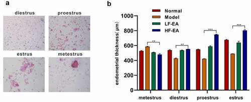
The thickness of endometrium in rats was observed by HE staining. The results showed that the thickness of endometrium in LF-EA group and HF-EA group was significantly higher than that in model group. The thickness of endometrium in model group was significantly lower than that in normal group. This suggested that electroacupuncture might improve endometrial receptivity by increasing endometrial thickness in rats.
Efficacies of different-frequency electroacupuncture treatment on rats’ implantation ability of blastocyst
Pinopode is a large, smooth membranous protrusion formed by endometrial epithelial cells. Microvilli on the surface of intimal epithelial cells disappeared during implantation and were replaced by a larger and smooth protuberance known as vesicle. Pinopode appeared about 1 week after ovulation. The presence lasted for a very short time and resolved after about 48 h. In normal and animal models, the number of pinopodes was positively correlated with the amount of blastocyst implantation. Fully developed pinopode was one of the most representative markers of morphological structure in the evaluation on the endometrium as well as in the embryo-positioned bed window. Six rats in each treatment group were randomly sacrificed with their uteri removed by laparotomy for electron microscope observation. Amount of pinopodes declined significantly in model treatment compared than normal treatment (). While amount of pinopodes were increased in LF-EA and HF-EA compared with model treatment, and in addition, more pinopodes were observed in HF-EA.
Figure 2. Effect of electroacupuncture on endometrial membrane capacitance in rats. (a) The pinopodes of endometrial epithelial cells were observed with electron microscopy. (b) Amount of blastocyst implantation in rats was calculated by observing the uterus of each group. Data are represented as mean ± SD (n ≥ 3 experiments). *p < 0.05, **p < 0.01, ***p < 0.001 and ****p < 0.0001 as determined using Student’s t-test (two groups) or one-way ANOVA, followed by Tukey’s test (more than two groups)
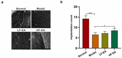
After accounting the number of implantation sites in the uterus on both sides of rats, we found that that number of model treatment was notably reduced in contrast to the control treatment; and it was inverse in LF-EA and HF-EA treatments that the amount of blastocyst implantation sites was greatly increased compared with model treatment ().
Efficacies of different-frequency electroacupuncture treatment on rats’ endometrial adhesion factors
We suspected that the adhesion molecules E-cadherin, β-catenin and CLDN1 in the present study might function dually. At the initial stage, it needs to be expressed on the cell surface to ensure adhesion, and at the later stage it begins to be down-regulated, resulting in epithelial cell dissociation and blastocyst invasion, and reduced expression of cell surface adhesion molecules.
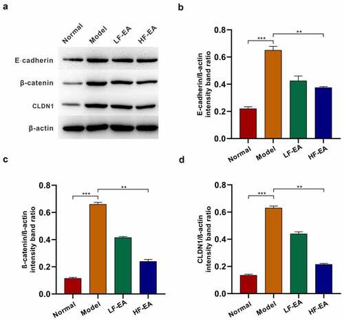
This study attempted to figure out the effects of different-frequency electroacupuncture on E-cadherin, β-catenin and CLDN1 in rats’ endometrium of different treatment groups, and expressions of E-cadherin, β-catenin and CLDN1were detected by immunofluorescence experiments. Then it turned out that expression of E-cadherin in model group was significantly higher than the normal; expression of β-catenin in model group increased signally than the normal; besides, expression of CLDN1 in model treatment group increased conspicuously compared with the normal; moreover, expression of CLDN1 decreased significantly in both LF-EA and HF-EA treatment groups (). The expressions of E-cadherin, β-catenin and CLDN1 were also detected by Western Blot experiments and the results were consistent with the immunofluorescence assays ().
Figure 3. Expressions of Claudin, E-cadherin and β-catenin in rat endometrium were detected by immunofluorescence. (a) The levels of E-cadherin in endometrium of rats were detected by immunofluorescence. (b) Expressions of β-catenin in endometrium of rats were detected by immunofluorescence. (c) Expressions of CLDN1 in endometrium of rats were detected by immunofluorescence. Data are represented as mean ± SD (n ≥ 3 experiments). *p < 0.05, **p < 0.01, ***p < 0.001 and ****p < 0.0001 as determined using Student’s t-test (two groups) or one-way ANOVA, followed by Tukey’s test (more than two groups)
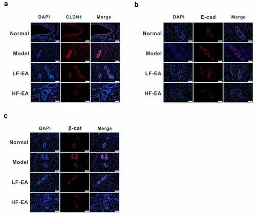
Figure 4. Electroacupuncture inhibited the expressions of E-cadherin, β-catenin and CLDN1 in the endometrium of rats. (a) Expressions of E-cadherin, β-catenin and CLDN1 in rats’ endometrium of each group were measured with Western bolt. (b) Protein quantitative map of E-cadherin in endometrium of rats in each group. (c) Quantitative protein mapping of β-catenin in endometrium of rats in each group. (d) Quantitative graph of CLDN1 protein in endometrium of rats
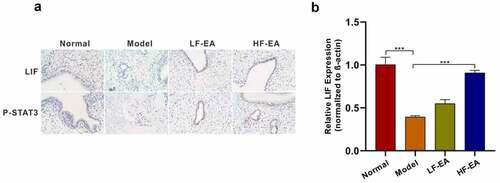
Efficacies of different-frequency electroacupuncture treatment on the LIF/ STAT3 signaling pathway in rat endometrium
Subsequently, an investigation was performed to verify the effects of different-frequency electroacupuncture remedy on LIF/STAT3 signaling pathway in rat endometrium; effects of LF-EA and HF-EA on LIF/STAT3 signaling pathway in rat endometrium have been verified with immunohistochemistry and qPCR. The findings showed that the expressions of LIF and P-STAT3 in model group were significantly lower than the normal group; either the expressions of LIF and P-STAT3 in the treatments underwent low frequency electroacupuncture or high frequency electroacupuncture was evidently higher than the model treatment (). The results of qPCR also indicated that the LIF expression in model group was obviously lower than the normal group, and the levels of LF-EA and HF-EA in model group were significantly higher than those in model group (). The results verified the impacts of different-frequency electroacupuncture remedy on LIF/ STAT3 signaling pathway.
Figure 5. Electroacupuncture increased levels of LIF/ STAT3 signaling pathway. (a) Both LIF and P-STAT3 expressions in rat endometrium were detected with immunohistochemical. (b) LIF expression in rat endometrium was detected with qPCR. Data are represented as mean ± SD (n ≥ 3 experiments). *p < 0.05, **p < 0.01, ***p < 0.001, and ****p < 0.0001 as determined using Student’s t-test (two groups) or one-way ANOVA, followed by Tukey’s test (more than two groups)
Discussion
Numerous recent research has been found on female infertility, including endometrial cancer, endometriosis and polycystic ovary syndrome [Citation40–43]. In vitro fertilization-embryo transfer (IVF-embryo transfer) is an assisted reproductive technique in which COH ensures the number and quality of follicles and improve the success rate of ART, but the implantation rate of embryos is only 20%-30% [Citation44]. In addition to the quality of the embryo, the present mechanism of embryo implantation also contributes to the failure of implantation. Increasing studies have shown that ovarian steroid hormones, cytokines, transcription factors and lipid messengers are involved in the implantation process through autocrine and paracrine forms [Citation21,Citation45,Citation46]. In particular, approximately 30% of embryo implantation failure is associated with low uterine receptivity [Citation47,Citation48]. Therefore, improved endometrial receptivity is expected to increase success rate of embryo implantation and lay a foundation for improving clinical pregnancy rate.
Previous studies have showed that electroacupuncture has good clinical effect on endometrial receptivity deficiency [Citation34]. However, the molecular mechanism of electroacupuncture treatment for endometrial receptivity is not fully elucidated. Frequency is one of the important parameters of electroacupuncture stimulation. Curative effects of electroacupuncture remedy with low frequency electricity have been widely reported, whereas those with high frequency electricity on endometrial receptivity remain to be fully studied.
To verify the effect of different frequencies of electroacupuncture on endometrial receptivity during the superovulatory cycle, we studied the effect of electroacupuncture on the implantation capacity of rat blastocysts. The results turned out that the blastocysts implantation number in HF-EA group increased significantly than model group. The above described results suggested that electroacupuncture remedy increased the number of endocytosis and implantation ability of blastocysts in endometrium, and the implantation effect of HF-EA group was better than that of LF-EA group.
As adhesion molecules, E-cadherin, β-catenin and CLDN1 are closely associated with endometrial epithelial cell adhesion, enabling epithelial cell separation and blastocyst invasion and are essential in embryo transplantation [Citation49]. The results suggested that E-cadherin, β-catenin and CLDN1 are involved in embryo transfer. Our study showed that expressions of E-cadherin, β- catenin and CLDN1 in endometrium of ovulation-promoting rats decreased significantly after treatment with LF-EA and HF-EA, which indicated that electroacupuncture could dissociate epithelial cells, increase the ability of blastocyst transplantation and improve the endometrium receptivity.
Both LIF and STAT3 are essential for rats’ blastocyst implantation [Citation50,Citation51]. LIF plays a crucial role in the process of blastocyst transplantation and can regulate endometrial receptivity and control blastocyst transplantation by promoting cell transplantation and differentiation. It also regulates phosphorylation of STAT3 and plays an important role in embryo transfer to endometrium. Immunohistochemistry and qPCR were carried out to investigate the effects of LF-EA and HF-EA on the expression levels of LIF and p-STAT3 proteins in COH rats. The results showed that the expressions of LIF and p-STAT3 in LF-EA and HF-EA treatment and normal treatment were significantly increased compared with model treatment. These results suggested that electroacupuncture could improve endometrial receptivity and enhance blastocyst transplantation ability in COH rats by enhancing LIF/STAT3 signaling pathway.
Conclusion
The assay findings revealed that electroacupuncture at different frequencies could increase the receptivity of endometrium in rats, and this effect of electroacupuncture with HF-EA was better than the one with LF-EA. Further verification indicated that electroacupuncture reduced the expression of adhesion molecules such as E-cadherin, β- catenin and CLDN1 and provided conditions for blastocyst implantation. Furthermore, it also demonstrated that electroacupuncture increased the level of LIF/STAT3 signaling pathway. However, more investigations are still necessary to explore the effects of different frequencies of electroacupuncture on other factors of endometrial receptivity.
Disclosure statement
No potential conflict of interest was reported by the author(s).
Additional information
Funding
References
- Alauddin M, Salker MS, Umbach AT, et al. Annexin A7 regulates endometrial receptivity. Front Cell Dev Biol. 2020;8: DOI:10.3389/fcell.2020.00770.
- Wang W, Yu Y. An update on the progress of transcriptomic profiles of human endometrial receptivity†. Biol Reprod. 2018;98(4):440–448.
- Altmäe S, Aghajanova L. Growth hormone and endometrial receptivity. Front Endocrinol (Lausanne). 2019;10(653):653.
- Paulson RJ. Sterility, Introduction: endometrial receptivity: evaluation, induction and inhibition. Fertil Steril. 2019;111(4):609–610.
- Teh W-T, McBain J, Rogers P. What is the contribution of embryo-endometrial asynchrony to implantation failure? J Assist Reprod Genet. 2016;33(11):1419-1430.
- Cortínez A, Decarvalho I, Vantman D, et al. Hormonal profile and endometrial morphology in letrozole-controlled ovarian hyperstimulation in ovulatory infertile patients. Fertil Steril. 2005;83(1):110–115.
- Paulson RJ, Sauer MV, Lobo RA. Potential enhancement of endometrial receptivity in cycles using controlled ovarian hyperstimulation with antiprogestins: a hypothesis. Fertil Steril. 1997;67(2):321.
- Xi J, Cheng J, Jin -C-C, et al. Electroacupuncture improves pregnancy outcomes in rats with thin endometrium by promoting the expression of pinopode-related molecules. Biomed Res Int. 2021;2021:6658321.
- Zhong Y-J, Zeng F-Z, Liu W-J, et al. Acupuncture in improving endometrial receptivity: a systematic review and meta-analysis. BMC Complement Altern Med. 2019;19(1):61.
- Zheng C-S, Luo D, Pan L-P, et al. Therapeutic effects on infertility of ovulation failure in the patients with kidney deficiency treated with abdominal acupuncture and periodic therapy of Chinese herbal medicine. Zhongguo Zhen Jiu. 2019;39:3.
- Liu Y, Pan L, Wang Y. [Effects of the combined therapy of heat sensitive moxibustion and acupoint injection on endometrial receptivity of hypdrosalphinx infertility in the patients after hysteroscopy and laparoscopy]. Zhongguo Zhen Jiu. 2018;038(1):22–26.
- Liu H-R, Fang X-Y, Wu H-G, et al. Effects of electroacupuncture on corticotropin-releasing hormone in rats with chronic visceral hypersensitivity. World J Gastroenterol. 2015;21(23):7181–7190.
- Ying W, Gehringer R, Mousa SA, et al. CXCL10 controls inflammatory pain via opioid peptide-containing macrophages in electroacupuncture. PLoS One. 2014;9(4):e94696.
- Jiang X, Tian Y, Xu L, et al. Inhibition of triple-negative breast cancer tumor growth by electroacupuncture with encircled needling and its mechanisms in a mice xenograft model. Int J Med Sci. 2019;16(12):1642–1651.
- Kim HY, Koo ST, Kim JH, et al. Electroacupuncture analgesia in rat ankle sprain pain model: neural mechanisms. Neurol Res. 2010;32(sup1):10–17.
- Li W, Kong L-H, Wang H, et al. High-frequency electroacupuncture evidently reinforces hippocampal synaptic transmission in Alzheimer′s disease rats. J Neural Regener Res. 2016;11(5):801–806.
- Xu W, OuYang S, Chi Z, et al. Effectiveness and safety of electroacupuncture in treating Parkinson disease: a protocol for systematic review and meta-analyses. Medicine (Baltimore). 2021;100(10):e25095.
- Lin YF, Liu X-H, Cui Z-Y, et al. Weakened effective connectivity related to electroacupuncture in stroke patients with prolonged flaccid paralysis: an EEG pilot study. Neural Plast. 2021;2021:6641506.
- Huo L-R, Liang X-B, Li B, et al. The cortical and striatal gene expression profile of 100 Hz electroacupuncture treatment in 6-hydroxydopamine-induced parkinson’s disease model. Evid Based Complement Alternat Med. 2012;2012:908439.
- Yu CC, Wang Y, Shen F, et al. High-frequency (50 Hz) electroacupuncture ameliorates cognitive impairment in rats with amyloid beta 1–42-induced Alzheimer’s disease. J Neural Regener Res. 2018;13(10):1833–1841.
- Shuai Z, Lian F, Li P, et al. Effect of transcutaneous electrical acupuncture point stimulation on endometrial receptivity in women undergoing frozen-thawed embryo transfer: a single-blind prospective randomised controlled trial. Acupuncture Med. 2015;33(1):9–15.
- Qu F, Wang -F-F, Wu Y, et al. Transcutaneous electrical acupoint stimulation improves the outcomes of in vitro fertilization: a prospective, randomized and controlled study. EXPLORE (NY). 2017;13(5):306–312.
- Anderson BJ, Haimovici F, Ginsburg ES, et al. In vitro fertilization and acupuncture: clinical efficacy and mechanistic basis. Altern Ther Health Med. 2007;13(3):38.
- Movaghar B, Askarian S. Expression of E-cadherin, leukemia inhibitory factor and progesterone receptor in mouse blastocysts after ovarian stimulation. Cell J. 2012;14(3):225–230.
- Rahnama F, Thompson B, Steiner M, et al. Epigenetic regulation of e-cadherin controls endometrial receptivity. Endocrinology. 2009;150(3):1466–1472.
- Zhou X-W, Wu B-F, Zhang Dan, et al. Loss of CDYL Results in Suppression of CTNNB1 and Decreased Endometrial Receptivity. Front Cell Dev Biol. 2020;8:105.
- Bellati F, Costanzi F, De Marco MP, et al. Low endometrial beta-catenin and cadherins expression patterns are predictive for primary infertility and recurrent pregnancy loss. Gynecological Endocrinol. 2019;35(8):1–5.
- Shirane A, Wada-Hiraike O, Tanikawa M, et al. Regulation of SIRT1 determines initial step of endometrial receptivity by controlling E-cadherin expression. Biochem Biophys Res Commun. 2012;424(3):604–610.
- Chung T-W, Park M-J, Lee H, et al. Enhancement of endometrial receptivity by cnidium officinale through expressing LIF and Integrins. Evid Based Complement Alternat Med. 2019;2019:7560631.
- Shariati M, Niknafs B, Seghinsara AM, et al. Administration of dexamethasone disrupts endometrial receptivity by alteration of expression of miRNA 223, 200a, LIF, Muc1, SGK1, and ENaC via the ERK1/2‐mTOR pathway. J Cell Physiol. 2019;234(11):19629–19639.
- Shokrzadeh N, Alivand MR, Abedelahi A, et al. Calcitonin administration improves endometrial receptivity via regulation of LIF, MUC-1 and MicroRNA Let-7a in Mice. J Cell Physiol. 2018;234(8):12989–13000.
- Nicola NA, Babon JJ. Leukemia inhibitory factor (LIF). Cytokine Growth Factor Rev. 2015;26(5):533–544.
- Heim MH. The jak-stat pathway: cytokine signalling from the receptor to the nucleus. J Recept Res. 1999;19(1–4):75–120.
- Chen W, Chen J, Xu M, et al. Electroacupuncture facilitates implantation by enhancing endometrial angiogenesis in a rat model of ovarian hyperstimulation. Biol Reprod. 2019;100(1):268–280.
- Nguyen DT, O’Hara M, Graneli C, et al. Humanizing miniature hearts through 4-flow cannulation perfusion decellularization and recellularization. Sci Rep. 2018;8(1):7458.
- Zhao X, Li J, Cheng S, et al. Study on the role of paclitaxel nano-drug delivery system in inhibiting intimal hyperplasia and improving vascular remodeling in abdominal aortic injury model. J Nanosci Nanotechnol. 2021;21(2):1385–1389.
- Zhao J, Shi Q, Zheng Y, et al. Insights into the mechanism of tyrosine nitration in preventing β-amyloid aggregation in alzheimer’s disease. Front Mol Neurosci. 2021;14:619836.
- Tawfeek MA, Eid MA, Hasan AM, et al. Assessment of leukemia inhibitory factor and glycoprotein 130 expression in endometrium and uterine flushing: a possible diagnostic tool for impaired fertility. BMC Women’s Health. 2012;12(1):10.
- Shi Y, Zou Y, Xiong Y, et al. Host Gasdermin D restrains systemic endotoxemia by capturing Proteobacteria in the colon of high-fat diet-feeding mice. Gut Microbes. 2021;13(1):1946369.
- Jing. L, Liang J, Xu M, et al. Identification of an eleven-miRNA signature to predict the prognosis of endometrial cancer. Bioengineered. 2021;12(1):4201–4216.
- Seow K-M, Chang Y-W, Chen K-H, et al. Molecular mechanisms of laparoscopic ovarian drilling and its therapeutic effects in polycystic ovary syndrome. Int J Mol Sci. 2020;21(21):8147.
- Rogers PAW, Adamson GD, Al-Jefout M, et al. Research priorities for endometriosis. Reprod Sci. 2017;24(2):202–226.
- Yingying. Y, Li G, He X, et al. MicroRNA-21 regulate the cell apoptosis and cell proliferation of polycystic ovary syndrome (PCOS) granulosa cells through target toll like receptor TLR8. Bioengineered. 2021;12(1):5789–5796.
- De Geyter C, Jorge CC, Kupka MS, et al. The European IVF-monitoring Consortium (EIM) ‡ for the European Society of Human Reproduction and Embryology (ESHRE). Human Reproduction. 2018;33(9):1586-1601.
- Mu Y-Y, Li Q, Cheng J, et al. Integrated miRNA-seq analysis reveals the molecular mechanism underlying the effect of acupuncture on endometrial receptivity in patients undergoing fertilization: embryo transplantation. Biotechnology. 2020;10(1):1–9.
- Penzias A, Bendikson K, Butts S, et al. Performing the embryo transfer: a guideline. Fertil Steril. 2017;107(4):882.
- Sharkey AM, Smith SK. The endometrium as a cause of implantation failure. Best Pract Res Clin Obstetrics Gynaecol. 2003;17(2):289–307.
- Bissonnette L, Drissennek L, Antoine Y, et al. Human S100A10 plays a crucial role in the acquisition of the endometrial receptivity phenotype. Cell Adhes. 2016;10(3):282–298.
- Sachiko M, Darcha C, Maleysson E, et al. Impaired down-regulation of E-cadherin and beta-catenin protein expression in endometrial epithelial cells in the mid-secretory endometrium of infertile patients with endometriosis. J Clin Endocrinol. 2010;95(7):3437–3445.
- Sun X, Bartos A, Whitsett JA, et al. Uterine deletion of gp130 or stat3 shows implantation failure with increased estrogenic responses. Mol Endocrinol. 2013;27(9):1492–1501.
- Stewart CL, Kaspar P, Brunet LJ, et al. Blastocyst implantation depends on maternal expression of leukaemia inhibitory factor. Nature. 1992;359(6390):76–79.