
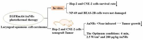
ABSTRACT
It reported that heat generated by near-infrared laser irradiation of gold nanorods (AuNRs) effectively inhibited tumor cells, and the conjugate of epidermal growth factor receptor monoclonal antibody (EGFRmAb) and gold nanorods could selectively binded to the surface of cancer cell membrane expressing EGFR. However, there are few research reports on EGFRmAb-AuNRs in laryngeal squamous cell carcinoma. Therefore, our study aimed to investigate the photothermal effect of EGFRmAb modified AuNRs in inducing tumor cell death in an animal model of laryngeal squamous cell carcinoma. We showed that the conjugates of EGFRmAb and AuNRs selectively entered laryngeal squamous cell carcinoma cells. We analyzed the parameters of laser irradiation by controlling the near-infrared to optimize the condition and procedure of targeted treatment in nude mice treated with EGFRmAb and AuNRs. In addition, we examined the safety of the combined therapy. Test results showed that EGFRmAb-AuNRs inhibited the growth of Hep-2 and CNE-2 cells but not normal epithelial cells, and the semi-inhibitor concentration of EGFRmAb in Hep-2 and CNE-2 cells was 4 pmol/ml and 2 pmol/ml, respectively. AuNRs injected into the tumor and irradiated by near-infrared laser effectively inhibited tumor growth in nude mice without toxic effect in mice. We further confirmed that the apoptosis and necrosis rates of tumor cells in mice were highest under 3 W/cm2 near-infrared laser irradiation and AuNRs minimum concentration of 280 μg/kg. In conclusion, we developed a new method of targeting EGFRmAb combined with AuNRs to achieve photothermal effect in the treatment of laryngeal squamous cell carcinoma.
Graphical abstract
Introduction
Laryngeal squamous cell carcinoma is an aggressive cancer characterized by metastatic potential and high mortality [Citation1]. In recent years, the incidence of laryngeal squamous cell carcinoma has increased significantly worldwide [Citation2]. Most of the patients are commonly diagnosed in the middle and late stages [Citation3]. Therefore, it is necessary to develop effective treatment model to improve the survival rates of patients.
Gold nanorods (AuNRs) are one of the most important noble metal nanomaterials easy to prepare and have good stability [Citation4]. The experiments in vitro showed that instantaneous heat generated by near-infrared laser irradiation of AuNRs was sufficient to destroy tumor cells effectively [Citation5]. In addition, AuNRs can encapsulate tumor-specific labels or antibodies on the surface of the particles to enhance the targeting of AuNRs to tumor tissues, so that AuNRs efficiently aggregate into tumor tissues [Citation6]. These characteristics make AuNRs an ideal tool for plasma photothermal therapy and targeted cancer therapy [Citation7]. AuNRs have been widely used in the treatment of head and neck tumors [Citation8].
AuNRs have a strong affinity with antibodies, proteins, and DNA fragments, and the resulting conjugates can further target cancer cells [Citation6,Citation9]. Epidermal growth factor receptor (EGFR) is a tyrosine kinase receptor overexpressed in head and neck malignant tumors [Citation10,Citation11]. Therefore, EGFR is considered to be an important biological therapeutic target [Citation12]. Some studies showed that the conjugates of EGFR monoclonal antibodies and AuNRs can selectively bind to the membrane surface of cancer cells expressing EGFR [Citation13–15]. EGFR monoclonal antibody (EGFRmAb) has been extensively studied as a functionalized antibody commonly used with AuNRs [Citation16,Citation17]. Our previous studies have shown that the functionalization of EGFRmAb did not change the optical properties of AuNRs, but increased the ability of AuNRs to map high expression of EGFR protein in laryngeal squamous cell carcinoma (Hep-2) cells. The photothermal effect of functionalized AuNRs can induce apoptosis of Hep-2 cells by controlling the irradiation rate and time of near-infrared [Citation13,Citation14]. However, few studies have focused on the optimal treatment mode of EGFRmAb-AuNRs for laryngeal squamous cell carcinoma.
Based on the above research report, we hypothesized that the combination of EGFRmAb and Aurs simultaneously improved the therapeutic efficiency of laryngeal squamous cell carcinoma by adjusting the near-infrared laser irradiation parameters. In this study, AuNRs were functionalized by EGFRmAb as modifier, and the temperature of photothermal interaction of AuNRs was controlled by adjusting the parameters of near-infrared laser irradiation. The cell lines of Hep-2, CNE-2, NP-69 and BEAS-2B were used to explore the safety of EGFRmAb-AuNRs. Furthermore, we used Hep-2 and CNE-2 nude mice transplanted tumor model to construct an ideal model of EGFRmAb-AuNRs targeting and aggregating tumor tissue. Based on this model, by controlling the parameters of near-infrared laser irradiation, the therapeutic mode of AuNRs photothermal therapy for laryngeal squamous cell carcinoma was optimized.
Materials and methods
Cell culture
NP-69 and BEAS-2B cell lines were purchased from Yunnan Oncology Research Institute and Hep-2 and CNE-2 cell lines were purchased from Chinese Academy of Sciences. BEAS-2B [Citation18], Hep-2 [Citation18] and CNE-2 [Citation19] cell lines were cultured in RPMI-1640 medium supplemented with 10% fetal bovine serum (FBS). NP-69 [Citation20] cell lines were cultured in DMEM-F12 medium supplemented with 10% FBS at 37°C in an incubator containing 5% CO2. DMEM-F12 and 1640 RPMI medium were purchased from GBICO (MA, USA). AuNRs were obtained from Beijing National Center for Nanotechnology (Beijing, China). EGFRmAb was purchased from Sigma-Aldrich (MO, USA); Merck KGaA (Darmstadt, Germany).
Animals
Two hundred female BALB/c-nude mice (age, 4–6 weeks; weight, 18–22 g, n = 5) were purchased from Fukang Biological Company Co. Ltd. (Beijing, China). Mice were exposed to constant temperature (25 ± 2°C) and humidity (45–50%) at the Laboratory Animal Center of Kunming Medical University. All experimental procedures were conducted in accordance with the institutional guidelines for the care and use of laboratory animals, and were approved by the Institutional Animal Care and Use Committee of Kunming Medical University.
MTT assay [Citation21]
Different concentrations of EGFRmAb-AuNRs were prepared by dissolving EGFRmAb in HEPEs buffer. Hep-2 and CNE-2 cells were cultured in logarithmic phase and then treated with 0.1 pmol/ml, 0.2 pmol/ml, 0.3 pmol/ml, 0.4 pmol/ml, 0.5 pmol/ml, 0.6 pmol/ml, 0.7 pmol/ml, 0.8 pmol/ml EGFRmAb-AuNRs, respectively. For BEAS-2B and NP-69 cells the concentration of EGFRmAb-AuNRs was adjusted to 1 pmol/ml, 2 pmol/ml, 3 pmol/ml, 4 pmol/ml, 5 pmol/ml, 6 pmol/ml, 7 pmol/ml and 8 pmol/ml. After 24 h, 48 h and 72 h near-infrared laser irradiation, 5 mg/ml MTT solution was added to each pore. Samples were incubated at 37°C for 4 h, then 150 μl DMSO was added to each pore and the plates were shaken for 10 minutes. OD was measured at 578 nm.
Transmission electron microscope [Citation22]
Transmission electron microscope was used to observe the apoptosis of cancer cells. The cells of each group were collected, washed with PBS, and centrifuged to prepare cell smears. They were fixed with 4% paraformaldehyde and 1% osmium acid, and dehydrated with gradient ethanol and acetone. The cell smear is immersed in acetone, and then the cell smear is embedded with an embedding agent. Use a microtome to cut the slices into 100 nm thick slices. Ultrathin sections were stained with uranyl acetate and lead citrate. The cell apoptosis was observed under a transmission electron microscope at an acceleration voltage of 80 kV.
Transplantation tumor model [Citation23]
Hep-2 and CNE-2 cells (2x10⁶/ml) at logarithmic growth stage were collected and injected into the right anterior axilla of nude mice. Behavior ability and growth of tumors were observed every other day in nude mice. One week after injection, sclerosis appeared subcutaneously, indicating the establishment of transplanted tumor model. Follow-up experiments were carried out when the size of tumors reached about 100–200 mm3. Nude mice were randomly divided into 6 groups: 1) Control group; 2) near-infrared (NIR) group; 3) AuNRs.it+ NIR group; 4) AuNRs.iv + NIR group; 5) EGFRmAb.iv group; 6) EGFRmAb-AuNRs.iv+ NIR group. The concentration of AuNRs injection was 560 μg/kg, and the main power of near-infrared laser irradiation was 3 W/cm2 for 8 min. The mice were weighed one hour before near-infrared laser irradiation. Subsequently, near-infrared laser irradiation was performed every other day and AuNRs was injected every 2 days. The length and diameter of tumors and the body weight of nude mice were recorded. GraphPad-Prism 8.0 was used to plot growth curve and weight change curve.
Statistical analysis
SPSS 18.0 software was used to analyze the data. Each independent experiment was repeated three times. Student’s t-test was used for comparison between two groups, and one-way ANOVA was used for comparison between multiple groups. P < 0.05 was considered to be significant.
Results
EGFRmAb-AuNRs inhibited the proliferation of tumor cells
In this study, we mainly explored the therapeutic effects of different concentrations of EGFRmAb and AuNRs on laryngeal squamous cell carcinoma by irradiation with different powers of near-infrared lasers, and studied them at the cellular and animal levels. EGFRmAb is a modified substance to realize the targeted functionalization of gold nanorods. The treatment plan is further improved and optimized by adjusting the NIR laser irradiation parameters, and the safety and efficacy of in vivo experiments, in order to provide an important theoretical basis for clinical trials.
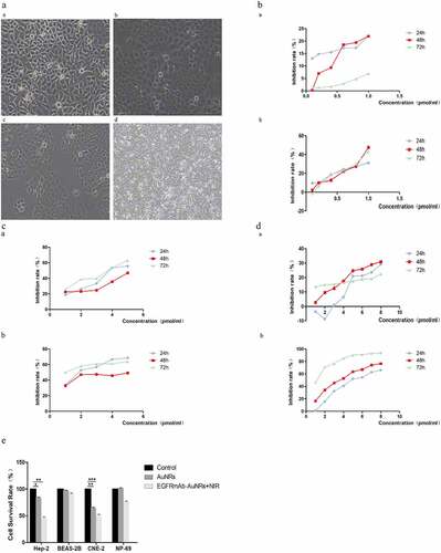
First, we explored the effect of EGFR MAB – Aurs on the proliferative activity of laryngeal squamous cell carcinoma cells HEP-2 and CNE-2 and normal epithelial cells. The morphology of Hep-2, CNE-2, NP – 69 and BEAS-2B cells was observed under microscope ()). Cell were treated with EGFRmAb-AuNRs of 0.1 pmol/ml, 0.2 pmol/ml, 0.3 pmol/ml, 0.4 pmol/ml, 0.5 pmol/ml, 0.6 pmol/ml, 0.7 pmol/ml, 0.8 pmol/ml for 6 h. Subsequently, the cells were irradiated with near-infrared laser irradiation for 24 h, 48 h and 72 h, and the cell viability was measured by MTT. The results showed that Hep-2 and CNE-2 cells did not reach half maximal inhibitory concentration (IC50) at multiple concentrations ()). The concentration gradient of EGFRmAb-AuNRs medium was adjusted to 1 pmol/ml, 2 pmol/ml, 3 pmol/ml, 4 pmol/ml and 5 pmol/ml. The result showed that the IC50 of Hep-2 and CNE-2 were 4 pmol/ml and 2 pmol/ml, respectively ()). NP-69 or BEAS-2B cells were treated with EGFRmAb-AuNRs at the concentration of 1 pmol/ml, 2 pmol/ml, 3 pmol/ml, 4 pmol/ml, 5 pmol/ml, 6 pmol/ml, 7 pmol/ml and 8 pmol/ml ()). The IC50 was about 5 pmol/ml in NP-69 cells and 8 pmol/ml in BEAS-2B cells. The survival rate of Hep-2, BEAS-2B, CNE-2 and NP-69 cells were 62.72%, 96.65%, 67.67% and 108.95% in the non-irradiated group, and were 47.41%, 88.57%, 48.24% and 76.29% in the irradiated group, respectively ()). Taken together, EGFRmAb-AuNRs inhibited the growth of Hep-2 and CNE-2 cells but not normal epithelial cells.
Figure 1. EGFRmAb-AuNRs inhibited the proliferation of laryngeal squamous cell carcinoma cells. (a)The morphology of Hep-2, CNE-2, NP-69 and BEAS-2B cells under a microscope. (b) The cell viability was measured by MTT after treatment with EGFRmAb-AuNRs and near-infrared laser irradiation for 24 h, 48 h and 72 h. (a) Hep-2 cells (b) CNE-2 cells. (c) The cell viability was measured by MTT after treatment with EGFRmAb-AuNRs and near-infrared laser irradiation for 24 h, 48 h and 72 h.(a) Hep-2 cells (b) CNE-2 cells. (d) The cell viability was measured by MTT after treatment with EGFRmAb-AuNRs and near-infrared laser irradiation for 24 h, 48 h and 72 h.(a) NP-69 cells (b) BEAS-2b cells. (e) The cell survival rates of Hep-2, BEAS-2B, CNE-2 and NP-69 were measured by CCK-8. Data were expressed as mean ± SD. *P < 0.05, **P < 0.01, ***P < 0.001.
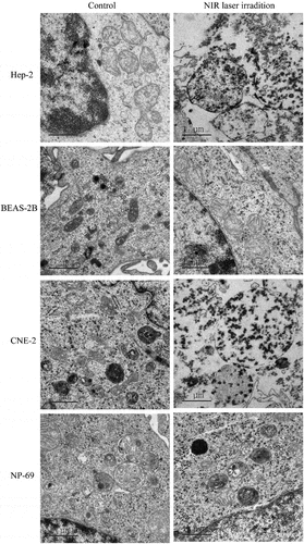
EGFRmAb-AuNRs conjugation increased apoptosis of tumor cells
The entry of EGFRmAb-AuNRs into Hep-2, CNE-2, BEAS-2B and NP-69 cells was observed under electron microscopy. As showed in , Hep-2 and CNE-2 in near-infrared laser irradiation group showed necrosis and apoptosis. Incomplete cell residues and some intact cells were observed. Moreover, there are many cell fragments attached to the whole cell by AuNRs. Most cancer cells showed necrosis such as swelling of mitochondria and endoplasmic reticulum, cell links breaking, nuclear chromatin edge collection, nuclear fragmentation and concentration, and a small number of apoptotic bodies were observed. A large number of AuNRs were observed in the mitochondria of the cells, and also in the cytoplasm and lysosomes. CNE-2 cells showed more cell fragments than Hep-2 cells. However, neither BEAS-2B nor NP-69 cells showed necrosis or apoptosis. These results revealed that functionalized AuNRs selectively induced cancer cell apoptosis and had no effect on normal cells, which demonstrated that the functionalized AuNRs was safe.
Figure 2. EGFRmAb-AuNRs conjugation increased laryngeal squamous cell carcinoma cancer cell apoptosis. The entry of EGFRmAb-AuNRs into Hep-2, CNE-2, BEAS-2B and NP-69 cells was observed under electron microscopy.
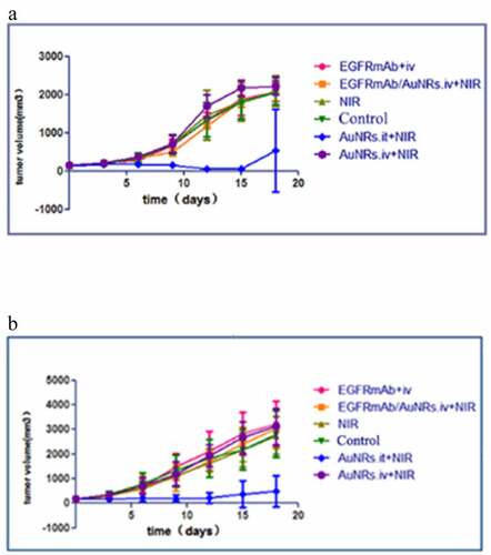
AuNRs with near-infrared photothermal therapy effectively reduced tumor growth in vivo
To confirm the effects of AuNRs on tumor growth in vivo, Hep-2 and CNE-2 cells were injected into the right anterior axilla of nude mice. There was no significant difference in body weight before and after the injection, and no mice died at the time of tumorigenesis. Furthermore, there was no swelling at the injection site. Seven days after inoculation, nodules larger than 5 mm in diameter formed subcutaneously, suggesting that the model was successfully established. After cell injection, the volume of tumors and the weight of mice were recorded every other day (). The growth curve of the tumors was measured every two days after treatment ()). The results showed that compared with the control group, tumor growth was significantly inhibited by direct injection of AuNRs into the tumors and near-infrared laser irradiation (AuNRs. it + NIR group). On the third day of near-infrared laser irradiation, obvious local necrosis and growth inhibition were observed. However, EGFRmAb-AuNRs.iv+NIR, NIR, EGFRmAb.iv and AuNRs.iv + NIR did not inhibit the growth of tumors, indicating that direct injection of AuNRs into tumors followed by near-infrared laser irradiation could effectively inhibit the growth of tumors, while tail vein injection of EGFRmAb-AuNRs followed by near-infrared laser irradiation had no significant effect on tumors in nude mice. The growth curve showed that the growth rate of CNE-2 transplanted tumors in nude mice was much faster than that of Hep-2 transplanted tumors.
Figure 3. AuNRs with near-infrared photothermal therapy effectively reduced laryngeal squamous cell carcinoma tumor growth in nude mice. The growth curve of the tumors was measured every two days after treatment. (a) Hep-2 transplanted tumor in nude mice (b) CNE-2 transplanted tumor in nude mice.
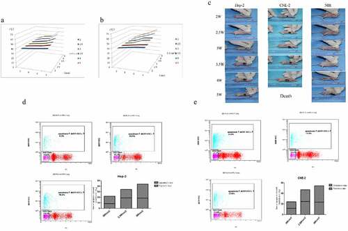
Table 1. Weight of Hep-2 tumor-bearing mice and tumor volume before treatment
Table 2. Weight of CNE-2 tumor-bearing mice and tumor volume before treatment
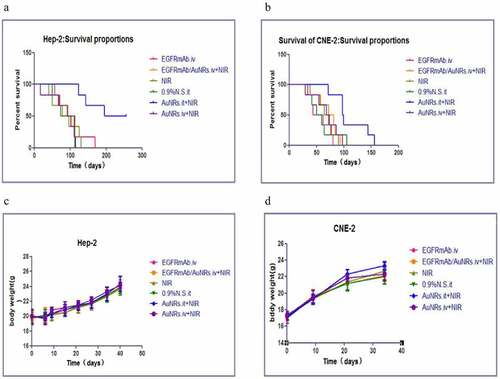
Improvement of survival in mice after photothermal treatment with EGFR-AuNRs
Survival probability is an index of therapeutic effect [Citation24]. The survival curves of Kaplan-Meier after treatment with EGFRmAb.iv, EGFRmAb-AuNRs.iv+NIR, 0.9% saline.it, AuNRs.it +NIR and AuNRs.iv +NIR were used in Hep-2 nude mice and CNE-2 nude mice, respectively ()). The survival of AuNRs.it +NIR group was significantly higher than that of the other five groups. Logarithmic rank test analysis showed that the survival rate of mice in AuNRs.iv + NIR group was significantly higher than that in other groups, indicating that near-infrared laser irradiation on the tumor site significantly prolonged the median survival time of mice. However, EGFRmAb-AuNRs.iv + NIR and AuNRs.iv + NIR treatment on the tumor-bearing mice significantly prolonged the median survival time of mice. There was no significant difference in the survival time between the two groups. The results revealed that NIR laser irradiation after AuNRs injection inhibited the growth of tumors, which improved their survival advantage. In AuNRs.it + NIR group, the survival time of the mice was significantly prolonged. To some extent, local injection of AuNRs showed no long-term toxicity to nude mice based on the use of weight changes as an indicator of survival improvement [Citation24,Citation25]. Therefore, we also weighed the mice that began to receive treatment ()). The results showed that the weight of mice in each group did not change significantly after 42 days of treatment, indicating that AuNRs had no toxic effect on mice in each group.
Figure 4. Improved survival of laryngeal squamous cell carcinoma transplanted mice after photothermal treatment with EGFR-AuNRs. (a and b) Kaplan-Meier survival curves of nude mice after treatment with EGFRmAb.iv, EGFRmAb-AuNRs.iv+NIR,0.9% saline.it, AuNRs.it +NIR and AuNRs.iv+NIR. (c and d) The weight of the nude mice in each group.
Optimizing near-infrared irradiation power and single irradiation time
In order to provide better clinical treatment, the power and time of near-infrared laser irradiation were optimized. Local temperatures over 6 minutes after 3.5 W/cm2, 4 W/cm2 and 5 W/cm2 irradiation reached 50–60°C. After 6 minutes of near-infrared laser irradiation with 2 W/cm2, 2.5 W/cm2 and 3 W/cm2, the local temperature was higher than 43°C (the lowest local temperature required by PTT effect) ()). Therefore, a single near-infrared laser irradiation time of Hep-2 and CNE-2 transplanted tumors were selected as 6 minutes. The apoptotic rate/necrotic rate of CNE-2 loaded mice in 2.5 W/cm2 group was 1.17 ± 0.21. However, the ratio of apoptotic/necrotic were 0.96 ± 0.28 and 0.84 ± 0.17 in 2 W/cm2 and 3 W/cm2 groups, which confirmed that the therapeutic effect of 2.5 W/cm2 group was better than 3 W/cm2 group. Thus, we optimized the irradiation power to 3 W/cm2 (high necrosis mode) and 2.5 W/cm2 (high apoptotic mode) ()). The ratio of apoptotic/necrosis of Hep-2 mice in 2.5 W/cm2 group was 1.30 ± 0.37, while the ratio of apoptotic /necrosis of 2 W/cm2 and 3 W/cm2 was 0.86 ± 0.36 and 0.78 ± 0.06, respectively. Under 3 W/cm2 power near-infrared laser irradiation, the ratio of apoptosis and necrosis was the highest.
Figure 5. Optimizing near-infrared irradiation power and single irradiation time. (a) The relationship between the surface temperature of Hep-2 tumor and irradiation power and irradiation time. (b) The relationship between the surface temperature of CNE-2 tumor and irradiation power and irradiation time. (c-e) The ratio of apoptotic/necrosis of Hep-2 nude mice and CNE-2 nude mice. *P < 0.05 vs 2 W/cm2 group.
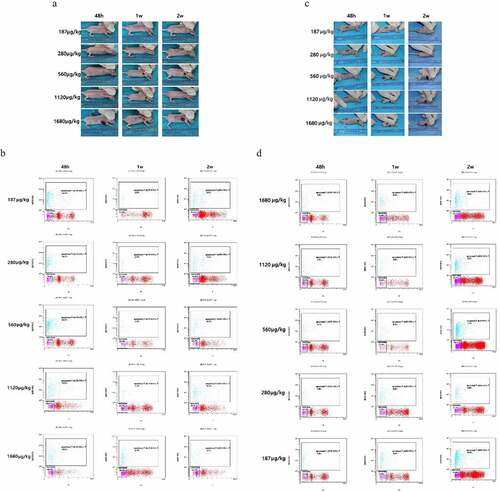
Dose optimization of intra-tumoral injection
Furthermore, we optimized the dose of intra-tumoral injection. Interestingly, when the dose of CNE-2 tumor-bearing mice was 187 μg/kg, the tumor continued to grow after 2 weeks of treatment. However, when the dose of Hep-2 tumor-bearing mice was 187 μg/kg, the tumor did not change significantly after 2 weeks of treatment ()). When the dose of intra-tumoral injection was 1680 μg/kg, a large area of necrosis occurred 48 h after near-infrared laser irradiation, even involving the skin around the tumor. Therefore, doses of 187 μg/kg and 1680 μg/kg were excluded. As shown in ), there was no significant difference in apoptotic rate among the groups, and the minimum concentration was 280 μg/kg.
Figure 6. Dose optimization of intra-tumoral injection. (a and c) Tumor inhibition after 48 hours, 1 week and 2 weeks of photothermal therapy with different concentrations of AuNRs. (b and d) TUNEL and flow cytometry of apoptosis of tumor at 48 h, 1 week and 2 weeks after treatment. *P < 0.05, **P < 0.01 vs 187 μg/mL group.
Discussion
Laryngeal squamous cell carcinoma is the most common head and neck cancer, which is characterized by high morbidity and mortality [Citation1,Citation26]. At present, the most difficult problem in the treatment of laryngeal squamous cell carcinoma is how to selectively target cancer cells without side effects on normal cells. EGFR is usually overexpressed and activated in tumor tissues, and plays an important role in the pathogenesis of laryngeal squamous cell carcinoma [Citation27]. Nevertheless, targeting therapy with monoclonal antibodies and kinase inhibitor has not significantly inhibit malignant tumors [Citation28]. AuNRs can penetrate biological tissues well, and the longest light penetration depth is up to 10 cm [Citation29]. In this context, near-infrared photothermal therapy plasma photothermal therapy using AuNRs as carriers showed promise. In this study, we combined EGFRmAb with AuNRs to target laryngeal squamous cell carcinoma cells.
In vitro, we irradiated cells with different concentrations of EGFRmAb-AuNRs and near-infrared laser, and then detected cell viability at different time points after irradiation by MTT method. EGFRmAb-AuNRs had no effect on normal cells at the effective killing concentration of photothermal therapy for tumor cells. Electron microscopy showed that a large number of AuNRs accumulated in the mitochondria, cytoplasm and lysosomes of tumor cells. After near-infrared laser irradiation, cell necrosis and apoptosis were observed. However, no AuNRs were observed in normal cells, and no necrosis and apoptosis were observed after near-infrared laser irradiation, suggesting that EGFRmAb-AuNRs were relatively safe. Furthermore, we established the models of Hep-2 and CNE-2 transplanted tumors in vivo. The results showed that tumor growth in CNE-2 tumor-bearing nude mice was faster than in Hep-2 tumor-bearing nude mice, and the median survival of CNE-2 tumor-bearing nude mice was significantly shorter than that of Hep-2 tumor-bearing mice. Taken together, plasma photothermal therapy selectively kills cancer cells to achieve the goal of cancer treatment, but the treatment cycle is long, and it is more suitable for the treatment of tumors near important tissues and organs with relatively slow development.
The rapid tumor growth in the near-infrared laser irradiation group showed that near-infrared laser irradiation at a certain power would not damage the cells, suggesting that we need to define a safe and effective irradiation power. Therefore, we need to optimize the power and duration of near-infrared laser irradiation. Previously, we showed that functionalized AuNRs significantly inhibited tumor growth and increased the ability of AuNRs to enter cells [Citation13]. However, in this study we failed to achieve tail vein injection of EGFRmAb-AuNRs to inhibit tumor growth. The reason may be the unstable binding of EGFRmAb and AuNRs, the failure of EGFRmAb-AuNRs to reach the effective concentration through systemic circulation, and low expression of EGFR in nude mice transplanted tumors.
Plasma photothermal therapy treatment of cancer is via photothermal inhibition of tumor cell growth [Citation30,Citation31]. There are two main ways of cancer cell death, necrosis and apoptosis [Citation32]. Interestingly, whether cancer cells undergo necrosis or apoptosis depends on the heat load produced by plasma photothermal therapy on cancer cells [Citation33]. Apoptosis is a programmed death, which causes cancer cell death but does not cause damage to healthy tissues [Citation34].
Regulating plasma photothermal therapy treatment modes, such as irradiation power, duration of irradiation and dosage of AuNRs injection, can lead to apoptotic death of cancer cells and effectively reduce side effects of cancer treatment in the clinical [Citation31,Citation35]. We successfully established Hep-2 and CNE-2 tumor models in nude mice. By optimizing irradiation power, single irradiation time and injection dose, we tried to induce apoptosis of tumor cells in nude mice. The single near-infrared irradiation duration for CNE-2 tumor model was optimized as 6 minutes. The near-infrared irradiation power was optimized as 3 W/cm2 (high necrosis mode), and 2.5 (high apoptotic mode), which could effectively inhibit the growth of nasopharyngeal carcinoma in early clinical stage and protect surrounding tissues and organs. The optimum irradiation power of Hep-2 mice was 3 W. Under this irradiation power, the necrosis rate and apoptosis rate of the tumors were the highest in all groups. In addition, some studies have reported that the near infrared radiation of gold nanorods could effectively inhibit the growth and metastasis of tumor cells. Gold nanorods are considered as an effective nanocarrier because of their large surface area and relatively simple means of surface functionalization, which can transport small molecule drugs, photosensitizers, nucleic acids (DNA and siRNA, etc.) into cells. Gold nanorods themselves can also be used as photothermal agents, which can achieve the purpose of treating tumors through their mediated photothermal effects, and have great potential in tumor treatment. At present, we have only studied the survival rate of laryngeal squamous cell carcinoma cell line and the apoptosis and necrosis of cancer cells in xenograft nude mice by near infrared irradiation. In the study, we have not observed the inhibitory effect of EGFRmAb on mouse tumors, which may be caused by the unstable binding between AuNRs and EGFRmAb, or the lack of accumulation of EGFRmAb in mice, or the inability of transplanted tumors in nude mice to express EGFR highly. In the future research, we will further study these possible reasons to improve the therapeutic effect of gold nanorods on tumors.
In conclusion, AuNRs photothermal therapy can kill tumor cells while avoiding damage to normal cells. Moreover, AuNRs injection combined with near-infrared laser irradiation could effectively inhibit Hep-2 and CNE-2 tumor model in nude mice, although the effect of tail vein injection was not obvious. Therefore, we developed a new method of targeting EGFR monoclonal antibody combined with nano gold to achieve photothermal effect in the treatment of laryngeal squamous cell carcinoma.
Conclusion
To sum up, we found that photothermal treatment with EGFRmAb/AuNRs inhibited the viability of laryngeal squamous cell carcinoma cells Hep-2 and CNE-2 without compromising the viability of normal cells. In xenografts nude mice, tumor injection of AuNRs photothermal therapy can significantly promote the apoptosis rate and necrosis rate of tumor cells. The optimal single NIR laser irradiation duration for plasma resonance photothermal therapy of CNE-2 and Hep-2 tumor-bearing nude mice was optimized to be 6 minutes, the optimal irradiation power was 3 W/cm2, and the optimal tumor injection dose was 280 μg/kg.
Highlights
EGFR/AuNRs photothermal therapy could kill head and neck squamous carcinoma cells while avoiding damage to normal cells.
The photothermal therapy with AuNRs injection had a significant effect on HEP-2 and CNE-2, and The inhibition rate of heP-2 transplanted tumor was 100%.
The optimal single NIR laser irradiation duration for plasma resonance photothermal therapy of CNE-2 and Hep-2 tumor-bearing nude mice were optimized to be 6 min, with the optimal irradiation power of 3 W/cm2 (high necrosis mode) and the optimal tumor injection dose of 280 μg/kg.
Availability of Data and Materials
All data and materials are available from the corresponding author on reasonable request.
Disclosure statement
No potential conflict of interest was reported by the author(s).
Additional information
Funding
References
- Zhu Y, Yan L, Zhu W, et al. MMP2/3 promote the growth and migration of laryngeal squamous cell carcinoma via PI3K/Akt‐NF‐κB‐mediated epithelial–mesenchymal transformation. J Cell Physiol. 2019;234(9):15847–15855.
- Li X, Wang HL, Peng X, et al. miR-1297 mediates PTEN expression and contributes to cell progression in LSCC. Biochem Biophys Res Commun. 2012;427(2):254–260.
- Zhj L, Gao W, Wb L, et al. The diagnostic value of methylated DNA in laryngeal squamous cell carcinoma: meta-analysis. Head Neck Oncol. 2013;5(2):15.
- Shi Z, Yu X, Wu A. Gold Nanorods for Biomedical Imaging and Therapy in Cancer. In: Dai Z. (eds) Advances in Nanotheranostics I. Springer Series in Biomaterials Science and Engineering, 6. Berlin, Heidelberg: Springer. 2016. https://doi.org/10.1007/978-3-662-48544-6_3.
- Mackey MA, Ali MR, Austin LA, et al. The most effective gold nanorod size for plasmonic photothermal therapy: theory and in vitro experiments. J Phys Chem B. 2014;118(5):1319–1326.
- Cabral RM, Baptista PV. Anti-cancer precision theranostics: a focus on multifunctional gold nanoparticles. Expert Rev Mol Diagn. 2014;14(8):1041–1052.
- Ali MR, Rahman MA, Wu Y, et al. Efficacy, long-term toxicity, and mechanistic studies of gold nanorods photothermal therapy of cancer in xenograft mice. Proc Natl Acad Sci U S A. 2017;114(15):E3110.
- Rao L, Bu LL, Ma L, et al. Platelet‐facilitated photothermal therapy of head and neck squamous cell carcinoma. Angew Chem. 2018;57(4):986–991.
- Ding AA, Chen YY, Wang CRC, et al. HER-2 antibody conjugated gold nano rod for in vivo photothermal therapy. IEEE Conference on Nanotechnology, 2008, 882–885. doi:10.1109/NANO.2008.264.
- Garousi J, Andersson KG, Mitran B, et al. PET imaging of epidermal growth factor receptor expression in tumours using 89Zr-labelled ZEGFR:2377 affibody molecules. Int J Oncol. 2016;48(4):1325–1332.
- Biscardi JS, Maa MC, Tice DA, et al. c-Src-mediated phosphorylation of the epidermal growth factor receptor on Tyr845 and Tyr1101 is associated with modulation of receptor function. J Biol Chem. 1999;274(12):8335–8343.
- Ming Z, Jiang M, Li W, et al. Bioinformatics analysis and expression study of fumarate hydratase in lung cancer. Thorac Cancer. 2014;5(6):543–549.
- Zhang S, Li Y, He X, et al. Photothermolysis mediated by gold nanorods modified with EGFR monoclonal antibody induces Hep-2 cells apoptosis in vitro and in vivo. Int J Nanomed. 2014;9:1931–1946.
- Zhang Y, He J, Wang Y, et al. Photothermal therapy with AuNRs and EGFRmAb-AuNRs inhibits subcutaneous transplantable hypopharyngeal tumors in nude mice. Int J Oncol. 2018;53(6):2647–2658.
- Ren YT, Chen Q, Hong Q, et al. Experimental comparison of photothermal conversion efficiency of gold nanotriangle and nanorod in laser induced thermal therapy. Nanomaterials. 2017;7(12):416.
- † El-Sayed IH, Xiaohua Huang A, ‡ El-Sayed, et al. Surface plasmon resonance scattering and absorption of anti-EGFR antibody conjugated gold nanoparticles in cancer diagnostics: applications in oral cancer. Nano Lett. 2005;5(5):829–834.
- El-Sayed IH, Huang X, El-Sayed MA. Selective laser photo-thermal therapy of epithelial carcinoma using anti-EGFR antibody conjugated gold nanoparticles. Cancer Lett. 2006;239(1):129–135.
- Liu F, Zhu Y, Qian Y, et al. Effects of gold nanorods modified with antiepidermal growth factor receptor monoclonal antibody on laryngeal cancer cells. Turk J Biol. 2018;42(2):144–151.
- Jiang L, Xu G, Li Z, et al. RNAi-mediated knockdown of CAIX enhances the radiosensitivity of nasopharyngeal carcinoma cell line, CNE-2. Onco Targets Ther. 2017;10:4701–4709.
- Li P, Li S, Yin D, et al. EGCG sensitizes human nasopharyngeal carcinoma cells to TRAIL-mediated apoptosis by activation NF-κB. Neoplasma. 2017;64(1):74–80.
- Zhao L, Sun H, Kong H, et al. The Lncrna-TUG1/EZH2 axis promotes pancreatic cancer cell proliferation, migration and EMT phenotype formation through sponging Mir-382. Cell Physiol Biochem. 2017;42(6):2145–2158.
- Huang F, Chen W, Peng J, et al. LncRNA PVT1 triggers Cyto-protective autophagy and promotes pancreatic ductal adenocarcinoma development via the miR-20a-5p/ULK1 axis. Mol Cancer. 2018;17(1):98.
- Li X, Ren H. Long noncoding RNA PVT1 promotes tumor cell proliferation, invasion, migration and inhibits apoptosis in oral squamous cell carcinoma by regulating miR‑150‑5p/GLUT‑1. Oncol Rep. 2020;44(4):1524–1538.
- Rao GN, Haseman JK. Influence of corn oil and diet on body weight, survival, and tumor incidences in F344/N rats. Nutr Cancer. 1993;19(1):21.
- Humphreys SR, Goldin A, Venditti JM, et al. The influence of amethopterin on the survival time of leukemic mice with respect to food intake, body weight changes, and tumor growth. J Natl Cancer Inst. 1958;20(1):211.
- Dequanter DD, Lothaire P, Zouaoui K, et al. Epidemiology and clinical characteristics of Larynx and Hypopharynx carcinoma: a comparative study in the hainaut and review of the literature. Acta Chir Belg. 2012;112(6):423–425.
- Demory ML, Boerner JL, Parsons SJ. The localization of the epidermal growth factor receptor at the mitochondria is regulated by c-Src and endocytosis. Cancer Res. 2006;66:543.
- Mamot C, Rochlitz C. Targeting the epidermal growth factor receptor (EGFR)–a new therapeutic option in oncology? Swiss Med Wkly. 2006;136(1–2):4.
- Zhou R, Wu ZW, Sun ZH, et al. Synthesis of long gold nanorods as an efficient photothermal agent in the second near-infrared window. J Nano Res. 2016;40:180–189.
- Dickerson EB, Dreaden EC, Huang X, et al. Gold nanorod assisted near-infrared plasmonic photothermal therapy (PPTT) of squamous cell carcinoma in mice. Cancer Lett. 2008;269(1):57–66.
- Ali MRK, Ali HR, Rankin CR, et al. Targeting heat shock protein 70 using gold nanorods enhances cancer cell apoptosis in low dose plasmonic photothermal therapy. Biomaterials. 2016;102:1–8.
- Moreover. Perspectives on the role of photodynamic therapy in the treatment of pancreatic cancer. Int J Photoenergy. 2011;2012(2012):101.
- Bonfil RD, Bustuoabad OD, Ruggiero RA, et al. Tumor necrosis can facilitate the appearance of metastases. Clin Exp Metastasis. 1988;6(2):121–129.
- Danial NN, Korsmeyer SJ. Cell death: critical control points. Cell. 2004;116(2):205–219.
- Ali RK, Ibrahim IM, Ali HR, et al. Treatment of natural mammary gland tumors in canines and felines using gold nanorods-assisted plasmonic photothermal therapy to induce tumor apoptosis. Int J Nanomed. 2016;11:4849–4863.