
Abstract
Objective
This study aimed to evaluate the safety and tolerability of a fixed-dose co-formulation of ciprofloxacin and celecoxib (PrimeC) in patients with amyotrophic lateral sclerosis (ALS), and to examine its effects on disease progression and ALS-related biomarkers.
Methods
In this proof of concept, open-label, phase IIa study of PrimeC in 15 patients with ALS, participants were administered PrimeC thrice daily for 12 months. The primary endpoints were safety and tolerability. Exploratory endpoints included disease progression outcomes such as forced vital capacity, revised ALS functional rating scale, and effect on algorithm-predicted survival. In addition, indications of a biological effect were assessed by selected biomarker analyses, including TDP-43 and LC3 levels in neuron-derived exosomes (NDEs), and serum neurofilaments.
Results
Four participants experienced adverse events (AEs) related to the study drug. None of these AEs were unexpected, and most were mild or moderate (69%). Additionally, no serious AEs were related to the study drug. One participant tested positive for COVID-19 and recovered without complications, and no other abnormal laboratory investigations were found. Participants’ survival compared to their predictions showed no safety concerns. Biomarker analyses demonstrated significant changes associated with PrimeC in neural-derived exosomal TDP-43 levels and levels of LC3, a key autophagy marker.
Interpretation
This study supports the safety and tolerability of PrimeC in ALS. Biomarker analyses suggest early evidence of a biological effect. A placebo-controlled trial is required to disentangle the biomarker results from natural progression and to evaluate the efficacy of PrimeC for the treatment of ALS.
Twitter handles: @NeurosenseT, @ShiranZimri
•What is the current knowledge on the topic? ALS is a severe neurodegenerative disease, causing death within 2–5 years from diagnosis. To date there is no effective treatment to halt or significantly delay disease progression.
•What question did this study address? This study assessed the safety, tolerability and exploratory efficacy of PrimeC, a fixed dose co-formulation of ciprofloxacin and celecoxib in the ALS population.
•What does this study add to our knowledge? This study supports the safety and tolerability of PrimeC in ALS, and exploratory biomarker analyses suggest early insight for disease related-alteration.
•How might this potentially impact the practice of neurology? These results set the stage for a larger, placebo-controlled study to examine the efficacy of PrimeC, with the potential to become a new drug candidate for ALS.
Summary for social media if published
Introduction
Amyotrophic lateral sclerosis (ALS) is a severe neurodegenerative disease characterized by progressive deterioration of motor neurons, leading to muscle paralysis, and ultimately death within 2–5 years from onset (Citation1).
The pathogenesis of ALS is not fully elucidated and involves multiple pathophysiological pathways, including neuroinflammation (Citation2), iron accumulation (Citation3), dysregulation of microRNA metabolism and aberrant RNA binding proteins (Citation4) among others. The multifactorial nature of the disease and the lack of a clear leading pathogenesis points to a combination of pathways, contributing to neurodegeneration in ALS – a multifactorial pathogenesis requiring multi-targeted therapy.
PrimeC is a combined formulation composed of unique doses of ciprofloxacin and celecoxib, which aim to synergistically inhibit the progression of ALS by addressing the three aforementioned pathologies.
Ciprofloxacin is a fluoroquinolone antibiotic which has been shown to bind the RNA-binding protein TRBP, facilitating its association with the RISC-loading complex, regulating microRNA processing (Citation5). Imbalance in these processes is observed in ALS and other neurodegenerative diseases (Citation6). Ciprofloxacin is also an iron chelator (Citation7). There are various examples of iron chelators reducing iron stores in the brain that are neuroprotective in neurodegenerative diseases (Citation8).
Celecoxib is a non-steroidal anti-inflammatory drug (NSAID) best known for its selective COX-2 inhibition, with additional COX-2-independent mechanisms responsible for its anti-inflammatory action (Citation9). Celecoxib was not beneficial in ALS when given at high doses as a single agent (Citation10).
However, pre- clinical studies combining celecoxib with ciprofloxacin showed a synergistic effect, with benefits to motor function, as well as the morphology of CNS cells (Citation11).
Here we report the findings from a proof of concept, open-label, phase IIa study that evaluated the safety, tolerability and exploratory target engagement markers of an oral administration of PrimeC in people with ALS. Indication for biological activity was evaluated by measuring selected biomarkers including serum neurofilament levels, which have been studied extensively in ALS (Citation12). Additionally, exosomal levels of two enzymes related to ALS pathology were measured: TAR-DNA binding protein 43 (TDP-43), which increases with disease progression (Citation13) and Microtubule-associated proteins 1A/1B light chain 3B (LC3) representing autophagy, which is impaired in various neurodegenerative disease, including ALS (Citation14).
Methods
Trial design and oversight
This was a 12-months, open-label, phase-IIa study aimed at evaluating the safety and tolerability of a fixed dose combination of celecoxib and ciprofloxacin (PrimeC immediate release, hereby referred to as PrimeC) given orally thrice daily. The study enrolled participants at Tel-Aviv Sourasky Medical Center (TASMC), Israel from November 2019 through January 2020. The study protocol was approved by the TASMC Helsinki Committee for Human Rights (0553-19-TLV) and is registered on clinicaltrials.gov NCT04165850. All participants provided written informed consent at screening. The trial was designed by and conducted through TASMC, in collaboration with NeuroSense Therapeutics.
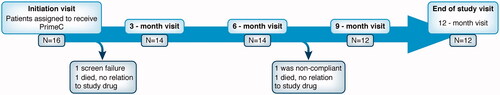
Participants were evaluated and monitored in the clinic every 3 months, and in between were evaluated by phone visits as described in .
Figure 1 NST002 Study participant flowchart. Participants’ disposition in an open-label 12 months of dosing study.
Participant selection criteria
Participants were diagnosed with either familial or sporadic ALS and fulfilled the World Federation of Neurology revised El Escorial criteria of laboratory-supported probable, clinically probable, or definite ALS (Citation15), had a disease duration of less than 36 months and were on a stable dose of edaravone and/or riluzole. Participants with a forced vital capacity (FVC) less than 50% predicted for age, height, weight and sex were excluded, as well as those unable to swallow capsules.
Trial procedures and outcomes
The primary outcomes of the study were the safety and tolerability of PrimeC administration, defined as the incidence and severity of treatment-emergent adverse events (TEAEs), as well as by monitoring the causes for treatment discontinuation and incidence of abnormal laboratory results. Safety assessments included quarterly in-clinic evaluations of AEs, as well as vital signs, electrocardiogram, and safety laboratory evaluations including blood count and full biochemistry.
Exploratory outcomes included FVC, expressed as percentage from predicted for age, height, weight and sex, measured with a Koko spirometer (nSpire Health, USA) and the revised ALS functional rating scale (ALSFRS-R) (Citation16), which consists of 12 questions across four subdomains of functions (bulbar, fine motor, gross motor, and respiratory) with each item scored on an ordinal scale from 0 (no function) to 4 (fully functional); total scores range from 0 to 48, with higher scores indicating better function.
Additional exploratory outcomes measured PrimeC target engagement by analyzing the levels of two key neuron-derived exosomal (NDE) ALS-pathology-related enzymes: TDP43 and LC3, and measurements of serum neurofilaments concentrations (neurofilament light and phosphorylated heavy chains (NFL and pNFH, respectively).
Biomarker assay
Sample collection
Clinical study: Blood samples from NST002 participants were collected and stored for biomarker analysis once pretreatment and twice during treatment at approximately 6 and 12 months. After collection, samples were centrifuged at room temperature at 1,000 RPM for 10 min and sera were stored at −80 °C.
Baseline study: Blood samples of broadly age and sex-matched ALS (N = 35) and healthy control (N = 25) samples were obtained from the NEALS biorepository, and basal levels of TDP-43 and LC3 in controls and people with ALS were compared. Samples were centrifuged at 1750G at room temperature for 10 min and stored at −80 °C.
Isolation and identification of neuron-derived exosomes
Serum samples were thawed on ice and were isolated using NeuroDex ExoSORTTM proprietary protocol for immunocapture of NDEs. Briefly, the isolation is based on precipitation of total serum NDEs followed by immunoaffinity isolation with two antibodies specific to neuronal surface markers. The isolated NDEs were lysed in a buffer that contains protease and phosphatase inhibitors and stored at -80C until assays. Importantly, the isolated NDEs were characterized according to MISEV (Citation17), including electron microscopy, nanoparticle tracking analysis, and western blot analysis (Citation18).
Immunoassays for LC3, and TDP43 quantification
LC3 and TDP-43 were quantitatively measured in the NDE protein extracts using Human LC3 DuoSet ELISA (R&D Systems, Cat. No DY8558-05), and ELISA kit for TDP43 (MyBioSource, Cat No MBS2019049) per manufacturer’s instructions. LC3 was measured in fivefold dilution (equivalent to 60ul original serum), and TDP43 was measured in 2.5-fold dilution (equivalent to 120 µl original serum). NDEs were analyzed for levels of CD81, a pan-exosomal marker, in order to quantify the exosomes extracted, to ensure similarity between samples.
Neurofilament analysis
The concentrations of NFL were assessed with the use of the Simoa NF-light advantage assay (Quanterix), and concentrations of pNFH with the use of the Simple Plex Ella immunoassay (Protein Simple) pNFH quantification. pNFH serum levels were measured through the ELLA microfluidic (Protein Simple) according to the manufacturer’s instructions.
Statistical analysis
Participants’ baseline characteristics were expressed as either mean (standard deviation) or number of cases (percentage). For each participant, we determined the prognostic risk profile using the ENCALS survival prediction model (Citation19) and the Origent survival model (Citation20–22), and calculated their probability to survive 12 months, conditional on being alive at screening. Participants were ranked based on their survival probability to assess the ‘expectedness’ of dropout due to death (i.e., were participants that died during follow-up also the ones with the worst ranked survival probability?). The ENCALS survival prediction model was originally established in order to be used as an inclusion criterion instead of arbitrary eligibility criteria, as well as to stratify randomization (Citation19), and has since been used in clinical trials to this end (Citation23). The Origent models have similar goals (Citation24).
In addition, AEs were coded to preferred terms from the MedDRA library (version 23.0) and reported as frequency (proportion). Tolerability was defined as the ability to complete the 12-month dosing period on the study drug.
On an exploratory basis, we evaluated the mean monthly change in clinical markers for disease progression. Linear mixed models (LME) were fitted with a fixed effect for time since baseline visit and a random slope for time and intercept per individual, allowing imputation of missing data. All models were based on restricted maximum likelihood (REML); 95% confidence intervals (95% CI) around estimates were based on the profile likelihood. To put the observed longitudinal trajectories into context, we compared the progression rate of the enrolled cohort to historical data obtained from the Pooled Resource Open-Access ALS Clinical Trials (PRO-ACT) database (Citation25). We used propensity matching (1 PrimeC participant to 2 PRO-ACT patients) to match on age, sex, symptom duration, vital capacity, diagnostic delay, site of onset and ΔFRS, ALSFRS-R total score and body mass index (Citation25). The mean difference in progression rates was estimated by adding an interaction term between time and cohort (PrimeC or PRO-ACT) in the LME.
Lastly, biomarker data were analyzed using a mixed model for repeated measures with time as a categorical factor. The mean change from baseline to month 12 was estimated and reported with 95% CI and p value. LME models were fitted using the R lme function (Citation26).
Results
Safety and tolerability
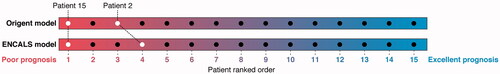
A total of 16 people living with ALS were screened for the study, of whom 15 began administration of PrimeC. One individual screen failed due to rapid cognitive decline and lack of family support that made future adherence to treatment doubtful. During the study, one participant was withdrawn by the investigator after 6 months due to poor adherence, one participant died approximately 2-and-a-half months after initiation of the study, and one participant died after 7 months (). Both these events were determined by the investigator as not related to the study drug. These participants were ranked by both the ENCALS and the Origent survival prediction models as having a low probability of survival ().
Figure 2 Survival of patients in clinical trial population compared to survival prediction models. For each patient, the probability to survive 12 months was calculated according to the Origent and ENCALS survival prediction models. Patients were subsequently ranked from low to high according to their predicted survival probability, with a low rank reflecting a poor survival probability. Both models were in agreement that both deaths occurred in the lower quartile of the population at high risk for death.
In total, 12 participants completed the 12 months PrimeC administration period (80%). Baseline demographic and disease characteristics are summarized in . Eighty percent of the participants received riluzole, two of them also received edaravone. The majority of participants (87%) had limb-onset ALS, and the average symptom duration from onset was 21 months. Average ALSFRS-R total score was 37 points, and average FVC (% predicted) was 89%.
Table 1 Patient characteristics at baseline.
10 participants (67%) experienced AEs. Most AEs occurred within the first 3 months of the study, and 33% of participants remained AE-free throughout the study. The majority of AEs (67%) were mild or moderate in intensity. Only four participants (27%) experienced an AE that was assessed by the Investigator as related to the study drug. None of these AEs were unexpected, and most were mild or moderate (69%).
Of the AEs related to study drug, the majority were gastrointestinal (flatulence, dyspepsia, nausea, abdominal pain, constipation). In spite of the gastrointestinal symptoms, these participants did not encounter major weight loss. Importantly, no Serious-AEs were related to the study drug. An overview of AEs is presented in . During the study, one participant tested positive for COVID-19 and recovered without complications and without being withdrawn from the study, and no other abnormal laboratory investigations were found.
Table 2 Overview of adverse events.
Clinical measures of disease progression
The mean deterioration of the participants in both ALSFRS-R and FVC during the course of 12 months was assessed as described in statistical methods. These results were matched to the PRO-ACT cohort (Citation25,Citation27,Citation28); characteristics are given in . The estimated mean rate of change of ALSFRS-R total score in trial participants was −0.84 points/month (95% CI; −1.17 to −0.52), whereas the FVC declined by −2.09% predicted/month (95% CI −3.22 to −0.98; ).
Table 3 Propensity matching (1:2) with PRO-ACT.
Table 4 Rate of decline PrimeC vs. PRO-ACT.
When compared to PRO-ACT cohort, a mean difference of 0.18 points/month in ALSFRS-R progression rate (ns, 95% CI −0.23 to 0.59) and 0.90 points/month in % predicted FVC (ns, 95% CI −0.52 to 2.32; ) was observed; representing a difference of 18% and 30% respectively.
ALS-related biomarkers
Examination of the key ALS-related protein-TDP43 showed that in baseline-study, levels of NDE TDP43 were increased in subjects with ALS compared to age and sex matched controls (median patients 297.6 vs. median controls 238.4, P = 0.002 Wilcoxon test; ). In the clinical study the levels of NDE-associated TDP-43 were significantly reduced over time (from baseline measurements to 12 months, with a mean change from baseline of −56.3 pg/ml, 95% CI −87.3 to −25.3, P = 0.002; ).
Figure 3 ALS-pathology related NDE biomarker levels in ALS vs. control samples. Wilcox test comparing neural-derived blood exosomal biomarkers: TDP-43 (A) and LC3 (B) between patients with ALS and healthy volunteers (N = 35 for ALS and 25 for control per group) as obtained in a baseline study.
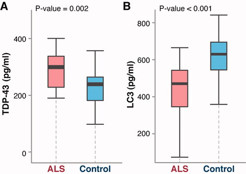
Figure 4 ALS-pathology related biomarker levels in PrimeC treated ALS patients over time. (A,B) Average ALS-pathology related serum NDE marker levels over time (pre in red, interim in light blue, and end of study in dark blue). (A) TDP-43 levels. (B) LC3 levels. (C,D) Average serum NFL (C) and pNFH (D) over time (pre in red, interim in light blue, and end of study in dark blue). P values tests whether the means at pre-treat are similar compared to the mean at end-of-treat, based on a mixed model for repeated measures.
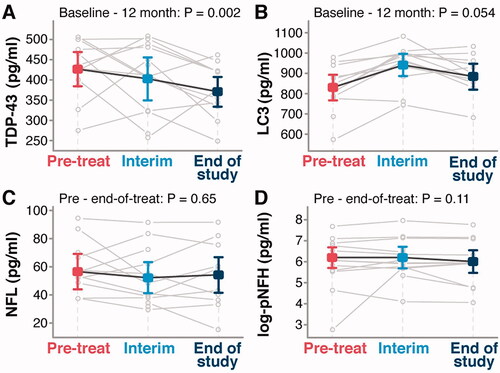
Levels of NDE LC3, a marker of autophagy (Citation14), were decreased in subjects with ALS, compared to controls in the baseline-study, (median patients 473.0 vs. median controls 630.6, P < 0.001 Wilcoxon test, ), in accordance to previous publications (Citation29), indicating impaired autophagy in ALS, and signaling a potential use as an ALS biomarker. In the clinical study, following dosing with PrimeC, LC3 levels in NDEs were significantly increased from baseline measurements to 12 months (mean change from baseline of 54.6 pg/ml, 95% CI 0.0 to 107.9, P = 0.054) ().
Lastly, levels of serum NFL and pNFH in the clinical study, remained stable over time (mean change from baseline −3.3 pg/ml 95% CI −13.8 to 7.3, P = 0.65), and mean change from baseline-0.18 log–pg/ml 95% CI −0.40 to 0.04, P = 0.11, for NFL and pNFH, respectively.
Discussion
This trial examined the safety and tolerability of PrimeC in 15 participants with ALS over 12 months. Trial results support the safety and tolerability of PrimeC in an ALS population. No Serious-AEs were related to the study drug and only four participants experienced AEs related to PrimeC. Analysis of clinical outcomes was comparative to matched historical controls from PRO-ACT. Importantly, examination of key biomarkers in the NDEs of longitudinal serum samples of trial participants showed significant effects on milestone aspects of the disease, such as TDP-43 accumulation and impaired autophagy (LC3), supporting the use of these biomarkers as a novel tool for measurement of disease progression and therapy-based effects.
The safety profiles of each of the components of PrimeC are well known, as these drugs have been in clinical use for many years. However, the safety of their combination has never been assessed. Therefore, this study was designed to test the safety profile of PrimeC in ALS, primarily through the assessment of AEs. During 12 months of dosing, most AEs were mild and transient and not related to the study drug. As expected, a few AEs affecting the gastrointestinal system were seen. No unexpected AEs arose from the use of the combination of both drugs chronically.
The study was underpowered to detect a significant effect on clinical measures due to a small sample size. However, to further corroborate the safety of PrimeC, we used both the ENCALS and Origent models of calculating survival prediction as virtual-control arms to predict the expectedness of survival for all participants. Reassuringly, neither indicated a negative effect of PrimeC on predicted participant survival.
An additional analysis was done by propensity matching with patients from the PRO-ACT database, the largest publicly available repository of ALS trial data (Citation25,Citation30). Trial participants showed a non-significant trend toward more moderate rates of deterioration on ALSFRS-R and FVC than records matched from the PRO-ACT database.
NDEs have been shown to cross the BBB and enter the blood circulation, enabling their easy isolation from blood samples. They carry neural molecular signatures echoing the content of cells from which they originated, providing valuable information on disease pathogenesis (Citation31). Therefore, NDEs may serve as sources of potential biomarkers in neurodegenerative disorders, including ALS (Citation32). To this end, the present study examined the biological-effect of PrimeC on exploratory NDEs ALS-related markers isolated from trial participants’ serum (Citation18,Citation32).
TDP-43 plays a key role in splicing and stability of RNA transcripts, micro-RNA processing, and other cellular functions (Citation33). Hyper-phosphorylated and ubiquitinated TDP-43 deposits act as inclusion bodies in the brain and spinal cord of people living with ALS (Citation33). Recent discovery of elevated plasma levels of TDP-43 in individuals with neurodegenerative diseases supports its possible utility as an in vivo biomarker to aid diagnosis and monitor therapy effects (Citation34). It has been demonstrated that TDP-43 is secreted from neuronal cells via an exosomal pathway (Citation35). Importantly, a recent longitudinal study in people with ALS showed that exosomal TDP-43 levels increased over time (Citation13). Therefore, the findings of the current study, which demonstrate decreased exosomal TDP-43 levels over time in trial participants, suggest a beneficial biological effect of PrimeC.
Impaired autophagy, represented in this study by the levels of LC3, is also one of the key phenomena in ALS-related pathophysiology (Citation29). Our findings showed a decrease in exosomal LC3 levels in ALS compared to control samples, were reinforced by a growing body of evidence that confirms decreased expression of LC3 in a number of ALS models, suggesting a decreased capacity for autophagy (Citation29). Importantly, PrimeC dosing showed increased longitudinal LC3 levels, suggesting possible biological activity. As celecoxib is a component of PrimeC, it is interesting to observe that celecoxib treatment can induce autophagy (Citation36).
Neurofilaments are emerging biomarkers, correlating with disease progression (Citation37). Nevertheless, the ability to detect a treatment-related effect of neurofilaments and its correlation to clinical changes is yet to be determined. While some studies were able to show a significant change in neurofilaments (Citation38), others have shown clinical efficacy without changes in neurofilament levels (Citation39). The present study did not show a significant change in neurofilament levels. Nevertheless, additional work, assessing the effect of PrimeC on neurofilaments is needed.
When examining the impact of the current study on the ALS drug development field, we suggest that virtual placebo models can be informative for the use of safety in early phase clinical trials.
Additionally, we present the use of a cassette of NDE-biomarkers as well as serum neurofilaments for the analysis of ALS-related effects, allowing better understanding and monitoring of key CNS-pathologies in a noninvasive manner. These markers can be used to validate disease progression, define homogeneous patient subgroups and observe therapy-based effects.
In conclusion, the present study supports the safety and tolerability of a unique combination of ciprofloxacin and celecoxib and shows its impact on key disease-related biomarkers in people with ALS. It is noteworthy that this trial was open-label, comparing the data generated to the PRO-ACT database, with its known limitations. Additionally, the trial was not powered to test an effect on clinical measures, though exploratory results using virtual controls were reassuring. A placebo-controlled trial is required to elucidate the biomarker results and to evaluate the efficacy of PrimeC for the treatment of ALS.
Author contributions
SSZ, AP, BA, JMS and VED contributed to the conception and design of the study. SSZ, AP, NRB, RPAvE, NB, BA, EE, EG, DB, DLE, JDB, SP and VED contributed to the acquisition and analysis of data. SSZ, AP, NRB, RPAvE, JDB and SP contributed to drafting the text and preparing the figures.
Acknowledgements
In memory of our colleague and friend, Shay Rishoni. PrimeC is our tribute to him. We thank the trial participants along with their families and caregivers, for choosing to participate in this study. We also thank Toby Ferguson for contributing to the analysis of the serum neurofilament, and Ariel Gordon, NeuroSense Therapeutics’ first investor, for providing the basis with which this work came to fruition. We extend our appreciation to Alon Ben-Noon and Dr. Ferenc Tracik, for their support and inputs on the manuscript, and to our unexpendable scientific advisory board members: Dr. Jeremy Shefner, Dr. Merit Cudkowicz, Dr. Orla Hardiman, Dr. Jinsy Andrews and Dr. Jeffery Rosenfeld, for their valued advice. Data used in the preparation of this article were obtained from the Pooled Resource Open-Access ALS Clinical Trials (PRO-ACT) Database. As such, the following organizations and individuals within the PRO-ACT Consortium contributed to the design and implementation of the PRO-ACT Database and/or provided data, but did not participate in the analysis of the data or the writing of this report: ALS Therapy Alliance, Knopp Biosciences, Neuraltus Pharmaceuticals, Inc., Neurological Clinical Research Institute, MGH, Northeast ALS Consortium, Novartis, Prize4Life Israel, Regeneron Pharmaceuticals, Inc., Sanofi, Teva Pharmaceutical Industries, Ltd., The ALS Association. We acknowledge the NEALS Biorepository for providing part of the biofluids from the healthy controls used in this study.
Declaration of interest
SSZ, AP and NRB are employees of NeuroSense Therapeutics (NST). RPAvE serves as statistical consultant and receives consultancy fees from NST. DLE and DB are employees of Origent Data Sciences. EE is a NeuroDex Ltd employee. JMS received compensation as a consultant for NST. NST owns patent rights to Cipro/Celecox combination that was used in this study. The other authors declare no conflicts of interest.
Additional information
Funding
References
- Brown RH, Jr., Al-Chalabi A. Amyotrophic lateral sclerosis. N Engl J Med. 2017;377:1602.
- Liu J, Wang F. Role of neuroinflammation in amyotrophic lateral sclerosis: cellular mechanisms and therapeutic implications. Front Immunol. 2017;8:1005.
- Ndayisaba A, Kaindlstorfer C, Wenning GK. Iron in neurodegeneration – cause or consequence? Front Neurosci. 2019;13:180.
- Donnelly CJ, Grima JC, Sattler R. Aberrant RNA homeostasis in amyotrophic lateral sclerosis: potential for new therapeutic targets? Neurodegener Dis Manag. 2014;4:417–37.
- Shan G, Li Y, Zhang J, Li W, Szulwach KE, Duan R, et al. A small molecule enhances RNA interference and promotes microRNA processing. Nat Biotechnol. 2008;26:933–40.
- Ricci C, Marzocchi C, Battistini S. MicroRNAs as biomarkers in amyotrophic lateral sclerosis. Cells. 2018;7:219.
- Badal S, Her YF, Maher LJ. 3rd. Nonantibiotic effects of fluoroquinolones in mammalian cells. J Biol Chem. 2015;290:22287–97.
- Nunez MT, Chana-Cuevas P. New perspectives in iron chelation therapy for the treatment of neurodegenerative diseases. Pharmaceuticals (Basel). 2018;11:109.
- Knopfová L, Smarda J. The use of Cox-2 and PPARγ signaling in anti-cancer therapies. Exp Ther Med. 2010;1:257–64.
- Cudkowicz ME, Shefner JM, Schoenfeld DA, Zhang H, Andreasson KI, Rothstein JD, et al. Trial of celecoxib in amyotrophic lateral sclerosis. Ann Neurol. 2006;60:22–31.
- Goldshtein H, Muhire A, Petel Legare V, Pushett A, Rotkopf R, Shefner JM, et al. Efficacy of ciprofloxacin/celecoxib combination in zebrafish models of amyotrophic lateral sclerosis. Ann Clin Transl Neurol. 2020;7:1883–97.
- Poesen K, Van Damme P. Diagnostic and prognostic performance of neurofilaments in ALS. Front Neurol. 2018;9:1167.
- Chen PC, Wu D, Hu CJ, Chen HY, Hsieh YC, Huang CC. Exosomal TAR DNA-binding protein-43 and neurofilaments in plasma of amyotrophic lateral sclerosis patients: a longitudinal follow-up study. J Neurol Sci. 2020;418:117070.
- Menzies FM, Fleming A, Caricasole A, Bento CF, Andrews SP, Ashkenazi A, et al. Autophagy and neurodegeneration: pathogenic mechanisms and therapeutic opportunities. Neuron 2017;93:1015–34.
- Brooks BR, Miller RG, Swash M, Munsat TL, World Federation of Neurology Research Group on Motor Neuron Diseases, World Federation of Neurology Research Group on Motor Neuron D. El Escorial revisited: revised criteria for the diagnosis of amyotrophic lateral sclerosis. Amyotroph Lateral Scler Other Motor Neuron Disord. 2000;1:293–9.
- Cedarbaum JM, Stambler N, Malta E, Fuller C, Hilt D, Thurmond B, et al. The ALSFRS-R: a revised ALS functional rating scale that incorporates assessments of respiratory function. BDNF ALS Study Group (Phase III). J Neurol Sci. 1999;169:13–21.
- Thery C, Witwer KW, Aikawa E, Alcaraz MJ, Anderson JD, Andriantsitohaina R, et al. Minimal information for studies of extracellular vesicles 2018 (MISEV2018): a position statement of the International Society for Extracellular Vesicles and update of the MISEV2014 guidelines. J Extracell Vesicles. 2018;7:1535750.
- Delgado-Peraza F, Nogueras-Ortiz CJ, Volpert O, Liu D, Goetzl EJ, Mattson MP, et al. Neuronal and astrocytic extracellular vesicle biomarkers in blood reflect brain pathology in mouse models of Alzheimer’s disease. Cells 2021;10:993.
- Westeneng HJ, Debray TPA, Visser AE, van Eijk RPA, Rooney JPK, Calvo A, et al. Prognosis for patients with amyotrophic lateral sclerosis: development and validation of a personalised prediction model. Lancet Neurol. 2018;17:423–33.
- Beaulieu D, Berry JD, Paganoni S, Glass JD, Fournier C, Cuerdo J, et al. Development and validation of a machine-learning ALS survival model lacking vital capacity (VC-Free) for use in clinical trials during the COVID-19 pandemic. Amyotroph Lateral Scler Frontotemporal Degener. 2021;22:22–32.
- Nicholson K, Chan J, Macklin EA, Levine-Weinberg M, Breen C, Bakshi R, et al. Pilot trial of inosine to elevate urate levels in amyotrophic lateral sclerosis. Ann Clin Transl Neurol. 2018;5:1522–33.
- Smith RA, Kaspar B, Svendsen C. Neurotherapeutics in the era of translational medicine. 1st ed. San Diego: Academic Press, Elsevier: Amsterdam; 2020.
- Gold J, Rowe DB, Kiernan MC, Vucic S, Mathers S, van Eijk RPA, et al. Safety and tolerability of Triumeq in amyotrophic lateral sclerosis: the Lighthouse trial. Amyotroph Lateral Scler Frontotemporal Degener. 2019;20:595–604.
- Berry JD, Taylor AA, Beaulieu D, Meng L, Bian A, Andrews J, et al. Improved stratification of ALS clinical trials using predicted survival. Ann Clin Transl Neurol. 2018;5:474–85.
- Atassi N, Berry J, Shui A, Zach N, Sherman A, Sinani E, et al. The PRO-ACT database: design, initial analyses, and predictive features. Neurology 2014;83:1719–25.
- Pinheiro J, Bornkamp B, Glimm E, Bretz F. Model-based dose finding under model uncertainty using general parametric models. Stat Med. 2014;33:1646–61.
- Zach N, Ennist DL, Taylor AA, Alon H, Sherman A, Kueffner R, et al. Being PRO-ACTive: what can a clinical trial database reveal about ALS? Neurotherapeutics. 2015;12:417–23.
- Kuffner R, Zach N, Norel R, Hawe J, Schoenfeld D, Wang L, et al. Crowdsourced analysis of clinical trial data to predict amyotrophic lateral sclerosis progression. Nat Biotechnol. 2015;33:51–7.
- Madill M, McDonagh K, Ma J, Vajda A, McLoughlin P, O'Brien T, et al. Amyotrophic lateral sclerosis patient iPSC-derived astrocytes impair autophagy via non-cell autonomous mechanisms. Mol Brain. 2017;10:22.
- Goutman SA, Brown MB, Glass JD, Boulis NM, Johe K, Hazel T, et al. Long-term Phase 1/2 intraspinal stem cell transplantation outcomes in ALS. Ann Clin Transl Neurol. 2018;5:730–40.
- Kalluri R, LeBleu VS. The biology, function, and biomedical applications of exosomes. Science 2020;367. DOI:10.1126/science.aau6977
- Hornung S, Dutta S, Bitan G. CNS-derived blood exosomes as a promising source of biomarkers: opportunities and challenges. Front Mol Neurosci. 2020;13:38.
- Sreedharan J, Blair IP, Tripathi VB, Hu X, Vance C, Rogelj B, et al. TDP-43 mutations in familial and sporadic amyotrophic lateral sclerosis. Science. 2008;319:1668–72.
- Majumder V, Gregory JM, Barria MA, Green A, Pal S. TDP-43 as a potential biomarker for amyotrophic lateral sclerosis: a systematic review and meta-analysis. BMC Neurol. 2018;18:90.
- Iguchi Y, Eid L, Parent M, Soucy G, Bareil C, Riku Y, et al. Exosome secretion is a key pathway for clearance of pathological TDP-43. Brain. 2016;139:3187–201.
- Huang S, Sinicrope FA. Celecoxib-induced apoptosis is enhanced by ABT-737 and by inhibition of autophagy in human colorectal cancer cells. Autophagy 2010;6:256–69.
- Oberstadt M, Claßen J, Arendt T, Holzer M. TDP-43 and cytoskeletal proteins in ALS. Mol Neurobiol. 2018;55:3143–51.
- Huang F, Zhu Y, Hsiao-Nakamoto J, Tang X, Dugas JC, Moscovitch-Lopatin M, et al. Longitudinal biomarkers in amyotrophic lateral sclerosis. Ann Clin Transl Neurol. 2020;7:1103–16.
- Paganoni S, Macklin EA, Hendrix S, Berry JD, Elliott MA, Maiser S, et al. Trial of sodium phenylbutyrate-taurursodiol for amyotrophic lateral sclerosis. N Engl J Med. 2020;383:919–30.