
Abstract
Objective
To elucidate the neuroprotective function of metformin in suppressing propofol-induced apoptosis of HT-22 cells.
Methods
HT-22 cells were treated with 0, 10 or 100 μmol/L propofol, followed by determination of their proliferative ability. Subsequently, changes in proliferation and apoptosis of propofol-treated HT-22 cells induced with metformin were assessed. Apoptosis-associated genes in HT-22 cells were detected by Western blot. At last, regulatory effects of Cav-1 on propofol and metformin-treated HT-22 cells were examined.
Results
Propofol treatment dose-dependently decreased proliferative ability and increased apoptosis ability in HT-22 cells, which were partially blocked by metformin administration. Upregulated Bcl-2 and downregulated Bax were observed in propofol-treated HT-22 cells following metformin administration. In addition, Cav-1 level in HT-22 cells was regulated by metformin treatment. Notably, metformin reversed propofol-induced apoptosis stimulation and proliferation decline in HT-22 cells via downregulating Cav-1.
Conclusion
In our study, we found that propofol could induce apoptosis of HT-22 cells and metformin could rescue the apoptosis effect regulated by propofol. Then, we found that metformin protects propofol-induced neuronal apoptosis via downregulating Cav-1.
Introduction
Propofol (2,6-diisopropylphenol) belongs to a type of short-acting intravenous anesthetics. Propofol is commonly used to induce and maintain anesthesia in adults owing to its rapid induction and recovery time.Citation1,Citation2 Since it has a good antiemetic effect, propofol is also widely applied in the induction and maintenance of pediatric and obstetric anesthesia.Citation3 Nevertheless, a certain dose of propofol may induce neurotoxicity as the nervous system is extremely sensitive to changes in internal and external environment.Citation4,Citation5 Recent studies reported that propofol administration would induce neuronal apoptosisCitation6,Citation7 and may impair learning, memory and cognitive function in children during brain development.Citation8 Therefore, it is necessary to clarify the mechanism underlying the neurotoxicity of propofol. So far, several mechanisms have been identified for explaining propofol-induced neurotoxicity, including mitochondrial dysfunction, translocation of apoptosis-inducing factors and the mTOR pathway.Citation9,Citation10 It is worth noting that propofol can regulate a variety of cellular pathways.Citation10
Apoptosis results from changes in plasma membrane phospholipids, cell shrinkage and nuclear DNA condensation and cleavage.Citation11 Apoptosis is regulated by abundant genes, especially the Bcl-2 family.Citation12 Among them, Bax, Bad and Bak exert pro-apoptotic effects, while Bcl-2 and Bcl-xL are anti-apoptotic genes.Citation13 The balance between expression levels of pro-apoptotic proteins (Bax, Bak, and Bad) and anti-apoptotic proteins (Bcl-2 and Bcl-xL) is critical for neuronal survival.Citation14 Apoptosis may be a crucial target during the process of propofol-induced neurotoxicity. Suppression of neuronal apoptosis could be a novel strategy for the treatment of propofol-induced neurotoxicity.
Metformin is a first-line antidiabetic drug. Metformin exerts hypoglycemic outcomes through reducing hepatic gluconeogenesis, increasing glucose utilization and improving insulin sensitivity.Citation15 Abundant evidence have suggested the potential function of metformin in anti-inflammation, anti-apoptosis and anti-oxidation in central nervous system diseases (ie, ischemic stroke, intracerebral hemorrhage and multiple sclerosis).Citation16 As a result, metformin is believed to exert the function of neuronal protection.
Caveolins is a membrane protein family composed of three members with 21–24 kDa. Caveolin-1 (Cav-1) is abundantly expressed in endothelial cells, and caveolin-3 (CAV-3) is mainly expressed in muscle cells.Citation17 Cav-1 is a structural protein responsible for maintaining vesicle morphology. It is involved in the disease progression of ischemia/reperfusion (I/R).Citation18–Citation20 A recent study demonstrated the extensive functions of Cav-1 in regulating multiple cellular behaviors.Citation21 A recent study reported that Cav-1 knockdown increases the therapeutic sensitivity of lung cancer to cisplatin-induced apoptosis.Citation22 Besides, β-Carotene induces apoptosis in esophageal squamous cell Carcinoma via the Cav-1/AKT/NF-κB signaling pathway.Citation23 The previous study showed that Cav-1was strongly associated with apoptosis. However, the role of Cav-1 in propofol-induced apoptosis remains unclear.
In this paper, we mainly explored the potential regulatory effects of metformin and Cav-1 on propofol-induced neurotoxicity.
Methods
Cell Culture of HT-22
Immortalized mouse hippocampal neuron HT-22 (CL-0595) cells were provided by Cell Bank, Shanghai. Cells were cultured in DMEM containing 10% FBS, 100 μ/mL penicillin and 100 ng/mL streptomycin (Invitrogen, Carlsbad, CA, USA). They were maintained at 37°C, 5% CO2. Cells were inoculated in 6-well plates with 1×10⁵ cells per well, and incubated with 0, 1, 10 or 100 μmol/L propofol for 24 hrs, respectively.
Transfection
HT-22 cells were treated with 100 μmol/L propofol and 10 μmol/L metformin for 24 h. Then, si-NC, si-Cav-1, pcDNA-NC and pcDNA-Cav-1 purchased from Invitrogen (Invitrogen, CA, USA) were transfected into HT-22 cells using Lipofectamine 3000. After transfection for 4–6 h, medium containing 10% FBS was replaced.
Cell Counting Kit-8 (CCK-8) Assay
Cells were seeded in the 96-well plate and cultured for 80% confluence. Twenty microliters of CCK-8 (Houston TX, USA) solution was added in each well and reacted at 37°C in dark for 2 h. Absorbance (A) at 450 nm was recorded at the appointed time points using the CCK-8 kit (Dojindo Laboratories, Kumamoto, Japan) for depicting the viability curves.
5-Ethynyl-2ʹ-Deoxyuridine (EdU) Assay
Cells were labeled with 100 μL of EdU (Ribobio, Guangzhou, China) reagent (50 μmol/L) per well for 2 h. After PBS washing, cells were fixed in 50 μL of fixation buffer, decolored with 2 mg/mL glycine and permeated with 100 μL of 0.5%Triton X-100 (Solarbio, Beijing, China). Cells were dyed with Apollo for 30 min, followed by Hoechst in dark for 10 min. EdU-positive cells, Hoechst-labeled cells and their merged images were determined under a fluorescent microscope.
Flow Cytometry
Cells were prepared into suspension with 1×10⁶/mL. One hundred microliters of suspension was added in each tube, incubated with 10 μL of AnnexinV, 380 μL of buffer and 10 μL of PI (propidium iodide). After 15-min incubation in dark, apoptosis was analyzed by a BD FACSCalibur flow cytometer (BD Bioscience, USA).
TdT-Mediated dUTP Nick-End Labeling (TUNEL)
For TUNEL assay (Roche, Shanghai, China), cells were subjected to 30-min fixation in 4% paraformaldehyde, followed by 30-min incubation in H2O2 to inactivate the endogenous enzyme. Cells were immersed in 0.2% Triton X-100 solution for 5 min to enhance cell membrane permeability, and further incubated with deoxynucleotide terminal transferase (rTdT) at 37°C for 1 h. The cell nucleus was stained brown. Each section was randomly selected for 5 fields. Apoptotic rate (TUNEL-positive ratio) was finally calculated (magnification 400×).
Western Blot
Cellular protein was extracted and quantified by BCA method. After electrophoresis, proteins were transferred on a PVDF membrane. Membranes were immersed in 5% skim milk for blocking the non-specific sites. Two hours later, membranes were reacted with primary and secondary antibodies. Band exposure was achieved by ECL and analyzed by Image Software. The antibodies purchased from Proteintech used in our research were as follows: Bax (50599-2-Ig, 1:1000), Bcl-2 (26593-1-AP, 1:1000), Cav-1 (16447-1-AP, 1:1000), Gapdh (60004-1-Ig, 1:1000).
Statistical Processing
SPSS 22.0 was used for data analyses. Data were expressed as mean ± standard deviation. Differences were analyzed by one-way ANOVA. P < 0.05 was considered as statistically significant.
Results
Propofol Administration Inhibited Proliferation and Induced Apoptosis in HT-22 Cells
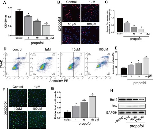
CCK-8 assay revealed a dose-dependent decline in the viability of HT-22 cells after propofol administration (). EdU assay further depicted the dose-dependently declined EdU-positive ratio in propofol-treated HT-22 cells (). After treatment of increased doses of propofol, the apoptotic rate gradually increased (). TUNEL-positive ratio was dose-dependently elevated by propofol treatment in HT-22 cells (). Apoptosis-associated genes were determined by Western blot. As data revealed, Bcl-2 was downregulated and Bax was upregulated in propofol-treated hippocampal neurons in a dose-dependent way ().
Figure 1 Propofol-induced apoptosis in HT-22 cells. (A) CCK-8 assay results showed viability in HT-22 cells treated with 0, 1, 10 and 100 μM propofol, respectively. (B and C) EdU assay results showed EdU-positive HT-22 cells treated with 0, 1, 10 and 100 μM propofol, respectively (B). Quantitative analysis of EdU-positive ratio (C). (D and E) Flow cytometry results showed distribution of apoptotic cells, necrotic cells and survival cells following the treatment of 0, 1, 10 and 100 μM propofol in HT-22 cells, respectively (D). Quantitative analysis of apoptosis rate (E). (F and G) TUNEL results showed TUNEL-positive cells following the treatment of 0, 1, 10 and 100 μM propofol in HT-22 cells, respectively (F). Quantitative analysis of TUNEL-positive rate (G). (H) Protein levels of Bcl-2 and Bax in HT-22 cells treated with 0, 1, 10 and 100 μM propofol, respectively (*p<0.05 compared to control group).
Metformin Treatment Reversed Propofol-Induced Apoptosis in HT-22 Cells
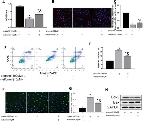
To elucidate the influence of metformin on HT-22 cells, they were administrated with metformin and propofol. Interestingly, the declined viability owing to propofol treatment was reversed following metformin administration (). Similarly, decreased EdU-positive ratio in propofol-treated HT-22 cells was partially blocked by metformin (). Decreased apoptotic rate was observed after metformin administration in propofol-treated HT-22 cells (). Compared with those treated with propofol, TUNEL-positive ratio decreased in HT-22 cells treated with both propofol and metformin (). As data revealed, Bcl-2 was downregulated and Bax was upregulated in propofol-treated hippocampal neurons which were reversed by metformin (). As a result, metformin effectively reversed propofol-induced proliferation inhibition and apoptosis stimulation in hippocampal neurons.
Figure 2 Metformin reversed propofol-induced apoptosis in HT-22 cells (A) CCK-8 assay results showed viability in propofol-induced HT-22 cells either treated with 10 μM metformin or not. (B and C) EdU assay results showed EdU-positive HT-22 cells with propofol induction, followed by 10 μM metformin treatment or not (B). Quantitative analysis of EdU-positive ratio (C). (D and E) Flow cytometry results showed distribution of apoptotic cells, necrotic cells and survival cells in propofol-induced HT-22 cells either treated with 10 μM metformin or not (D). Quantitative analysis of apoptosis rate (E). (F and G) TUNEL results showed TUNEL-positive cells in propofol-induced HT-22 cells either treated with 10 μM metformin or not (F). Quantitative analysis of TUNEL-positive rate (G). (H) Protein levels of Bcl-2 and Bax in propofol-induced HT-22 cells either treated with 10 μM metformin or not (*p<0.05 compared to control group; &p<0.05, compared to propofol (100μM) group).
Metformin Regulated Cav-1 Level
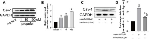
Western blot analysis uncovered that the protein level of Cav-1 dose-dependently upregulated in propofol-treated HT-22 cells (). Furthermore, metformin treatment downregulated Cav-1 level in propofol-treated HT-22 cells (). Hence, metformin was able to regulate propofol-induced Cav-1 upregulation.
Figure 3 Metformin treatment regulated Cav-1 level. (A and B). Protein level of Cav-1 in HT-22 cells treated with 0, 1, 10 and 100 μM propofol, respectively (A). Grey value analysis of Cav-1 (B). (C and D). Protein level of Cav-1 in propofol-induced HT-22 cells either treated with 10 μM metformin or not (C). Grey value analysis of Cav-1 (D) (*p<0.05 compared to control group; &p<0.05, compared to propofol (100μM) group).
Propofol-Induced Apoptosis in HT-22 Cells by Upregulating Cav-1
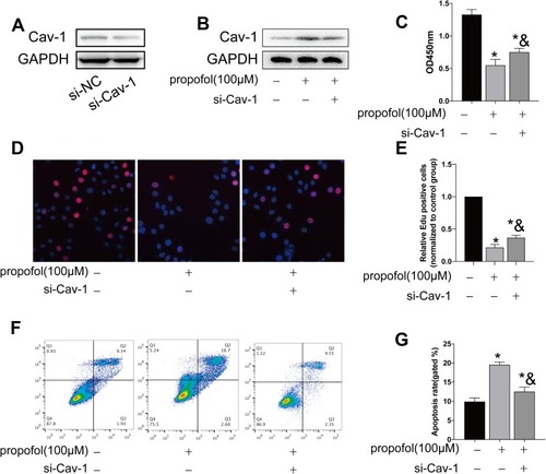
To further explore the function of propofol in inducing neuronal apoptosis, we constructed si-Cav-1 and pcDNA-Cav-1. Transfection efficacy of si-Cav-1 was determined in HT-22 cells by Western blot (). The protein level of Cav-1 was markedly upregulated after propofol treatment, which was then downregulated by transfection of si-Cav-1 (). CCK-8 assay showed that knockdown of Cav-1 partially reversed the declined viability in HT-22 cells treated with propofol (). EdU assay obtained a similar result (). The increased apoptotic rate in propofol-treated HT-22 cells was slightly reduced after transfection of si-Cav-1 ().
Figure 4 Propofol-induced apoptosis in HT-22 cells through upregulating Cav-1. (A) Transfection efficacy of si-Cav-1 in HT-22 cells. (B) Protein level of Cav-1 in propofol-induced HT-22 cells transfected with si-NC or si-Cav-1. (C) CCK-8 assay results showed viability in propofol-induced HT-22 cells transfected with si-NC or si-Cav-1. (D and E) EdU assay results showed EdU-positive cells in propofol-induced HT-22 cells transfected with si-NC or si-Cav-1 (D). Quantitative analysis of EdU-positive ratio (E). (F and G) Flow cytometry results showed distribution of apoptotic cells, necrotic cells and survival cells in propofol-induced HT-22 cells transfected with si-NC or si-Cav-1 (F). Quantitative analysis of apoptosis rate (G) (*p<0.05 compared to control group; &p<0.05, compared to propofol (100μM) group).
Metformin Reversed Propofol-Induced Neuronal Apoptosis Through Downregulating Cav-1
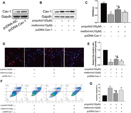
Next, transfection efficacy of pcDNA-Cav-1 was verified in HT-22 cells (). Transfection of pcDNA-Cav-1 inhibited the upregulated protein level of Cav-1 in propofol-treated HT-22 cells (). Metformin reversed the declined viability and EdU-positive ratio in propofol-treated HT-22 cells, which were further reduced after overexpression of Cav-1 (–). In the meantime, overexpression of Cav-1 enhanced apoptosis in propofol-treated neurons even after metformin treatment (). The above data proved that Cav-1 was responsible for protective effects of metformin on propofol-induced neurotoxicity.
Figure 5 Metformin reversed propofol-induced apoptosis in HT-22 cells through downregulating Cav-1. (A) Transfection efficacy of pcDNA-Cav-1 in HT-22 cells. (B) Protein level of Cav-1 in propofol-induced HT-22 cells transfected with pcDNA-NC or pcDNA-Cav-1 with either metformin treatment or not. (C) CCK-8 assay results showed viability in propofol-induced HT-22 cells transfected with pcDNA-NC or pcDNA-Cav-1 with either metformin treatment or not. (D and E) EdU assay results showed EdU-positive cells in propofol-induced HT-22 cells transfected with pcDNA-NC or pcDNA-Cav-1 with either metformin treatment or not (D). Quantitative analysis of EdU-positive ratio (E). (F and G) Flow cytometry results showed distribution of apoptotic cells, necrotic cells and survival cells in propofol-induced HT-22 cells transfected with pcDNA-NC or pcDNA-Cav-1 with either metformin treatment or not (F). Quantitative analysis of apoptosis rate (G) (*p<0.05 compared to control group; &p<0.05, compared to propofol (100μM) group).
Discussion
Increasing evidence have proved that propofol administration is able to induce developmental neurotoxicity, thereafter leading to long-term cognitive and learning abnormalities. Hence, the safety usage of propofol in pediatric anesthesia is well concerned.Citation24 Fredriksson et alCitation25 proposed that propofol administration dose-dependently aggravates the apoptotic degree in neurons of young rats, which could impair the long-term learning and memory ability of rats. In 7-day-old neonatal SD rats, propofol treatment markedly induces apoptosis in hippocampal neurons.Citation26 Our results revealed that propofol treatment dose-dependently decreased proliferative ability and increased apoptosis in HT-22 cells.
Apoptosis is of significance in maintaining homeostasis and normal development. Nevertheless, excessive apoptosis leads to adverse biological consequences. For instance, uncontrolled apoptosis is closely linked to ischemic heart disease, AIDS and Alzheimer’s disease, Parkinson’s disease and other neurodegenerative diseases.Citation27 Cell apoptosis has been well concerned in biology and medical researches.
Metformin has been verified as a safe and effective oral hypoglycemic agent extensively used in the treatment of diabetes.Citation28,Citation29 In nervous system, metformin stimulates the growth of newly formed hippocampal neurons and improves spatial learning and memory in mice.Citation30,Citation31 Through activating the S1P1-dependent ERK1/2 pathway, metformin protects rabbits from sevoflurane-induced neuronal apoptosis.Citation32 Besides, metformin another AMPK activator AICAR activates AKT S473 which provides survival signal for cells by inhibiting apoptotic signaling pathways.Citation33 In this analysis, metformin treatment protected propofol-induced proliferation inhibition and apoptosis stimulation in HT-22 cells. Therefore, we have proved the protective effect of metformin on propofol-induced neurotoxicity.
Cav-1 is a 22 kDa plasma membrane scaffold protein.Citation34 A previous study reported that Cav-1 regulates Caspase 3-mediated apoptosis pathway by downregulating Survivin.Citation35 Cav-1 deficiency has protective effects on both intracellular and extracellular apoptosis.Citation36 In addition, through interacting with Ras, Cav-1 promotes the activation of the Ras/Raf/ERK pathway, an important pathway participating in cell proliferation and apoptosis.Citation37–Citation40 Here, Cav-1 level was upregulated in propofol-treated HT-22 cells, which was downregulated following metformin treatment. In particular, knockdown of Cav-1 suppressed propofol-induced apoptosis in HT-22 cells. Rescue experiments further confirmed that overexpression of Cav-1 abolished the anti-apoptosis function of metformin in propofol-treated HT-22 cells. Collectively, metformin protected propofol-induced neurotoxicity through Cav-1.
Conclusions
In our study, we found that propofol treatment dose-dependently decreased proliferative ability and increased apoptosis in HT-22 cells. Besides, metformin could rescue the apoptosis effect of propofol induced in HT-22. In addition, we found that propofol induced the apoptosis of HT-22 by up-regulating Cav-1 and metformin protects propofol-induced neuronal apoptosis via downregulating Cav-1.
Disclosure
The authors report no conflicts of interest in this work.
References
- TrapaniG, AltomareC, LisoG, et al. Propofol in anesthesia. Mechanism of action, structure-activity relationships, and drug delivery. Curr Med Chem. 2000;7:249–271. doi:10.2174/092986700337533510637364
- KotaniY, ShimazawaM, YoshimuraS, et al. The experimental and clinical pharmacology of propofol, an anesthetic agent with neuroprotective properties. CNS Neurosci Ther. 2008;14(2):95–106. doi:10.1111/j.1527-3458.2008.00043.x18482023
- BerckerS, BertB, BittigauP, et al. Neurodegeneration in newborn rats following propofol and sevoflurane anesthesia. Neurotox Res. 2009;16(2):140–147. doi:10.1007/s12640-009-9063-819526290
- OlneyJW, FarberNB, WozniakDF, et al. Environmental agents that have the potential to trigger massive apoptotic neurodegeneration in the developing brain. Environ Health Perspect. 2000;108(Suppl 3):383–388. doi:10.1289/ehp.00108s3383
- KahramanS, ZupSL, McCarthyMM, et al. Gabaergic mechanism of propofol toxicity in immature neurons. J Neurosurg Anesthesiol. 2008;20(4):233–240. doi:10.1097/ANA.0b013e31817ec34d18812886
- CattanoD, YoungC, StraikoMM, et al. Subanesthetic doses of propofol induce neuroapoptosis in the infant mouse brain. Anesth Analg. 2008;106(6):1712–1714. doi:10.1213/ane.0b013e318172ba0a18499599
- CreeleyC, DikranianK, DissenG, et al. Propofol-induced apoptosis of neurones and oligodendrocytes in fetal and neonatal rhesus macaque brain. Br J Anaesth. 2013;110(Suppl 1):i29–i38. doi:10.1093/bja/aet17323722059
- MellonRD, SimoneAF, RappaportBA. Use of anesthetic agents in neonates and young children. Anesth Analg. 2007;104:509–520. doi:10.1097/sa.0b013e31815c102217312200
- TaoT, LiCL, YangWC, et al. Protective effects of propofol against whole cerebral ischemia/reperfusion injury in rats through the inhibition of the apoptosis-inducing factor pathway. Brain Res. 2016;1644:9–14. doi:10.1016/j.brainres.2016.05.00627163721
- NohHS, ShinIW, HaJH, et al. Propofol protects the autophagic cell death induced by the ischemia/reperfusion injury in rats. Mol Cells. 2010;30(5):455–460. doi:10.1007/s10059-010-0130-z20821058
- BredesenDE. Neural apoptosis. Ann Neurol. 1995;38(6):839–851. doi:10.1002/ana.4103806048526456
- WhiteE. Life, death, and the pursuit of apoptosis. Genes Dev. 1996;10(1):1–15. doi:10.1101/gad.10.1.18557188
- HackerG, VauxDL. Apoptosis. A sticky business. Curr Biol. 1995;5(6):622–624. doi:10.1016/s0960-9822(95)00126-67552172
- SmaleG, NicholsNR, BradyDR, et al. Evidence for apoptotic cell death in Alzheimer’s disease. Exp Neurol. 1995;133(2):225–230. doi:10.1006/exnr.1995.10257544290
- SchulteJM, RothausCS, AdlerJN. Clinical decisions. Management of type 2 diabetes–polling results. N Engl J Med. 2014;370(1):e2. doi:10.1056/NEJMclde080107824382086
- NathN, KhanM, PaintliaMK, et al. Metformin attenuated the autoimmune disease of the central nervous system in animal models of multiple sclerosis. J Immunol. 2009;182(12):8005–8014. doi:10.4049/jimmunol.099006019494326
- PatelHH, MurrayF, InselPA. Caveolae as organizers of pharmacologically relevant signal transduction molecules. Annu Rev Pharmacol Toxicol. 2008;48(1):359–391. doi:10.1146/annurev.pharmtox.48.121506.12484117914930
- FridolfssonHN, PatelHH. Caveolin and caveolae in age associated cardiovascular disease. J Geriatr Cardiol. 2013;10:66–74. doi:10.3969/j.issn.1671-5411.2013.01.01123610576
- KangJW, LeeSM. Impaired expression of caveolin-1 contributes to hepatic ischemia and reperfusion injury. Biochem Biophys Res Commun. 2014;450(4):1351–1357. doi:10.1016/j.bbrc.2014.06.13124997335
- DasM, GherghiceanuM, LekliI, et al. Essential role of lipid raft in ischemic preconditioning. Cell Physiol Biochem. 2008;21(4):325–334. doi:10.1159/00012939118441521
- RothbergKG, HeuserJE, DonzellWC, et al. Caveolin, a protein component of caveolae membrane coats. Cell. 1992;68(4):673–682. doi:10.1016/0092-8674(92)90143-z1739974
- LiuY, FuY, HuX, et al. Caveolin-1 knockdown increases the therapeutic sensitivity of lung cancer to cisplatin-induced apoptosis by repressing Parkin-related mitophagy and activating the ROCK1 pathway. J Cell Physiol. 2019. doi:10.1002/jcp.29033
- MukhopadhyayS, ChatterjeeA, KoganD, et al. 5-Aminoimidazole-4-carboxamide-1-beta-4-ribofuranoside (AICAR) enhances the efficacy of rapamycin in human cancer cells. Cell Cycle. 2015;14(20):3331–3339. doi:10.1080/15384101.2015.108762326323019
- BosnjakZJ, LoganS, LiuY, et al. Recent insights into molecular mechanisms of propofol-induced developmental neurotoxicity: implications for the protective strategies. Anesth Analg. 2016;123(5):1286–1296. doi:10.1213/ANE.000000000000154427551735
- FredrikssonA, PontenE, GordhT, et al. Neonatal exposure to a combination of n-methyl-d-aspartate and gamma-aminobutyric acid type a receptor anesthetic agents potentiates apoptotic neurodegeneration and persistent behavioral deficits. Anesthesiology. 2007;107(3):427–436. doi:10.1097/01.anes.0000278892.62305.9c17721245
- HanD, JinJ, FangH, et al. Long-term action of propofol on cognitive function and hippocampal neuroapoptosis in neonatal rats. Int J Clin Exp Med. 2015;8(7):10696–10704.26379861
- KannanK, JainSK. Oxidative stress and apoptosis. Pathophysiology. 2000;7(3):153–163. doi:10.1016/s0928-4680(00)00053-510996508
- YuanX, WeiW, BaoQ, et al. Metformin inhibits glioma cells stemness and epithelial-mesenchymal transition via regulating yap activity. Biomed Pharmacother. 2018;102:263–270. doi:10.1016/j.biopha.2018.03.03129567539
- FuX, PanY, CaoQ, et al. Metformin restores electrophysiology of small conductance calcium-activated potassium channels in the atrium of gk diabetic rats. BMC Cardiovasc Disord. 2018;18(1):63. doi:10.1186/s12872-018-0805-529636010
- GeX-H, ZhuG-J, GengD-Q, et al. Metformin protects the brain against ischemia/reperfusion injury through pi3k/akt1/jnk3 signaling pathways in rats. Physiol Behav. 2017;170:115–123. doi:10.1016/j.physbeh.2016.12.02128017679
- LiJ, DengJ, ShengW, et al. Metformin attenuates Alzheimer’s disease-like neuropathology in obese, leptin-resistant mice. Pharmacol Biochem Behav. 2012;101(4):564–574. doi:10.1016/j.pbb.2012.03.00222425595
- YueH, HuB, LuoZ, et al. Metformin protects against sevoflurane-induced neuronal apoptosis through the s1p1 and erk signaling pathways. Exp Ther Med. 2019;17(2):1463–1469. doi:10.3892/etm.2018.709830680029
- ZhuX, ZhangY, LiQ, et al. beta-carotene induces apoptosis in human esophageal squamous cell carcinoma cell lines via the Cav-1/AKT/NF-kappaB signaling pathway. J Biochem Mol Toxicol. 2016;30(3):148–157. doi:10.1002/jbt.2177326733226
- FujimotoT, KogoH, NomuraR, et al. Isoforms of caveolin-1 and caveolar structure. J Cell Sci. 2000;113(Pt 19):3509–3517. doi:10.1023/A:100562942763010984441
- JinY, KimHP, ChiM, et al. Deletion of Caveolin-1 protects against oxidative lung injury via up-regulation of heme oxygenase-1. Am J Respir Cell Mol Biol. 2008;39(2):171–179. doi:10.1165/rcmb.2007-0323OC18323531
- WangX, WangY, KimHP, et al. Carbon monoxide protects against hyperoxia-induced endothelial cell apoptosis by inhibiting reactive oxygen species formation. J Biol Chem. 2007;282(3):1718–1726. doi:10.1074/jbc.M60761020017135272
- KortumRL, FernandezMR, Costanzo-GarveyDL, et al. Caveolin-1 is required for kinase suppressor of ras 1 (ksr1)-mediated extracellular signal-regulated kinase 1/2 activation, h-rasv12-induced senescence, and transformation. Mol Cell Biol. 2014;34(18):3461–3472. doi:10.1128/MCB.01633-1325002533
- LiuP, YingY, AndersonRG. Platelet-derived growth factor activates mitogen-activated protein kinase in isolated caveolae. Proc Natl Acad Sci U S A. 1997;94(25):13666–13670. doi:10.2307/437909391083
- LiW, HuangR, ShettyRA, et al. Transient focal cerebral ischemia induces long-term cognitive function deficit in an experimental ischemic stroke model. Neurobiol Dis. 2013;59:18–25. doi:10.1016/j.nbd.2013.06.01423845275
- LinL, SuZ, LebedevaIV, et al. Activation of Ras/Raf protects cells from melanoma differentiation-associated gene-5-induced apoptosis. Cell Death Differ. 2006;13(11):1982–1993. doi:10.1038/sj.cdd.440189916575407