
Abstract
In vivo responses to gold nanoparticles (GNPs) vary not only according to the size, shape, surface charge, and capping agent of GNPs but also according to the animal model, the route of administration, and the exposure frequency and duration. We illustrate here the changes in some hematologic parameters, in the hepatic and renal functions, and in the histopathology of solid organs after multiple intraperitoneal injections of 18 nm GNPs in adult male Syrian golden hamsters. We scored the histopathological changes in the liver and kidneys to grade the deleterious effects. Multiple intraperitoneal injections of 18 nm GNPs in hamsters were nonlethal in the short term but resulted in macrocytosis and hypochromasia, leukocytosis, neutrophilia, lymphocytosis, and monocytosis. The hepatic and renal functions showed nonsignificant changes; however, histopathological examination showed hepatic and renal alterations ranging from mild to marked degeneration, with occasional necrosis of hepatocytes and tubular epithelium.
Background
Materials at the nanoscale (1–100 nm) differ from their larger counterparts in being more reactive due to a relatively large surface-to-diameter ratio. Gold nanoparticles (GNPs) have unique physical and chemical properties, such as biocompatibility and ease of preparation and modification,Citation1,Citation2 and unique optical properties arising from the surface plasmon oscillation of free electrons.Citation3 GNP properties are convenient for biomedical applications, such as drug and gene delivery,Citation4,Citation5 DNA detection,Citation6 bioimaging,Citation7–Citation9 and photothermal therapy of cancers.Citation10,Citation11
GNP safety data derived from studies on tissue cultures may not reflect the real picture in vivo.Citation12 Available in vivo data are contradictory. Many reports concluded that GNPs do not produce toxicity in laboratory animals.Citation13–Citation15 Others claimed that GNPs induce morbidity and mortality when injected in the same animals.Citation16 GNP-induced changes are attributed to their sizes able to cross the biological barriers.Citation17–Citation19 The particle surface chemistry and charge also have important roles in the induced changes due to their immunogenicity and effects on bioclearance.Citation13,Citation15,Citation20–Citation24 The route of administration has a major impact on GNP-induced effects. Intravenous route was shown to be the safestCitation12,Citation25 followed by subcutaneous application.Citation24 On the other hand, intraperitoneal (I/P) administration of GNPs showed a moderate toxicity,Citation12,Citation16 while the oral route was the most toxic.Citation12,Citation26
Eighteen-nanometer GNPs were found to have a high retention rate in living organisms after intraesophageal administration with a high accumulation rate in solid organs, such as the brain and heart.Citation27 It was hypothesized that the specific curvature and surface structure of the 18 nm GNPs alter the structure and function of single adsorbed proteins or select proteins, increasing the probability of intestinal epithelial penetration for the 18 nm GNPs compared to other GNP sizes. Also, 24 hours after intravenous injection in mice, 18 nm GNPs showed a high retention in blood cells compared to serum, indicating their partial binding to blood cells. It showed indeed >90% accumulation in the liver. Single 18 nm GNPs were found in hepatocytes and endothelial cells indicating little agglomeration in the blood.Citation28
Syrian golden hamsters (Mesocricetus auratus) are widely used to model cancers, especially cancers of the upper aerodigestive tract.Citation29 The pattern of the upper aerodigestive tree, and esophageal cancers produced in hamsters resembles that seen in human smokers and suggests that this model may serve as a system for testing chemopreventive or chemotherapeutic agents for tumors in these areas.Citation30
As far as we know, no quantitative or semiquantitative biodistribution study of the 18 nm GNPs after in vivo I/P administration in hamsters exists. Thereby, the aim of this study was to investigate the effects of repeated I/P injection of 18 nm sized GNPs on some hematologic parameters, on the hepatic and renal functions and on the histopathology of solid organs in healthy adult male Syrian golden hamsters receiving 30 ppb of GNPs daily for 14 consecutive days (a total dose of 420 ppb per animal). The observed effects were assessed in a semiquantitative manner to serve as a baseline, where the effects of intralesional administrations of GNPs in a hamster tumor model that we developed are compared with those of systemic injections of GNPs in the same model. This work is part of a project aiming to assess the safety and efficacy of GNPs in the diagnosis and photo-thermal therapy of chemically induced oral cancers.
Materials and methods
GNPs’ preparation and characterization
GNPs were prepared by citrate reduction of gold(III) chloride trihydrate following the Frens modification of the Turkevich method.Citation31 Before the reduction process, all glassware was cleaned in aqua regia (three parts HCl and one part HNO3), rinsed with deionized H2O, and then dried. An aqueous solution of gold(III) chloride trihydrate (≥99.9%; Sigma-Aldrich Co., St Louis, MO, USA) was brought to boiling and stirred continuously. A solution of 38.8 mM sodium citrate tribasic dehydrate (≥98%; Sigma-Aldrich Co.) was added quickly, resulting in a change in solution color from pale yellow to black to deep red. A 50:2 ratio of tetrachloroauric acid to citrate was used in order to obtain the particles of ~18 nm size. GNPs were washed three times with deionized water to remove the nonreacted precursors. The prepared nanoparticles were then characterized using the ultraviolet-visible (UV-Vis) absorption spectrophotometer (Cary 5000; Agilent, Santa Clara, CA, USA), the particle size analyzer (Malvern Zetasizer Nano ZSPXRD; Malvern Instruments, Malvern, UK), and the transmission electron microscopy (TEM) (Tecnai; FEI, Eindhoven, the Netherlands). Absorption spectra were recorded using a double beam UV-Vis spectrophotometer. The absorption spectra of the diluted solutions of prepared GNPs in aqueous medium were recorded within the appropriate scan range (400–850 nm). The spectrum of the pure solvent was taken as a calibrating reference. Measurements were performed at room temperature. The morphology of GNPs and their particle sizes were examined under TEM operating at an accelerating voltage of 80 kV. A drop from a dilute sample solution was deposited on an amorphous carbon-coated copper grid and left to evaporate at room temperature forming a monolayer. Analysis of the particle size diameters of prepared GNPs was estimated using the software program Gaten over several shots of TEM images for the target sample. The purity of GNPs was assessed by energy-dispersive X-ray spectroscopy on TEM. In order to calculate the volume to be injected in each animal, the concentration of gold in solution was determined by atomic absorption spectroscopy (SpectrAA 220; Varian Medical Systems).
In vivo animal experiments
A total of 20 adult male Syrian golden hamsters were purchased from the Holding Company for Biological Products and Vaccines (VACSERA), Helwan, Cairo, Egypt. The hamsters were of nearly uniform age (10–12 weeks) and weight (100–150 g). The animal care protocol was in compliance with the Humane Care for Animals Act, the guidelines of The Canadian Council of Animal Care, and the EU Directive 2010/63/EU for animal experiments. The study design was approved by the Animal Care Committee at the Faculty of Veterinary Medicine, Cairo University, Egypt. Hamsters were fed on dry, 26% protein pellets and tap water ad libitum. They were housed two to three hamsters per cage in a temperature-controlled and well-ventilated breeding area in a 12-hour light/dark cycle. The environmental conditions were monitored twice daily. Hamsters were kept under observation for 2 weeks before starting the experiment to ensure full conditioning and healthy state. They were randomly divided into two groups: the control group, consisting of ten hamsters, kept under similar environmental conditions to the injected group, also consisting of ten animals. In the injected group, each animal received one I/P injection of 0.5 mL of GNPs (30 ppb) daily for 14 consecutive days. At the end of the experiment, animals were anesthetized diethyl ether (SD Fine-Chem Ltd, Chennai, India). Blood was collected from the retro-orbital venous plexus in tubes containing disodium ethylene diamine tetra-acetic acid (El Nasr Pharmaceutical Chemicals Company, ADWIC, Nasr Pharma, Qalyub, Egypt) for complete blood count and in plain vacutainers to yield serum by centrifugation (3,000 rpm for 10 minutes). Serum was kept at −20°C for biochemical tests. Animals were euthanized by intracardiac injection of overdose of Mepacaine L (each 1 mL contains mepivacaine HCl 20.0 mg and levonordefin HCl 0.06 mg; Alexandria Co., Pharmaceuticals, Alexandria, Egypt). Solid organs were dissected and taken for histopathological examination.
Complete blood count
Red blood cell (RBC) and white blood cell counts were performed using an improved Neubauer hemocytometer. Packed cell volume was estimated by the microhematocrit technique. Hemoglobin concentration was determined colorimetrically using the cyanmethemoglobin method. Differential leukocytic count was performed on the stained blood smear according to Feldman et al.Citation32
Biochemical tests
Hepatic and renal function tests were measured using diagnostic kits (Spectrum Diagnostics, Obour City, Egypt) according to the manufacturer’s recommendations. Total proteins were measured as described by Weichselbaum,Citation33 albumin was measured as described by Dumas and Biggs,Citation34 ALT and AST were measured as described by Reitman and Frankel,Citation35 urea was measured as described by Searcy et al,Citation36 and creatinine was measured as described by Fabiny and Ertingshausen.Citation37
Tissue distribution
Tissue distribution was assessed by flame atomic absorption spectroscopy (SpectrAA 220; Varian Medical Systems). Tissue samples were prepared according to Kehoe et alCitation38 where known amounts of tissues are weighed and digested by a mixture of perchloric and nitric acid (1:3) while heating at 200°C.
Statistical analysis of the biochemical tests
Data were presented as mean ± SD. Independent samples Student’s t-test was used to compare mean between groups after assuring normal distribution of the data using Shapiro–Wilk test. The significance level was set as P-value ≤0.05. Statistical analysis was performed using the SPSS software version 16.
Histopathological examination
After taking the blood samples and euthanizing the laboratory animals from both groups, the liver, kidneys, spleen, and lungs were carefully dissected and collected as whole organ specimens from all experimental animals. Organs were fixed in neutral buffered formalin (10%) then dehydrated, with sequential exposure to grades of ethanol (70%, 80%, 90%, 95%, and 100%). Dehydration was followed by clearing the samples in two changes of Xylene. Samples were impregnated with two changes of molten paraffin wax, then embedded, and blocked out. Paraffin sections of 5 μm thickness were cut using a rotatory microtome (Leica RM 2235, Leica Biosystems, Nussloch, Germany), stained with hematoxylin and eosin according to Bancroft et al,Citation39 and examined using an optical microscope (Olympus BX 50; Olympus Corporation, Tokyo, Japan). Histopathological diagnosis was performed according to the Standardized System of Nomenclature and Diagnostic Criteria.Citation40
Scoring system
Semiquantitative scoring to determine the extent of injury in the liver and kidneys was done using a four-digit numerical scoring system where 0 indicates no change and 1–4 indicate increasing severity according to Mann et al.Citation41
Statistical analysis of the scoring
The obtained scores were presented as mean ± standard deviation. Statistical significance between the different groups was analyzed using the analysis of variance test (SPSS 10.0) followed by Duncan’s multiple range test.Citation42
Results
GNP characterization
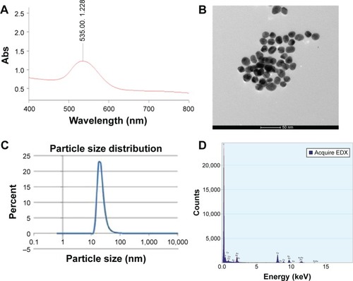
The prepared GNPs showed a maximum extinction of 1.228 at 535 nm by UV-Vis spectrophotometer (). TEM images showed that the examined particles (n=100) have a spherical shape with a mean particle diameter of 18 nm (±1.1 nm) (). Particle size distribution assessed by zetasizer showed peak percentage (22.9%) at 18 nm diameter (). The energy-dispersive X-ray spectroscopy showed peaks corresponding to Au element at 2.12 keV, 9.44 keV, and 11.4 keV, confirming the existence of GNPs. In addition, acceptable peaks for C at 0.277 keV and for Cu at 0.93 keV, 8.04 keV, and 8.9 keV were observed owing to the composition of copper grid used in the TEM imaging process ().
Figure 1 GNPs characterization.
Notes: (A) Peak absorption of prepared GNPs (1.2) at 535 nm wavelength. (B) TEM images showing gold nanospheres with average 18 nm diameter (magnification, 97,000×). (C) Particle size distribution by zeta sizer showing peak percent (22.9%) at 18 nm diameter. (D) EDX showing peaks corresponding to Au element at 2.12 keV, 9.44 keV, and 11.4 keV, confirming the existence of GNPs.
Abbreviations: EDX, energy-dispersive X-ray spectroscopy; GNPs, gold nanoparticles; TEM, transmission electron microscopy; Abs, absorbance.
Clinical signs
No hamster mortality occurred during the 14 days of I/P injection of GNPs. Furthermore, no abnormal clinical signs or behaviors were detected in the injected or in the control groups. No changes were noticed in the fur, and there was no discharge. Necropsy at the end of the experiment did not show any macroscopic changes in the organs of the injected group.
Erythrogram
Results of the erythrogram showed significantly higher mean packed cell volume and mean corpuscular volume in the injected group (P≤0.05). On the other hand, the mean cell hemoglobin concentration in the injected group was significantly lower than in the control group as shown in .
Table 1 Mean ± SD of clinical hematology, clinical chemistry, and tissue distribution data
Leukogram
Leukocytosis was evident in hamsters of the injected group. Neutrophilia, lymphocytosis, and monocytosis occurred. On the contrary, no difference was noticed in the eosinophil and basophil counts between the injected and control groups. Results of the leukogram are shown in .
Liver function tests
None of the assessed parameters (total proteins, albumin, globulin, albumin/globulin ratio, ALT, and AST) showed statistically significant differences between the injected and control groups as shown in .
Kidney function tests
There was no statistically significant difference in the mean values of urea and creatinine levels in serum between the injected and control groups as shown in .
Tissue distribution
Results of tissue distribution demonstrated that, in the GNP-injected hamsters, gold accumulated mainly in the liver (7.85±0.55 μg Au/g tissue) followed by the spleen (6.47±0.54 μg/g) and heart (6.37±0.58 μg/g). Pulmonary and renal tissues showed less concentration of gold (4.02±0.64 μg/g and 3.74±0.46 μg/g, respectively). On the other hand, tissues of control animals showed absence of any residue of gold as illustrated in .
Histopathological examination
Histopathological examination of different organs, namely the liver, kidneys, lungs, heart, and spleen, from animals of the control group did not reveal any structural abnormality. Hamsters of the injected group showed normal structure of the spleen, lungs, and heart except for mild-to-moderate congestion of blood vessels, whereas hepatic and renal tissues showed variable histopathological alterations of varying severity and extent as follows:
Liver
Mild-to-moderate and marked focal and diffuse vacuolar degeneration of hepatocytes together with Kupffer cell hyperplasia/upregulation and congestion of the central and portal blood vessels were commonly observed in almost all cases (). Mild-to-moderate perivascular and portal monolymphocytic cell infiltration () and variable sized sporadic foci of hepatic necrosis scattered throughout the hepatic parenchyma along with replacement of the necrotic tissue with mononuclear cells were commonly seen (). Occasional paracentral areas of coagulative necrosis of hepatocytes with mononuclear cell infiltration () and/or foci of coagulative necrosis of hepatocytes associated with sinusoidal leukocytosis () were also observed.
Figure 2 Liver and kidneys of hamsters of the injected group.
Notes: (A) Liver of a hamster of the injected group showing marked vacuolar degeneration of hepatocytes and congestion of hepatic blood vessels (hematoxylin and eosin stain [H&E], magnification, ×20). (B) Liver of a hamster of the injected group showing perivascular mononuclear cell infiltration (H&E, ×20). (C) Liver of a hamster of the injected group showing sporadic foci of hepatic necrosis scattered throughout the hepatic parenchyma along with replacement of the necrotic tissue with mononuclear cells (H&E, ×20). (D) Liver of a hamster of the injected group showing paracentral area of coagulative necrosis of hepatocytes with mononuclear cell infiltration (H&E, ×20). (E) Liver of a hamster of the injected group showing focal areas of hepatic necrosis and sinusoidal leukocytosis (H&E, ×20). (F) Kidney of a hamster of the injected group showing marked vacuolar degeneration and necrosis of tubular epithelium associated with thickening of the tubular and glomerular basement membranes, swelling of glomerular tuft, congestion of intertubular blood vessels, and intertubular hemorrhage (H&E, ×40).
![Figure 2 Liver and kidneys of hamsters of the injected group.Notes: (A) Liver of a hamster of the injected group showing marked vacuolar degeneration of hepatocytes and congestion of hepatic blood vessels (hematoxylin and eosin stain [H&E], magnification, ×20). (B) Liver of a hamster of the injected group showing perivascular mononuclear cell infiltration (H&E, ×20). (C) Liver of a hamster of the injected group showing sporadic foci of hepatic necrosis scattered throughout the hepatic parenchyma along with replacement of the necrotic tissue with mononuclear cells (H&E, ×20). (D) Liver of a hamster of the injected group showing paracentral area of coagulative necrosis of hepatocytes with mononuclear cell infiltration (H&E, ×20). (E) Liver of a hamster of the injected group showing focal areas of hepatic necrosis and sinusoidal leukocytosis (H&E, ×20). (F) Kidney of a hamster of the injected group showing marked vacuolar degeneration and necrosis of tubular epithelium associated with thickening of the tubular and glomerular basement membranes, swelling of glomerular tuft, congestion of intertubular blood vessels, and intertubular hemorrhage (H&E, ×40).](/cms/asset/cdfe39dc-a10f-49c5-abee-3c238addac31/dijn_a_102919_f0002_c.jpg)
Kidneys
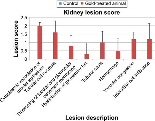
Renal cortex in almost all hamsters of the injected group revealed vacuolar degeneration and necrosis of tubular epithelium associated with thickening of the tubular and glomerular basement membranes, congestion of intertubular blood vessels, and intertubular hemorrhage (). Intraluminal casts of proteinaceous materials were commonly observed. Marked vacuolar degeneration and necrosis of tubular epithelium together with an increased mesangial matrix and hyalinization of glomerular tuft and tubular basement membrane were observed in two cases (). Multifocal areas of monolymphocytic interstitial cell infiltrates were commonly observed in most cases ().
Figure 3 Kidneys of hamsters of the injected group.
Notes: (A) Kidney of a hamster of the injected group showing marked vacuolar degeneration and necrosis of tubular epithelium together with an increased mesangial matrix and hyalinization of glomerular tuft and tubular basement membrane (hematoxylin and eosin stain [H&E], magnification, ×40). (B) Kidney of a hamster of the injected group showing focal interstitial mononuclear cell infiltration (H&E, ×40).
![Figure 3 Kidneys of hamsters of the injected group.Notes: (A) Kidney of a hamster of the injected group showing marked vacuolar degeneration and necrosis of tubular epithelium together with an increased mesangial matrix and hyalinization of glomerular tuft and tubular basement membrane (hematoxylin and eosin stain [H&E], magnification, ×40). (B) Kidney of a hamster of the injected group showing focal interstitial mononuclear cell infiltration (H&E, ×40).](/cms/asset/5fb3a3d5-d9d6-4fc9-ae7d-21e5ed50a087/dijn_a_102919_f0003_c.jpg)
Scoring system
Semiquantitative scoring showed that the overall lesions in the livers and kidneys did not exceed the moderate changes (– and and ).
Table 2 Scoring of histopathological lesions in the liver of 18 nm GNP-injected hamsters
Table 3 Scoring of histopathological lesions in the kidney of 18 nm GNP-injected hamsters
Table 4 Mean ± SD scores of lesions in livers of 18 nm GNPs- injected hamsters
Table 5 Mean ± SD scores of lesions in kidneys of 18 nm GNPs-injected hamsters
Figure 4 Scoring of hepatic histopathological lesions after I/P injection of 18 nm GNPs in Syrian golden hamsters for 14 consecutive days.
Abbreviations: GNPs, gold nanoparticles; I/P, intraperitoneal.
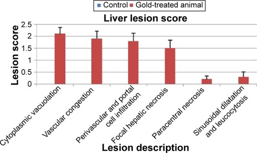
Figure 5 Scoring of renal histopathological lesions after I/P injection of 18 nm GNPs in Syrian golden hamsters for 14 consecutive days.
Abbreviations: GNPs, gold nanoparticles; I/P, intraperitoneal.
Discussion
Nanotechnology has recently emerged as a promising approach in the diagnosis and treatment of a variety of diseases. GNPs have been suggested as an auxiliary tool in eradicating tumors in the context of photothermal therapy.Citation10 However, the translation of such techniques into comprehensive therapeutic modalities is still pending. One reason is that the safety issues of GNPs and the questions over their metabolic fate are still unresolved and need further investigations at a preclinical level.Citation6,Citation43
Our study was done on Syrian golden hamsters. We are using these small animals to model the treatment of tumors of the upper aerodigestive tract by gold nanophotothermolysis. We detected several hematologic changes and some histopathological changes in the microscopic pictures of the liver and kidneys after I/P injection with 18 nm GNPs.
The erythrogram indicated the presence of macrocytosis and hypochromasia (high mean corpuscular volume and low mean cell hemoglobin concentration) in the RBCs of 18 nm GNP-injected hamsters. This is consistent with a previous study done by Zhang et alCitation12 and might be attributed to the ability of the 18 nm GNPs to cross the fenestrated endothelial membrane of the bone marrow (50–100 nm),Citation44,Citation45 thus interfering with the normal erythropoiesis resulting in immature RBCs. Since Lasagna-Reeves et alCitation46 and Simpson et alCitation13 reported that GNPs <5 nm in diameter have no effect on RBCs’ count and morphology, disagreement with our results may be attributed to different doses and duration of exposure to GNPs and more importantly to different sizes of GNPs.
The leukocytosis observed in GNP-injected hamsters is in agreement with Simpson et al,Citation22 Zhang et al,Citation47 and Sengupta et al.Citation48 The elevation in total leukocytic count might be attributed to the immunogenic effect of GNPsCitation15 or to their ability to trigger an inflammatory response by stimulating the release of cytokines.Citation49 Previously reported leukocytosis after injection of 10 nm and 60 nm particles in mice was attributed to an inflammatory response.Citation50 The monocytosis in the 18 nm GNP-injected hamsters can be regarded as an immune response attributed to the role of monocytes in the removal of xenobiotics, such as GNPs, from the circulation.Citation27
The lack of significant changes in the hepatic and renal function tests is in agreement with many reportsCitation14,Citation46,Citation48,Citation51 and in contradiction with others.Citation15,Citation47 In these latter reports, different routes of administration and different doses and sizes of GNPs were applied.
Eighteen-nanometer GNPs were accumulated in significant amounts in the liver and spleen and to lesser degrees in the lungs, kidneys, and heart following the I/P injection. These results are consistent with a previous report by Lipka et al.Citation20 Although the spleen was proved to be the dominant target organ for the 30 nm particles,Citation50 we found that the primary target for the 18 nm GNPs was the liver, and we did not find any remarkable histopathological alterations in the spleens of the 18 nm GNP-injected hamsters.
The microscopic lesions shown in the hepatic and renal tissues of hamsters exposed to GNPs are in agreement with Terentyuk et alCitation52 and Das et al.Citation15 Hepatocytes’ degeneration and necrosis might be attributed to an enhanced defense mechanism against foreign particles, intoxication, hemodynamic changes, or alteration in inflammation and apoptosis-related genes. The determination of the exact cause needs further studies.
The presence of vacuolar degeneration in the renal tissue might be attributed to increased intracellular water due to disturbed ions and fluid homeostasis. Other renal lesions can be attributed to the toxic effects of GNPs during clearance by renal tissue.
Conclusion
The repeated administration of 30 ppb of 18 nm GNPs for 14 consecutive days in hamsters induced macrocytosis, hypochromasia, leukocytosis, neutrophilia, lymphocytosis, and monocytosis. I/P administration of 18 nm GNPs in hamsters had no effect on plasma proteins, liver, and renal function tests. However, some hepatic and renal tissue alterations were observed ranging from mild to marked degeneration and necrosis of hepatocytes and tubular epithelium.
Acknowledgments
Financial support from the Science and Technology Development Fund, Egypt, through the Project # 1012 is gratefully acknowledged. Science and Technology Development Fund does not object to the decision to submit this article for publication.
Disclosure
The authors report no conflicts of interest in this work.
References
- DanielMCAstrucDGold nanoparticles: assembly, supramolecular chemistry, quantum-size-related properties, and applications toward biology, catalysis, and nanotechnologyChem Rev200410429334614719978
- KimTLeeKGongMSJooSWControl of gold nanoparticle aggregates by manipulation of interparticle interactionLangmuir2005219524952816207031
- HuangWQianWJainPKEl-SayedMAThe effect of plasmon field on the coherent lattice phonon oscillation in electron-beam fabricated gold nanoparticle pairsNano Lett200773227323417760479
- BaoQYGengDDXueJWGlutathione-mediated drug release from Tiopronin-conjugated gold nanoparticles for acute liver injury therapyInt J Pharm201344611211823416166
- CondeJLarguinhoMCordeiroAGold-nanobeacons for gene therapy: evaluation of genotoxicity, cell toxicity and proteome profiling analysisNanotoxicology2014852153223642008
- LiuCCYeungCYChenPHYehMKHouSYSalmonella detection using 16S ribosomal DNA/RNA probe-gold nanoparticles and lateral flow immunoassayFood Chem20131412526253223870991
- MurphyCJGoleAMStoneJWGold nanoparticles in biology: beyond toxicity to cellular imagingAcc Chem Res2008411721173018712884
- GongTOlivoMDinishUSGohDKongKVYongKTEngineering bioconjugated gold nanospheres and gold nanorods as label-free plasmon scattering probes for ultrasensitive multiplex dark-field imaging of cancer cellsJ Biomed Nanotechnol2013998599123858962
- ZhangJLiCZhangXIn vivo tumor-targeted dual-modal fluorescence/CT imaging using a nanoprobe co-loaded with an aggregation-induced emission dye and gold nanoparticlesBiomaterials20154210311125542798
- El-SayedIHHuangXEl-SayedMASelective laser photo-thermal therapy of epithelial carcinoma using anti-EGFR antibody conjugated gold nanoparticlesCancer Lett200623912913516198049
- LinJWangSHuangPPhotosensitizer-loaded gold vesicles with strong plasmonic coupling effect for imaging-guided photothermal/photodynamic therapyACS Nano201375320532923721576
- ZhangXDWuHYWuDToxicologic effects of gold nanoparticles in vivo by different administration routesInt J Nanomedicine2010577178121042423
- SimpsonCAAgrawalACBalinskiAHarknessKMCliffelDEShort-chain PEG mixed monolayer protected gold clusters increase clearance and red blood cell countsACS Nano201153577358421473648
- SungJHJiJHParkJDSubchronic inhalation toxicity of gold nanoparticlesPart Fibre Toxicol201181621569586
- DasSDebnathNMitraSDattaAGoswamiAComparative analysis of stability and toxicity profile of three differently capped gold nanoparticles for biomedical usageBiometals2012251009102222752843
- ChenYSHungYCLiauIHuangGSAssessment of the in vivo toxicity of gold nanoparticlesNanoscale Res Lett2009485886420596373
- ChoWSChoMJeongJSize-dependent tissue kinetics of PEG-coated gold nanoparticlesToxicol Appl Pharmacol201024511612320193702
- TedescoSDoyleHBlascoJRedmondGSheehanDOxidative stress and toxicity of gold nanoparticles in Mytilus edulisAquat Toxicol201010017818620382436
- AbdelhalimMAGold nanoparticles administration induces disarray of heart muscle, hemorrhagic, chronic inflammatory cells infiltrated by small lymphocytes, cytoplasmic vacuolization and congested and dilated blood vesselsLipids Health Dis20111023322151883
- LipkaJSemmler-BehnkeMSperlingRABiodistribution of PEG-modified gold nanoparticles following intratracheal instillation and intravenous injectionBiomaterials2010316574658120542560
- NiidomeTYamagataMOkamotoYPEG-modified gold nanorods with a stealth character for in vivo applicationsJ Control Release200611434334716876898
- SimpsonCAHuffmanBJGerdonAECliffelDEUnexpected toxicity of monolayer protected gold clusters eliminated by PEG-thiol place exchange reactionsChem Res Toxicol2010231608161620715858
- GirgisEKhalilWKEmamANMohamedMBRaoKVNanotoxicity of gold and gold-cobalt nanoalloyChem Res Toxicol2012251086109822486372
- SimpsonCASallengKJCliffelDEFeldheimDLIn vivo toxicity, biodistribution, and clearance of glutathione-coated gold nanoparticlesNanomedicine2013925726322772047
- KimJHKimJHKimKWKimMHYuYSIntravenously administered gold nanoparticles pass through the blood-retinal barrier depending on the particle size, and induce no retinal toxicityNanotechnology20092050510119923650
- HillyerJFAlbrechtRMGastrointestinal persorption and tissue distribution of differently sized colloidal gold nanoparticlesJ Pharm Sci2001901927193611745751
- SchlehCSemmler-BehnkeMLipkaJSize and surface charge of gold nanoparticles determine absorption across intestinal barriers and accumulation in secondary target organs after oral administrationNanotoxicology20126364621309618
- HirnSSemmler-BehnkeMSchlehCParticle size-dependent and surface charge-dependent biodistribution of gold nanoparticles after intravenous administrationEur J Pharm Biopharm20117740741621195759
- VairaktarisESpyridonidouSPapakostaVThe hamster model of sequential oral oncogenesisOral Oncol20084431532418061531
- EstensenRDAndersonWRGalbraithARA method of producing carcinoma in upper aerodigestive tree and esophagus of the Syrian golden hamster using wounding and instillation of N-methylnitrosoureaCancer Epidemiol Biomarkers Prev2007161644165017684140
- FrensGControlled nucleation for the regulation of particle size in monodisperse gold suspensionNat Phys Sci19732412022
- FeldmanBVZinklJGJianNCSchalm’s Veterinary HematologyPhiladelphiaLea and Fibiger2000
- WeichselbaumTEAn accurate rapid method for determination of protein in small amounts of blood, serum and plasmaAm J Clin Pathol194674021027099
- DumasBTBiggsHGStandard Methods of Clinical ChemistryNew YorkAcademic Press1972
- ReitmanSFrankelSA colorimetric method for the determination of serum GOT and GPTAm J Clin Pathol1957285363
- SearcyRLReardonJEForemanJAEstimation of ureaAm J Med Technol19673315206037908
- FabinyDLErtingshausenGAutomated reaction-rate method for determination of serum creatinineClin Chem1971176967005562281
- KehoeDFSullivanDMSmithRLDetermination of gold in animal tissue by graphite furnace atomic absorption spectrophotometryJ Assoc Off Anal Chem198871115311553240973
- BancroftDJCookCHStirlingRWTurnerDRManual of Histopathological Techniques and Their Diagnostic ApplicationsEdinburghChurchill Livingston1994
- Standardized System of Nomenclature and Diagnostic Criteria (SSNDC) Guides [webpage on the Internet]Society of Toxicologic Pathology Available from: http://www.toxpath.org/ssndc.aspAccessed September 17, 2015
- MannPCVahleJKeenanCMInternational harmonization of toxicologic pathology nomenclature: an overview and review of basic principlesToxicol Pathol2012407S13S22637736
- DuncanDBMultiple range and multiple F testsBiometrics195511142
- CaruthersSDWicklineSALanzaGMNanotechnological applications in medicineCurr Opin Biotechnol200718263017254762
- PorterCJMoghimiSMIllumLDavisSSThe polyoxyethylene/polyoxypropylene block co-polymer poloxamer-407 selectively redirects intravenously injected microspheres to sinusoidal endothelial cells of rabbit bone marrowFEBS Lett199230562661633861
- AlexisFPridgenEMolnarLKFarokhzadOCFactors affecting the clearance and biodistribution of polymeric nanoparticlesMol Pharm2008550551518672949
- Lasagna-ReevesCGonzalez-RomeroDBarriaMABioaccumulation and toxicity of gold nanoparticles after repeated administration in miceBiochem Biophys Res Commun201039364965520153731
- ZhangXDWuDShenXLiuPXFanFYFanSJIn vivo renal clearance, biodistribution, toxicity of gold nanoclustersBiomaterials2012334628463822459191
- SenguptaJDattaPPatraHKDasguptaAKGomesAIn vivo interaction of gold nanoparticles after acute and chronic exposures in experimental animal modelsJ Nanosci Nanotechnol2013131660167023755571
- DwivediPDTripathiAAnsariKMShankerRDasMImpact of nanoparticles on the immune systemJ Biomed Nanotechnol2011719319421485866
- ZhangXDWuDShenXSize-dependent in vivo toxicity of PEG-coated gold nanoparticlesInt J Nanomedicine201162071208121976982
- HuangXLZhangBRenLIn vivo toxic studies and biodistribution of near infrared sensitive Au-Au(2)S nanoparticles as potential drug delivery carriersJ Mater Sci Mater Med2008192581258817665103
- TerentyukGSMaslyakovaGNSuleymanovaLVCirculation and distribution of gold nanoparticles and induced alterations of tissue morphology at intravenous particle deliveryJ Biophotonics2009229230219434616