
Abstract
Small interfering RNA (siRNA) as a new therapeutic modality holds promise for cancer treatment, but it is unable to cross cell membrane. To overcome this limitation, nanotechnology has been proposed for mediation of siRNA transfection. Selenium (Se) is a vital dietary trace element for mammalian life and plays an essential role in the growth and functioning of humans. As a novel Se species, Se nanoparticles have attracted more and more attention for their higher anticancer efficacy. In the present study, siRNAs with polyethylenimine (PEI)-modified Se nanoparticles (Se@PEI@siRNA) have been demonstrated to enhance the apoptosis of HepG2 cells. Heat shock protein (HSP)-70 is overexpressed in many types of human cancer and plays a significant role in several biological processes including the regulation of apoptosis. The objective of this study was to silence inducible HSP70 and promote the apoptosis of Se-induced HepG2 cells. Se@PEI@siRNA were successfully prepared and characterized by various microscopic methods. Se@PEI@siRNA showed satisfactory size distribution, high stability, and selectivity between cancer and normal cells. The cytotoxicity of Se@PEI@siRNA was lower for normal cells than tumor cells, indicating that these compounds may have fewer side effects. The gene-silencing efficiency of Se@PEI@siRNA was significantly much higher than Lipofectamine 2000@siRNA and resulted in a significantly reduced HSP70 mRNA and protein expression in cancer cells. When the expression of HSP70 was diminished, the function of cell protection was also removed and cancer cells became more sensitive to Se@PEI@siRNA. Moreover, Se@PEI@siRNA exhibited enhanced cytotoxic effects on cancer cells and triggered intracellular reactive oxygen species, and the signaling pathways of p53 and AKT were activated to advance cell apoptosis. Taken together, this study provides a strategy for the design of an anticancer nanosystem as a carrier of HSP70 siRNA to achieve synergistic cancer therapy.
Introduction
Small interfering RNA (siRNA) is a synthetic double-stranded RNA composed of 21–23 nucleotides, which leads to a reduction in the level of messenger RNA (mRNA) by suppressing the expression of the target gene.Citation1,Citation2 siRNA holds great promise as a powerful research tool and represents a novel method for cancer therapy.Citation3,Citation4 However, the therapeutic applications of siRNA have been limited by its strong negative charge, instability under nuclease, and low transfection efficiency, which lead to rapid enzymatic degradation and inability to cross the cell membrane.Citation5,Citation6 Recently, different kinds of gene vectors, including lipids, peptides, iron oxide, silica, gold nanoparticles, and cationic polymers have been investigated widely as carriers for siRNA delivery.Citation7–Citation9 The major challenge of effective delivery systems is to avoid potentially toxic side effects and low transfection efficiency. As one of the most traditional polycationic polymers, polyethylenimine (PEI) has been developed to protect the siRNA release from endosomes to achieve gene delivery.Citation10–Citation13 Heat shock proteins (HSPs) are named according to their molecular weight, as three major members of the HSP family, HSP27, HSP70, and HSP90 are related to human cancer.Citation14 HSP70 is normally at low levels, but it is inducible under protein damage and overexpressed in different tumor types.Citation15 Because of its ability to induce antitumor immune responses, HSP70 provides an innovative approach for cancer immunotherapy.Citation16 Serum level of HSP70 could be a sensitive marker for the diagnosis of hepatocellular carcinoma.Citation17 As a potential target for tumor treatment, many HSP70 inhibitors were synthesized based on the HSP70 N-terminal nucleotide that may reduce the viability of resistant cancer cells.Citation18 In addition, HSP70 regulates apoptosis signaling pathways for cell growth and survival, such as Bcl-2 and p53.Citation19 Here, we attempt to clarify the role of HSP70 in HepG2 cells by detecting the effects of specific downregulation of HSP70 protein using siRNA approaches.
Selenium (Se) is an important trace element and necessary for mammalian life; it plays a crucial role in human health and regulates many crucial cellular functions.Citation20,Citation21 Cancer nanotechnology has opened up a new horizon for cancer diagnosis, and Se nanoparticles (SeNPs) have exhibited higher anticancer efficacy and lower side effects compared to inorganic and organic selenocompounds.Citation22 SeNPs with their small size and higher surface-to-volume ratio are attracting increasing attention for their excellent bioavailability.Citation23 As special Se species with application potential as drug carriers, SeNPs have attracted much attention in drug discovery.Citation24 Liu et alCitation25 reported the use of SeNPs as carriers of 5-fluorouracil to enhance anticancer efficacy. SeNPs’ conjugation of transferrin could increase their anticancer activity as reported by Huang et al.Citation26 SeNPs functionalized with polyethylene glycol were designed to overcome drug resistance in hepatocellular carcinoma HepG2 cells by Zheng et al.Citation27 SeNPs were used with surface decoration by Gracilaria lemaneiformis polysaccharide to achieve enhanced anticancer efficacy by Jiang et al.Citation28 SeNPs are superior because they are degradable in vivo and can be used as nutrients for many kinds of normal cells or as antiproliferative agents for many kinds of cancer cells.Citation29 Functionalized SeNPs could be internalized by cancer cells through endocytosis, which induces apoptotic cell death by triggering mitochondria-mediated apoptosis pathways.Citation30,Citation31 In the present study, we demonstrated the rational design and synthesis of PEI-conjugated SeNPs for siRNA delivery to achieve cancer cell apoptosis. HSP70 mRNA is immediately degraded through complementary pairing with HSP70 siRNA in the RNA-induced silencing complex. HepG2 cells are thereby depleted of HSP70, and their protective function and cellular sensitivity to cell death induced by SeNPs are increased. This study may provide an effective rational template for chemotherapy and gene therapy technology.
Materials and methods
Materials
Branched PEI with a molecular weight of 25 kDa, Na2SeO3, coumarin-6, thiazolyl blue tetrazolium bromide (MTT), propidium iodide, bicinchoninic acid kit, 4′,6′-diamidino-2-phenyindole (DAPI), 2′,7′-dichlorofluorescein diacetate, and caspase-3 substrate (Ac-DEVD-AMC) were obtained from Sigma-Aldrich (St Louis, MO, USA). Terminal transferase dUTP nick end labeling (TUNEL) assay kit and Annexin-V-FLUOS staining kit were purchased from Roche Applied Science (Basel, Switzerland). Vitamin C was purchased from Guangzhou chemical reagent factory (Guangzhou, People’s Republic of China). HSP70 siRNA was purchased from Santa Cruz Biotechnology (Dallas, TX, USA). LO2 (normal human liver cell line) was purchased from Shanghai Institute of Biochemistry and Cell Biology (Shanghai, People’s Republic of China). HepG2 cell line was purchased from American Type Culture Collection (ATCC® CCL-136™; Manassas, VA, USA) and incubated in Dulbecco’s Modified Eagle’s Medium (DMEM) (HyClone, Thermo Fisher Scientific Inc., Waltham, MA, USA) containing antibiotic and fetal bovine serum (Gibco Life Technologies, Grand Island, NY, USA) (10%), 100 units/mL penicillin, and 50 units/mL streptomycin (HyClone, Thermo Fisher Scientific Inc.) in a humidified incubator at 37°C with 5% CO2 atmosphere.
Preparation and characterization of siRNAs with PEI-modified Se nanoparticles (Se@PEI@siRNA)
Se@PEI@siRNA nanoparticles were prepared as follows: Briefly, 2 mL of 50 mM vitamin C solution was added drop-wise into 0.25 mL 0.1 M Na2SeO3 under magnetic stirring for half an hour at room temperature at a final volume of 25 mL. The synthesized SeNPs were centrifuged to remove the untreated chemicals. Then, 2 mL of SeNP solution was added to 3.792 mg PEI and 12 μL of 10 μM siRNA. After stirring for 1 hour, the Se@PEI@siRNA complex was purified by centrifugation at 10,000 rpm for 10 minutes and redispersed in Milli-Q water. The morphology, particle zeta potential, and size distribution of SeNPs, Se@PEI, and Se@PEI@siRNA were characterized by transmission electron microscope and Zetasizer Nano ZS particle analyzer (Malvern Instruments Limited, Manchester, UK).
Determination of siRNA complexation by gel retardation assay
The encapsulation degree between siRNA and Se@PEI was assessed by agarose gel electrophoresis.Citation32,Citation33 Se@PEI@siRNA were loaded onto 1% agarose gels with GoldView at 100 V for 30 minutes in Tris/borate/ethylenediaminetetraacetic acid buffer. Retardation of siRNA mobility was visualized by ultraviolet light and gel photography.
In vitro siRNA release
Se@PEI@siRNA were dispersed in pH 7.4 and pH 5.3. The release rate of siRNA was detected by Eppendorf Biophotometer Plus (Hamburg, Germany) (the wavelength of siRNA was 260 nm). In vitro siRNA release of Se@PEI@ siRNA was measured as previously reported.Citation34
Cell culture and cell viability assay
The HepG2 cells were incubated in DMEM containing antibiotic and fetal bovine serum (10%), 100 units/mL penicillin, and 50 units/mL streptomycin in a humidified incubator at 37°C with 5% CO2 atmosphere. The cytotoxicity of Se@ PEI@siRNA was determined using MTT assay as previously described.Citation35–Citation37 Briefly, HepG2 cells were seeded in 96-well culture plates at a density of 4×10⁴ cells per well at 37°C for 24 hours. Then, the cells were treated with Se@PEI@siRNA at different concentrations for different periods of time. After treatment, 20 μL/well of MTT solution (5 mg/mL in phosphate-buffered saline [PBS]) was added and incubated for another 5 hours. The medium was removed and replaced with 150 μL/well dimethyl sulfoxide to dissolve the formazan crystals. The color intensity of the formazan solution, which reflects the cell viability, was measured at 570 nm using a microplate spectrophotometer (VersaMax Molecular Devices Corporation, CA, USA). This study was conducted in accordance with the Declaration of Helsinki, and with approval from the Ethics Committee of Guangzhou Women and Children’s Medical Center.
Intracellular localization and cellular uptake of Se@PEI@siRNA
Endocytosis is one of the most important pathways for cellular uptake of extracellular nanoparticles.Citation38–Citation40 To examine the intracellular translocation of the nanoparticles, the localization of Se@PEI@siRNA in HepG2 cells was tracked by simultaneous staining of cell nucleus and lysosome by DAPI and LysoTracker (Thermo Fisher Scientific). Treated cells cultured on a cover glass in 6-well plates were incubated until 70% confluence with different concentrations of coumarin-6-loaded Se@PEI@ siRNA for various periods of time. The cells were then washed three times with PBS and examined under a fluorescence microscope. The localization of coumarin-6-labeled Se@ PEI@siRNA in HepG2 cells was monitored by DAPI and LysoTracker Red as previously described.Citation41 The mechanisms of cellular uptake of Se@PEI@siRNA in HepG2 cells were investigated by endocytosis inhibitors.Citation42
Flow cytometric analysis
Flow cytometry was used to characterize the effect of Se@PEI@siRNA on cell cycle distribution as previously reported.Citation43 The cells incubated with Se@PEI@siRNA were collected and centrifuged at a speed of 1,500 rpm for 10 minutes. The harvested cells were fixed with 70% ethanol at −20°C overnight followed by propidium iodide staining. The proportion of cells in G0/G1, S, and G2/M phases was represented as a DNA histogram. Apoptotic cells with hypodiploid DNA content were measured by quantifying the sub-G1peak in the cell cycle pattern.
TUNEL assay and DAPI staining
DNA fragmentation was examined with fluorescence staining by the TUNEL apoptosis detection kit according to the manufacturer’s instructions.Citation44 Briefly, HepG2 cells cultured in chamber slides were fixed with 3.7% formaldehyde and then permeabilized with 0.1% Triton X-100 in PBS. The HepG2 cells were labeled with TUNEL reaction mixture for 1 hour. For nuclear staining, the cells were incubated with 1 μg/mL of DAPI for 15 minutes at 37°C. The cells were rinsed with PBS and then observed under a fluorescence microscope (Nikon Eclipse 80i, Tokyo, Japan).
Caspase-3 activity
Caspase activity was determined using a fluorometric method as described previously.Citation45 Harvested cell pellets were suspended in cell lysis buffer and incubated on ice for 1 hour. After centrifugation at 11,000× g for 30 minutes, supernatants were collected and immediately measured for protein concentration and caspase activity. For determination of caspase activity, cell lysates were added in 96-well plates and then incubated with specific caspase-3 substrates (Ac-DEVD-AMC) for 1 hour at 37°C. Caspase-3 activity was determined by fluorescence intensity with the excitation and emission wavelengths set at 380 and 460 nm, respectively.
The efficacy of silencing the HSP70 mRNA by real-time quantitative PCR
The expression of HSP70 was quantitatively detected using real-time quantitative polymerase chain reaction (Q-PCR) technique.Citation46 The following primers were used in Q-PCR experiments. The sequences of primers were as follows: HSP70 primers (forward: 5′-CAGAUUGGAGCAUUUGUGUdTdT-3′, reverse: 5′-ACACAAAUGCUCCAAUCUGdTdT-3′), scrambled siRNA (forward: 5′-AAGAUUCGAGCGUGUGAGUdTdT-3′, reverse: 5′-CCACACAUGCUACAGUCUGdTdT-3′), β-actin primers (forward: 5′-GATATCGCCGCGCTCGTCGTC-3′, and reverse: 5′-TTCTGACCCATGCCCACCATCAC-3′). Scrambled siRNA and Lipofectamine 2000 were used as control to confirm the specificity of the cytotoxic effect of HSP70 knockdown.
Determination of reactive oxygen species generation
Reactive oxygen species (ROS) accumulation induced by Se@ PEI@siRNA-treated HepG2 cells was estimated by staining cells with dichlorofluorescein fluorescence assay as previously described.Citation37 Briefly, the cells were harvested and suspended in PBS containing 10 mM of 2′,7′-dichlorofluorescein diacetate. After incubation at 37°C for 30 minutes, the stained cells were collected and resuspended in PBS. Then, the ROS level was determined by measuring the fluorescence intensity using a microplate reader with excitation and emission wavelengths set at 500 and 529 nm, respectively.
Western blotting analysis
The effects of Se@PEI@siRNA on the expression levels of various intracellular proteins in HepG2 cells were detected by Western blotting as previously reported.Citation47,Citation48 HepG2 cells with Se@PEI@siRNA treatment for 24 hours were incubated with lysis buffer to obtain total cellular proteins. The protein concentration was examined by bicinchoninic acid assay. An equal amount of protein was electrophoresed in 12% tricine gels and blocked with 5% non-fat milk in Tris-buffered saline Tween-20 buffer for 1 hour. The membranes were incubated with primary antibodies at 1:1,000 dilutions overnight at 4°C with continuous agitation. Then, the membranes were incubated with secondary antibodies conjugated with horseradish peroxides at 1:1,000 dilutions for 2 hours at room temperature, followed by washing three times with Tris-buffered saline Tween-20 buffer. The bolts were developed with enhanced chemiluminescence reagent using an enhanced chemiluminescence kit that examined the target proteins on the X-ray film.
Statistical analysis
All the data are presented as mean ± standard deviation (SD). Differences between the two groups were evaluated using two-tailed Student’s t-test. One-way analysis of variance was used in multiple-group comparisons. These analyses were carried out by SPSS 13.0 (SPSS Inc., Chicago, IL, USA). A difference of P<0.05 (*) or P<0.01 (**) was considered statistically significant.
Results and discussion
Characterization of Se@PEI@siRNA
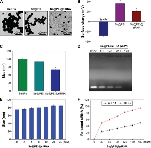
In this study, we demonstrated a simple method to synthesize functionalized SeNPs through self-assembly of functional ligands on the surface of nanoparticles. SeNPs were capped with PEI and siRNA to form more compact and stable globular nanocomposites (). The morphology of as-prepared Se@PEI@siRNA was first characterized by transmission electron microscope. Compared with SeNPs and Se@PEI, Se@PEI@siRNA presented uniform and monodisperse spherical particles as shown in . The zeta potential () of Se@PEI@siRNA with positive charge explains why Se@PEI@siRNA entered more easily into the cell. Regarding size distribution of different nanoparticles were shown in . The binding capacity to HSP70 siRNA was further analyzed by agarose gel electrophoresis as shown in , the binding capability of Se@PEI to HSP70 siRNA was enhanced with the weight ratio. When the weight ratio was over 40:1, almost all the HSP70 siRNAs were restrained with the nanocomposites. As shown in , the result revealed that Se@PEI@siRNA remained stable at least for 30 days. The small size of Se@PEI@siRNA with zeta potential contributed to the highly stability of the nanostructures and the ability to cross the cell membrane. The binding capacity to HSP70 siRNA was further analyzed by agarose gel electrophoresis as shown in ; the binding capability of Se@PEI to HSP70 siRNA was enhanced with the weight ratio. When the weight ratio was over 40:1, almost all the HSP70 siRNAs were restrained with the nanocomposites. The siRNA release capacity from Se@PEI@siRNA was detected at pH 7.4 and 5.3 (). The extremely rapid release rate of siRNA from Se@PEI@siRNA at pH 5.3 is due to the acidolysis of nanoparticles. This hydrolysis behavior of siRNA in response to acid pH.
Figure 1 Synthetic route of Se@PEI@siRNA.
Abbreviations: PEI, polyethylenimine; SeNPs, selenium nanoparticles; Se@PEI@ siRNA, small interfering RNAs with PEI-modified selenium nanoparticles; VC, vitamin C.
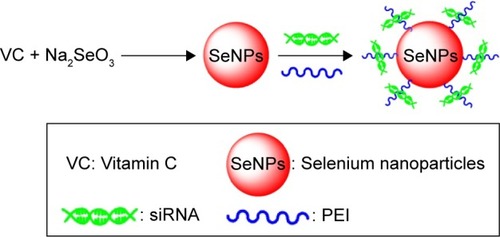
Figure 2 Characterization of Se@PEI@siRNA.
Notes: (A) TEM images of SeNPs, Se@PEI, and Se@PEI@siRNA, scale bar 200 nm. (B) Zeta potentials of SeNPs, Se@PEI, and Se@PEI@siRNA. (C) Size distribution of SeNPs, Se@PEI, and Se@PEI@siRNA. (D) Agarose gel retardation assay of Se@PEI/siRNA complexes at various ratios. Naked siRNA was used as control. (E) Stability of Se@PEI@siRNA in 10% fetal bovine serum. (F) In vitro release profiles of siRNA from Se@PEI@siRNA at pH 7.4 and 5.3. Bars with different characters are statistically different at P<0.05 (*) level.
Abbreviations: SeNP, selenium nanoparticle; Se@PEI@siRNA, small interfering RNAs with polyethylenimine-modified SeNPs; TEM, transmission electron microscope.
In vitro anticancer activity of Se@PEI@siRNA
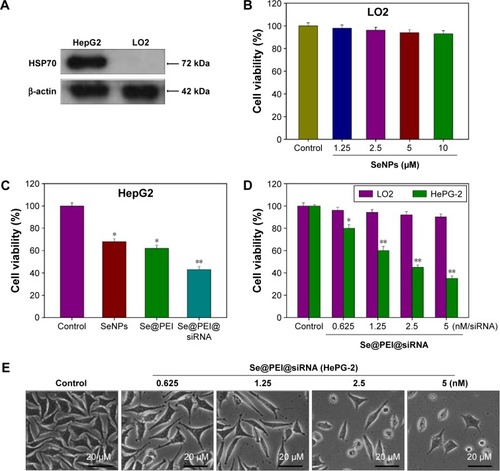
The expression level of HSP70 protein in cancer and normal cells was detected by Western blotting. As shown in , the expression level of HSP70 in HepG2 cells was significantly higher than in LO2 cells, suggesting HSP70-guided selectivity among HepG2 and LO2 cells. The anticancer activity of SeNPs and Se@PEI@siRNA was evaluated by MTT assay, which determines the cells’ survival rate and reflects their growth states. As shown in , SeNPs showed nontoxicity as gene delivery systems. Se@PEI@siRNA effectively suppressed HepG2 cells’ growth in a dose-dependent manner, but exhibited low cytotoxicity toward LO2 cells. As shown in , HepG2 cells treated with different concentrations of Se@ PEI@siRNA showed cytoplasmic shrinkage, reduction in cells numbers, and loss of cell-to-cell contact. The results suggest that Se@PEI@siRNA effectively inhibited the proliferation of HepG2 cells.
Figure 3 Effects of Se@PEI@siRNA on the growth of HepG2 cells as examined by MTT assay.
Notes: (A) HSP70 expression in HepG2 and LO2 cells. Western blotting was applied to determine the expression level of HSP70. (B–D) Antitumor activity of Se@PEI@ siRNA on HepG2 cells and normal cells was measured by MTT assay. (E) Morphological changes in HepG2 cells observed by phase-contrast microscopy (magnification, ×40). Bars with different characters are statistically different at P<0.05 (*) or P<0.01 (**) level.
Abbreviations: HSP, heat shock protein; LO2, normal human liver cell line; MTT, thiazolyl blue tetrazolium bromide; SeNP, selenium nanoparticle; Se@PEI@siRNA, small interfering RNAs with polyethylenimine-modified SeNPs.
Intracellular localization and cellular uptake of Se@PEI@siRNA
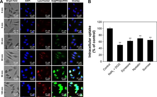
Endocytosis is one of the most important pathways for cellular uptake of extracellular nanoparticles. As shown in , the colocalization of Se@PEI@siRNA and lysosomes was found in HepG2 cells, and increased in a time-dependent manner. Se@PEI@siRNA escaped from lysosomes after 60 minutes and were transported into the cytosol and distributed in the cells after 90 minutes. To further dissect the Se@PEI@siRNA endocytosis mechanism, we used different endocytosis inhibitors to treat the cells before the addition of Se@PEI@siRNA. As shown in , treatments of NaN3 and 2-deoxyglucose markedly inhibited the internalization of Se@PEI@siRNA to 50% of the control. Dynasore and nystatin decreased the Se@PEI@siRNA internalization to 62% and 70% of the control, respectively. Moreover, sucrose induced the uptake of Se@PEI@siRNA to 65%. This result demonstrates that lysosome is the target organelle of Se@ PEI@siRNA. Se@PEI@siRNA showed time-dependent cell-penetration for cancer therapy.
Figure 4 Localization and uptake pathways of coumarin-6-loaded Se@PEI@siRNA in HepG2 cells.
Notes: (A) The cells were treated with coumarin-6-loaded Se@PEI@siRNA for different periods of time and were observed under a fluorescent microscope stained with LysoTracker (red fluorescence, lysosome) and DAPI (blue fluorescence, nucleus) (magnification, ×100). (B) Intracellular uptake of Se@PEI@siRNA in HepG2 cells with different endocytosis-inhibited conditions. Significant difference between treatment and control group is indicated at P<0.01 (**) level.
Abbreviations: DAPI, 4′,6′-diamidino-2-phenyindole; DOG, 2-deoxyglucose; Se@PEI@siRNA, small interfering RNAs with polyethylenimine-modified selenium nanoparticles; min, minutes.
Induction of cancer cell apoptosis by Se@PEI@siRNA
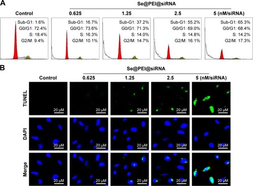
Flow cytometry and TUNEL-DAPI were employed to examine the anticancer mechanisms of Se@PEI@siRNA. As shown in , the sub-G1 apoptotic cell population significantly increased in the DNA histogram in a dose-dependent manner. For instance, Se@PEI@siRNA significantly increased the apoptotic cell population from 1.6% (control) to 55.2% at concentrations of 2.5 nM (siRNA), while no significant change in cell cycle distribution was observed. DNA fragmentation is an important biochemical hallmark of cell apoptosis. As shown in , HepG2 cells exhibited typical apoptotic features with increased concentrations of Se@PEI@siRNA, such as DNA fragmentation and nuclear condensation.
Figure 5 Se@PEI@siRNA-induced apoptosis in HepG2 cells.
Notes: (A) The cell cycle distribution after different treatments was analyzed by quantifying DNA content using flow cytometry analysis. (B) Representative photomicrographs of DNA fragmentation and nuclear condensation as detected by TUNEL-DAPI costaining assay. All results were representative of three independent experiments (magnification, ×100).
Abbreviations: DAPI, 4′,6′-diamidino-2-phenyindole; Se@PEI@siRNA, small interfering RNAs with polyethylenimine-modified selenium nanoparticles; TUNEL, terminal transferase dUTP nick end labeling.
Induction of caspase-mediated poly (ADP-ribose) polymerase (PARP) cleavage by Se@PEI@siRNA
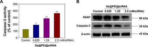
PARP, a DNA repair enzyme, is downstream of caspase family proteins in apoptosis pathways. Caspase-3 has been identified as a key mediator of apoptosis in mammalian cells. Caspase-3 and the subsequent cleavage of PARP were examined by Western blotting to evaluate their involvement and contribution to cell apoptosis. As shown in , the treatment of HepG2 cells with different concentrations of Se@PEI@siRNA significantly increased the activity of caspase-3 in a dose-dependent manner. As shown in , the expression level of caspase-3 and PARP were downregulated with a rise in concentration. The results show that Se@PEI@siRNA significantly enhanced the activation of caspase-3 and the cleavage of downstream effect PARP. Taken together, these results confirm that the synthetic nanosystem inhibits cancer cell growth through inducing cell apoptosis.
Figure 6 Induction of PARP cleavage and caspase-3 activity by Se@PEI@siRNA in HepG2 cells.
Notes: (A) Cells were treated with Se@PEI@siRNA for 24 hours, and caspase-3 activity was measured using synthetic fluorogenic substrate. (B) The expression of PARP and caspase-3 by Western blot; β-actin was used as loading control. Bars with different characters are statistically different at P<0.05 (*) or P<0.01 (**) level.
Abbreviations: PARP, poly (ADP-ribose) polymerase; Se@PEI@siRNA, small interfering RNAs with polyethylenimine-modified selenium nanoparticles.
Suppression of HSP70 mRNA and protein expression by Se@PEI@siRNA
Inhibition of gene expression has been found to be an effective strategy to suppress cancer growth. In this study, the efficacy of silencing HSP70 gene with Se@PEI@siRNA in HepG2 cells was examined by Q-PCR and Western blotting. As shown in , the level of HSP70 mRNA expression was reduced in the cells exposed to Se@PEI@ siRNA. Compared with Se@PEI@scramble siRNA and Lipofectamine 2000@siRNA, the higher silencing efficiency of Se@PEI@siRNA could be due to its positive charge and cancer-targeted delivery. The expression of HSP70 was evaluated by Western blotting. As shown in , Se@ PEI@siRNA effectively inhibited the expression of HSP70 in HepG2 cells. Both Q-PCR and Western blotting prove that Se@PEI@siRNA exhibit the ability to silence HSP70 at mRNA and protein levels, which may control the cell fate by regulation of downstream effectors.
Figure 7 Suppression of HSP70 mRNA and protein expression in HepG2 cells among Se@PEI@scramble siRNA, Se@PEI@siRNA, and Lipofectamine 2000@siRNA.
Notes: (A) Suppression of HSP70 mRNA levels was quantified by Q-PCR. (B) The expression of HSP70 was evaluated by Western blot. Bars with different characters are statistically different at P<0.05 (*) or P<0.01 (**) level.
Abbreviations: HSP, heat shock protein; mRNA, messenger RNA; Q-PCR, quantitative polymerase chain reaction; Se@PEI@siRNA, small interfering RNAs with polyethylenimine-modified selenium nanoparticles.

Induction of ROS generation by Se@PEI@siRNA
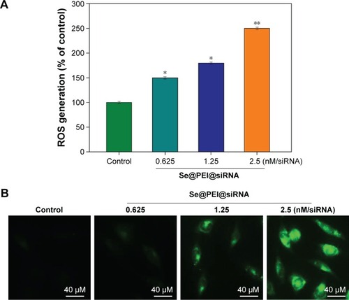
ROS generation has been regarded as a critical regulator of cell apoptosis, especially that induced by anticancer drugs. Mitochondria are the energy factories of cellular activities, but a variety of factors can result in damage to the structure and function of mitochondria and further induce cell apoptosis. ROS were generated from mitochondria that were attacked by Se@PEI@siRNA. Therefore, in this study, ROS generation was determined by DCF fluorescence assay to reveal its role in the action mechanisms of Se@PEI@siRNA. As shown in , the nanosystem increased the intracellular ROS generation in a dose-dependent manner in HepG2 cells. For instance, Se@PEI@siRNA increased the intracellular ROS generation to 180% and 250% at concentrations of 2.5 and 5 nM, respectively. The stronger fluorescent intensity of DCF was also found in HepG2 cells in . These results indicate the involvement of ROS in the anticancer action of Se@PEI@siRNA.
Figure 8 ROS overproduction induced by Se@PEI@siRNA in HepG2 cells.
Notes: (A) Changes of intracellular ROS generation. ROS levels were detected by DCF fluorescence intensity. (B) HepG2 cells were preincubated with 10 μM DCF for 30 minutes and then treated with Se@PEI@siRNA (magnification, ×100). Bars with different characters are statistically different at P<0.05 (*) or P<0.01 (**) level.
Abbreviations: DCF, dichlorofluorescein; ROS, reactive oxygen species; Se@PEI@siRNA, small interfering RNAs with polyethylenimine-modified selenium nanoparticles.
Activation of ROS-mediated signaling pathways by Se@PEI@siRNA
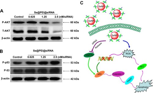
Intracellular ROS overproduction could trigger DNA damage and result in cell apoptosis through the regulation of AKT and p53 signaling pathways. Due to the detection of ROS overproduction in cells exposed to Se@PEI@siRNA, we tried to examine the effects on the ROS-mediated downstream pathways by using Western blotting. As shown in , treatment with Se@PEI@siRNA effectively suppressed the expression levels of total and phosphorylated AKT in HepG2 cells. Similarly, for the p53 signaling pathway, Se@PEI@ siRNA significantly increased the expression levels of p-p53 protein and total p53 (). Taken together, these results support the fact that the nanosystem induces cancer cell apoptosis by regulation of ROS-mediated AKT and p53 signaling pathways ().
Figure 9 Activation of intracellular apoptotic signaling pathways by Se@PEI@siRNA in HepG2 cells.
Notes: (A) Phosphorylation status expression levels of AKT pathways. (B) Activation of p53 signaling pathway. (C) The main signaling pathway of apoptosis induced by Se@PEI@siRNA.
Abbreviations: HSP, heat shock protein; PARP, poly (ADP-ribose) polymerase; ROS, reactive oxygen species; SeNP, selenium nanoparticle; Se@PEI@siRNA, small interfering RNAs with polyethylenimine-modified selenium nanoparticles.
Conclusion
In summary, PEI-modified SeNPs loaded with HSP70 siRNA have been successfully fabricated in the present study. As the expression of HSP70 was diminished, the cell protective function was also removed, and cancer cells became more sensitive to Se@PEI@siRNA. Se@PEI@siRNA exhibited enhanced cytotoxic effects on human cancer cells through the silencing of HSP70 expression. The gene-silencing efficiency of Se@PEI@siRNA was significantly much higher than Lipofectamine 2000@siRNA. The nanosystem entered cancer cells by endocytosis for intracellular delivery. Moreover, the selective delivery of HSP70 siRNA using the nanosystem resulted in a significantly reduced HSP70 mRNA and protein expression in the cancer cells through the regulation of ROS-mediated downstream signaling pathways, including AKT and p53 pathways. Taken together, this study provides a strategy for design of a nanosystem as a carrier of siRNA for cancer therapy.
Disclosure
The authors report no conflicts of interest in this work.
Acknowledgments
This work was supported by the National Natural Science Foundation of China (No: 309726300), China Postdoctoral Science Foundation (2015M582366), and the Guangdong Natural Science Foundation (No: 10151012001000002).
References
- LeeMYParkSJParkKKimKSLeeHHahnSKTarget-specific gene silencing of layer-by-layer assembled gold-cysteamine/siRNA/PEI/HA nanocomplexACS Nano2011586138614721739990
- LiTShenXChenYPolyetherimide-grafted Fe3O4@SiO2 nano-particles as theranostic agents for simultaneous VEGF siRNA delivery and magnetic resonance cell imagingInt J Nanomedicine2015104279429126170664
- GodinhoBMOgierJRQuinlanAPEGylated cyclodextrins as novel siRNA nanosystems: correlations between polyethylene glycol length and nanoparticle stabilityInt J Pharm20144731–210511224992319
- GandhiNSTekadeRKChouguleMBNanocarrier mediated delivery of siRNA/miRNA in combination with chemotherapeutic agents for cancer therapy: current progress and advancesJ Control Release201419423825625204288
- WebbJABardhanREmerging advances in nanomedicine with engineered gold nanostructuresNanoscale2014652502253024445488
- YangHLiYLiTMultifunctional core/shell nanoparticles cross-linked polyetherimide-folic acid as efficient Notch-1 siRNA carrier for targeted killing of breast cancerSci Rep20144707225400232
- PengXCChenJChengTPLGA modified Fe3O4 nanoclusters for siRNA deliveryMater Lett201281102104
- ZouSScarfoKNantzMHHeckerJGLipid-mediated delivery of RNA is more efficient than delivery of DNA in non-dividing cellsInt J Pharm20103891–223224320080162
- LiHZhengXKorenVVashistYKTsuiTYHighly efficient delivery of siRNA to a heart transplant model by a novel cell penetrating peptide-dsRNA binding domainInt J Pharm2014469120621324768403
- LiJChengDYinTCopolymer of poly(ethylene glycol) and poly(L-lysine) grafting polyethylenimine through a reducible disulfide linkage for siRNA deliveryNanoscale2014631732174024346086
- MorryJNgamcherdtrakulWGuSDDermal delivery of HSP47 siRNA with NOX4-modulating mesoporous silica-based nanoparticles for treating fibrosisBiomaterials201566415226196532
- Meneksedag-ErolDTangTUludagHProbing the effect of miRNA on siRNA-PEI polyplexesJ Phys Chem20151191754755486
- IsraelLLLelloucheEOstrovskySAcute in vivo toxicity mitigation of PEI-coated maghemite nanoparticles using controlled oxidation and surface modifications toward siRNA deliveryACS App Mater Interfaces20157281524015255
- WangCZhangYGuoKHeat shock proteins in hepatocellular carcinoma: Molecular mechanism and therapeutic potentialInt J Cancer201613881824183426853533
- WuPKHongSKVeerankiSA mortalin/HSPA9-mediated switch in tumor-suppressive signaling of Raf/MEK/extracellular signal-regulated kinaseMol Cell Biol201333204051406723959801
- ShevtsovMANikolaevBPYakovlevaLY70-kDa heat shock protein coated magnetic nanocarriers as a nanovaccine for induction of anti-tumor immune response in experimental gliomaJ Control Release2015220Pt A32934026522072
- GehrmannMCervelloMMontaltoGHeat shock protein 70 serum levels differ significantly in patients with chronic hepatitis, liver cirrhosis, and hepatocellular carcinomaFront Immunol2014530725071768
- ZengYCaoRZhangTLiSZhongWDesign and synthesis of piperidine derivatives as novel human heat shock protein 70 inhibitors for the treatment of drug-resistant tumorsEur J Med Chem201597193125935385
- KongFWangHGuoJHsp70 suppresses apoptosis of BRL cells by regulating the expression of Bcl-2, cytochrome C, and caspase 8/3In Vitro Cell De Biol Anim2016525568575
- AhmadMSYasserMMSholkamyENAliAMMehanniMMAnticancer activity of biostabilized selenium nanorods synthesized by Streptomyces bikiniensis strain Ess_amA-1Int J Nanomedicine2015103389340126005349
- ZhengWYinTChenQCo-delivery of Se nanoparticles and pooled SiRNAs for overcoming drug resistance mediated by P-glycoprotein and class III beta-tubulin in drug-resistant breast cancersActa Biomater20163119721026612416
- YuBZhangYZhengWFanCChenTPositive surface charge enhances selective cellular uptake and anticancer efficacy of selenium nanoparticlesInorg Chem201251168956896322873404
- KumarGSKulkarniAKhuranaAKaurJTikooKSelenium nanoparticles involve HSP-70 and SIRT1 in preventing the progression of type 1 diabetic nephropathyChem Biol Interact2014223C12513325301743
- KongLYuanQZhuHThe suppression of prostate LNCaP cancer cells growth by selenium nanoparticles through Akt/Mdm2/AR controlled apoptosisBiomaterials201132276515652221640377
- LiuWLiXWongYSSelenium nanoparticles as a carrier of 5-fluorouracil to achieve anticancer synergismACS Nano2012686578659122823110
- HuangYHeLLiuWSelective cellular uptake and induction of apoptosis of cancer-targeted selenium nanoparticlesBiomaterials201334297106711623800743
- ZhengSLiXZhangYPEG-nanolized ultrasmall selenium nanoparticles overcome drug resistance in hepatocellular carcinoma HepG2 cells through induction of mitochondria dysfunctionInt J Nanomedicine201273939394922915845
- JiangWFuYYangFGracilaria lemaneiformis polysaccharide as integrin-targeting surface decorator of selenium nanoparticles to achieve enhanced anticancer efficacyACS Appl Mater Interfaces2014616137381374825073123
- TranPASarinLHurtRHWebsterTJDifferential effects of nano-selenium doping on healthy and cancerous osteoblasts in coculture on titaniumInt J Nanomedicine2010535135820517480
- LiuTZengLJiangWFuYZhengWChenTRational design of cancer-targeted selenium nanoparticles to antagonize multidrug resistance in cancer cellsNanomedicine201511494795825680543
- FengYSuJZhaoZDifferential effects of amino acid surface decoration on the anticancer efficacy of selenium nanoparticlesDalton Trans20144341854186124257441
- LinGZhuWYangLDelivery of siRNA by MRI-visible nano-vehicles to overcome drug resistance in MCF-7/ADR human breast cancer cellsBiomaterials201435359495950725155545
- LiYZhangJWangBShenYOuahabACo-delivery of siRNA and hypericin into cancer cells by hyaluronic acid modified PLGA-PEI nanoparticlesDrug Dev Ind Pharm201642573774626472259
- XiaoYJaskula-SztulRJavadiACo-delivery of doxorubicin and siRNA using octreotide-conjugated gold nanorods for targeted neuroendocrine cancer therapyNanoscale20124227185719323070403
- YangFTangQZhongXSurface decoration by Spirulina polysaccharide enhances the cellular uptake and anticancer efficacy of selenium nanoparticlesInt J Nanomedicine2012783584422359460
- LiYLiXWongYSThe reversal of cisplatin-induced nephro-toxicity by selenium nanoparticles functionalized with 11-mercapto-1-undecanol by inhibition of ROS-mediated apoptosisBiomaterials201132349068907621864903
- LiYHLiXLZhengWJFanCDZhangYBChenTFFunctionalized selenium nanoparticles with nephroprotective activity, the important roles of ROS-mediated signaling pathwaysJ Mater Chem B201314663656372
- HeLChenTYouYA cancer-targeted nanosystem for delivery of gold(III) complexes: enhanced selectivity and apoptosis-inducing efficacy of a gold(III) porphyrin complexAngew Chem Int Ed Engl20145346125321253625220408
- HeLZHuangYYZhuHLCancer-targeted monodisperse mesoporous silica nanoparticles as carrier of ruthenium polypyridyl complexes to enhance theranostic effectsAdv Funct Mater2014241927542763
- FuXYangYLiXRGD peptide-conjugated selenium nano-particles: antiangiogenesis by suppressing VEGF-VEGFR2-ERK/AKT pathwayNanomedicine Epub201634
- WuHLLiXLLiuWSurface decoration of selenium nano-particles by mushroom polysaccharides-protein complexes to achieve enhanced cellular uptake and antiproliferative activityJ Mater Chem2012221996029610
- CaoWZhengWChenTRuthenium polypyridyl complex inhibits growth and metastasis of breast cancer cells by suppressing FAK signaling with enhancement of TRAIL-induced apoptosisSci Rep20155915725778692
- LiXLChenTWongYSInvolvement of mitochondrial dysfunction in human islet amyloid polypeptide-induced apoptosis in INS-1E pancreatic beta cells: An effect attenuated by phycocyaninInt J Biochem Cell Biol201143452553421163363
- YuBLiXLZhengWJFengYXWongYSChenTFpH-responsive cancer-targeted selenium nanoparticles: a transformable drug carrier with enhanced theranostic effectsJ Mater Chem B201423354095418
- LiXLXuGChenTPhycocyanin protects INS-1E pancreatic beta cells against human islet amyloid polypeptide-induced apoptosis through attenuating oxidative stress and modulating JNK and p38 mitogen-activated protein kinase pathwaysInt J Biochem Cell Biol20094171526153519166964
- LiJMWangYYZhaoMXMultifunctional QD-based co-delivery of siRNA and doxorubicin to HeLa cells for reversal of multidrug resistance and real-time trackingBiomaterials20123392780279022243797
- ZhouLDuanXMZengSCodelivery of SH-aspirin and curcumin by mPEG-PLGA nanoparticles enhanced antitumor activity by inducing mitochondrial apoptosisInt J Nanomedicine2015105205521826316750
- ZhangYBLiXLHuangZZhengWJFanCDChenTFEnhancement of cell permeabilization apoptosis-inducing activity of selenium nanoparticles by ATP surface decorationNanomedicine201391748422542821