
Abstract
Cardiospermum halicacabum is widely used in traditional medicine. Previous studies have focused on the aerial parts, while the seeds have been poorly investigated. This work aimed to analyse the chemical composition of extracts from aerial parts and seeds obtained using Naviglio and Soxhlet (PN, PS, and SN, SS, respectively), the inhibitory properties against tyrosinase, acetylcholinesterase (AChE), and butyrylcholinesterase (BChE) and the antioxidant effects. PN total extract showed significant anti-tyrosinase activity (IC50 value of 10.8 µg/mL). After partitioning with n-hexane, an HPLC method for analysing chemical constituents was established. Apigenin, luteolin, and apigenin-7-O-glucoside are the predominant constituents. SN n-hexane fraction was the most active inhibitor of BChE (IC50 of 57.9 µg/mL). Gas chromatography-mass spectrometry analysis revealed fatty acids, including eicosanoic acid, methyl 11-eicosenoate and oleic acid, as the major constituents. These findings suggest the potentiality of both seeds and aerial parts of C. halicacabum in the treatment of neurological disorders.
Introduction
Cardiospermum halicacabum L. is a plant that belongs from the Sapindaceae family wide spread in tropical and sub-tropical Asia and Africa that has been used in traditional medicine for a long time. In Chinese medicine it is used in the treatment of rheumatism, lumbago, nervous diseases, as a demulcent in orchitis and in dropsyCitation1. Indian system of medicine recommends C. halicacabum leaves for rheumatism, chronic bronchitis, stiffness of limbs and snakebiteCitation2. A toxicological evaluation of C. halicacabum revealed that the drug is safe and is not toxic up to 40 g/kg in ratsCitation2. Previous studies reported the potential antioxidant, sedative effect on the central nervous system, diuretic, emetic, antipyretic, antirheumatic, laxative and emmenagogue properties of C. halicacabumCitation3–5. Flavonoids, triterpenoids, glycosides, fatty acids and volatile ester have been reported as main classes of secondary metabolites for this plant speciesCitation6–9. Generally, all these studies are recognized on the aerial parts of C. halicacabum, while there are few research studies on the seeds of the plant.
The cellular oxidative stress is due to an increase in the levels of reactive oxygen species, which are produced as a result of many biochemical reactions and are considered primary causes of oxidative damage as: protein denaturation, mutagenesis and lipid peroxidation in aerobic cells. It is believed that oxidative stress contributes to the development of a vast number of diseases, including cardiovascular diseases, and neurodegenerative diseasesCitation10,Citation11.
Neurodegenerative diseases are undoubtedly an increasing problem in the health sciences, given the increase of life expectancy and occasional vicious life style. Parkinson’s disease (PD) and Alzheimer’s disease (AD) are the two most common neurodegenerative disorders. PD is a common, progressive, debilitating disease with substantial physical, psychological and social implications. Pharmacological management is complex and should be individualised according to the needs of the patient. In early disease, treatment is generally highly effective, but medication becomes increasingly inadequate in controlling motor fluctuations and dyskinesias as the disease progresses. Non-motor symptoms, especially depression and dementia, require a holistic, multidisciplinary approach to maximise quality of life for patients and their carersCitation12. In the literature, oxidative stress has been proposed as one of the possible etiologic agents in PD and several pathways have been identified for the oxidative stress induced by the oxidation of dopamine, including the production of toxic reactive oxygen species or the formation of highly reactive quinone speciesCitation13,Citation14. On these premises, tyrosinase, a copper-containing enzyme widely distributed in microorganisms, animals and plants that catalyses two distinct reactions of melanin synthesis, has been proposed to take part in the oxidative chemistry related to PD. Greggio et al.Citation15 demonstrated that this enzyme is ubiquitously expressed in the human brain, although at low levels, and its activity was highest in the substantia nigra.
AD is associated with a steady loss of attention and memory, which has been correlated with the impairment of brain cholinergic neurotransmission. Some pathogenic events, such as genetic alterations, β-amyloid deposition in senile plaques and brain vessels, neurofibrillary tangles due to hyper-phosphorilation of tau proteins, synaptic loss, neurotransmitter deficits, neurotrophic alterations, neuroinflammatory processes, accelerated neuronal death due to excitotoxic reactions, alterations in calcium homeostasis, and free radical formation, are involved in AD cause and progression. Moreover, carbonic anhydrases (CAs), a group of metallo-enzymes involved in numerous physiological and pathological processes, are down-regulated in ADCitation16. So, CA activators might constitute a new approach for the treatment of AD and other conditions in need of achieving memory therapyCitation17,Citation18.
Acetylcholinesterase (AChE) inhibitors are currently the drugs of choice, although only symptomatic and palliative, for the treatment of AD. In late AD stage, levels of AChE have declined by up to 85% and butyrylcholinesterase (BChE) represents the predominant cholinesterase in brainCitation19. BChE, primarily associated with glial cells but also with specific neuronal pathways, cleaves acetylcholine in a manner similar to AChE to terminate its physiological action. Such studies have targeted BChE as a new approach to intercede in the progression of AD. Therefore, one of the methods that has proven successful in the treatment of AD is the use of inhibitors of AChE and BChE to supplement the acetylcholine level. Several reports on screening of AChE and BChE inhibitors from natural sources have been made from our laboratoryCitation20–23.
Although the aerial parts of C. halicacabum has shown some important biological effects, there are not studies focusing on their effects on the inhibition of cholinesterase and tyrosinase. Thus, the objective of the present study was to investigate for the first time the modulation capacity of C. halicacabum on AChE, BChE and tyrosinase activity. In particular, herein we describe our studies on the analysis by high performance liquid chromatography equipped with UV-Vis diode array detector (HPLC-DAD), gas chromatography-flame ionization detector (GC-FID) and gas chromatography-mass spectrometry (GC-MS) of chemical constituents of aerial parts and seeds extracted with two different methods (Naviglio extractor and Soxhlet apparatus) and the biological activities correlate to diseases with high social impact (inhibition of enzymes involved in the etiopathogenesis of neurodegenerative diseases such as AD and PD).
Moreover, we evaluated the protection provided by C. halicacabum against oxidative damage through different in vitro models (DPPH, ABTS, FRAP and β-carotene bleaching test).
Materials and methods
Plant material
The aerial parts and seeds of C. halicacabum were collected in July 2012 in Modica (Sicily, Italy), and authenticated by Dr N.G. Passalacqua, Natural History Museum of Calabria and Botanic Garden, University of Calabria, Italy. A voucher specimen (CLU 22013) was deposited at the Herbarium of University of Calabria.
Chemicals and reagents
Methanol, n-hexane, water, and acetonitrile were obtained from VWR (Milan, Italy). Sodium phosphate buffer, acetylthiocholine iodide (ATCI), 5,5′-dithiobis(2-nitrobenzoic-acid) (DTNB), butyrylthiocholine iodide (BTCI), Folin-Ciocalteau reagent, AlCl3, physostigmine, acetylcholinesterase (AChE) from Electrophorus electricus (EC 3.1.1.7, Type VI-S), butyrylcholinesterase (BChE) from equine serum (EC 3.1.1.8), butylated hydroxytoluene (BHT), 2,2-diphenyl-1-picrylhydrazyl (DPPH), Tween 20, β-carotene, quercetin, rutin, apigenin, apigenin-7-O-glucoside, caffeic acid, p-coumaric acid, ferulic acid, and chlorogenic acid were obtained from Sigma-Aldrich S.p.A. (Milan, Italy).
Extraction procedures
The aerial parts and seeds of C. halicacabum were subjected to extraction by:
Naviglio extractor (Mod. 2 litres capacity, Nuova Estrazione S.a.s., Naples, Italy); plant materials were put in a porous bag and then it was introduced in the extraction chamber; 2 L of methanol were added. To allow the total recovery of the extract, 30 extractive cycles of 4 min each for a total of 2 h (2 min in static phase and 2 min in dynamic phase) were performed;
Soxhlet apparatus with methanol as solvent. Combined methanol solutions were concentrated under reduced pressure and dried.
In order to operate a separation of lipophilic compounds, the total extract was solubilised with methanol and extracted with n-hexane. The n-hexane solutions were combined and dried to obtain the n-hexane fraction. Data are reported in .
Table 1. Extractive yield (%) of C. halicacabum samples.
Total phenol and flavonoid content
The extracts of C. halicacabum were tested for their total phenol content by using the Folin-Ciocalteau methodCitation20. In this procedure, the extract was mixed with 0.2 mL Folin–Ciocalteau reagent, 2 mL of distilled water and 1 mL of 15% Na2CO3. After 2 h incubation at room temperature, the absorbance was read at 765 nm (UV-Vis Jenway 6003 spectrophotometer). Chlorogenic acid was used as a standard and the total phenol content was expressed as milligrams of chlorogenic acid equivalents per grams of plant materials.
The total flavonoid content was determined using a method based on the formation of a flavonoid-aluminium complexCitation20. The extract (1 mL) and distilled water (4 mL) were added to a volumetric flask. At zero time, 0.3 mL of 5% (w/v) sodium nitrite was added to the flask. After 5 min, 0.6 mL of 10% (w/v) AlCl3 was added and then at 6 min 2 mL of 1 mol/L NaOH was also added to the mixture, followed by the addition of 2.1 mL distilled water. Absorbance at 510 nm was immediately read. Quercetin was chosen as a standard and the total flavonoid content was expressed as milligrams of quercetin equivalents per grams of plant materials.
HPLC-DAD fingerprint profile
HPLC analysis of C. halicacabum was carried out using high performance liquid chromatography (Jasco) equipped with UV-Vis diode array detector (DAD) MD-910, two pumps PU-980, and a C18 reverse phase column (Phenomenex Luna 5 µm, 250 × 4.60 mm). The method involved the use of a binary gradient with mobile phases containing: (A) 0.1% formic acid in water (0.1%, v/v) and (B) acetonitrile. The flow rate was 1.0 ml/min, and the injection volume was 20 µL. The solvent gradient elution program was as follows: 2 min 100% A, 55 min from 100% A to 100% B, 3 min from 100% B to 100% A. The monitoring wavelength was 210–400 nm. The analytical data was evaluated using Borwing data processing software. Quercetin, luteolin, rutin, apigenin, apigenin-7-O-glucoside, caffeic acid, p-coumaric acid, ferulic acid and chlorogenic acid were used as standard and their content was calculated from the integrated peak area of the sample and the corresponding standard.
GC and GC-MS analysis
Cardiospermum halicacabum n-hexane fractions were analysed by using a Hewlett-Packard 6890 gas chromatograph equipped with an HP-5 MS capillary column (30 m length, 0.25 mm i.d., 0.25 µm film thickness) and interfaced with a Hewlett Packard 5973 Mass Selective. Ionization of the sample components was performed in electron impact mode (EI, 70 eV). Helium was used as carrier gas. The analytical conditions were as follow: oven temperature was 5 min isothermal at 50 °C, then 50–250 °C at a rate of 5 °C/min; then held isothermal for 10 min. The injector and detector temperatures were 250 °C and 280 °C, respectively. Extracts were dissolved in acetone (ca. 1 mg/mL) and aliquots of 1 µL were injected. Constituents were tentatively identified by gas chromatography by comparison of their retention times with those of the literature or with those of authentic compounds available in our laboratory. Further tentative identification was made by comparison of their mass spectra with those stored in Wiley 138, Wiley 275 and NIST 98 libraries. Fractions were analysed also by a Shimadzu GC17A gas chromatograph system. An SE-52 capillary column (30 m with an internal diameter of 0.25 mm and a film thickness of 0.25 µm) was used with nitrogen as the carrier gas. GC oven temperature and conditions were as described above. The quantification of the components was performed on the basis of their GC peak areas and the percentages of the characterized components were as given in . Component relative concentrations were calculated based on GC peak areas without using correction factors.
Determination of effect on DPPH radical
The effect of samples on the DPPH radical was estimated as described previouslyCitation24. An aliquot of 1.5 mL of 0.25 mmol/L DPPH solution in ethanol and 12 µL of samples at concentrations ranging from 25 to 150 µg/mL were mixed. The solution was shaken and kept in the dark for 30 min. The bleaching of DPPH was determined by measuring the absorbance at 517 nm against a blank without DPPH. Ascorbic acid was used as positive control. The DPPH radicals scavenging activity was calculated according to the following equation: Scavenging activity = [(A0 − A1/A0) × 100], where A0 is the absorbance of the control (blank, without extract) and A1 is the absorbance in the presence of the extract.
Determination of ABTS radical inhibition
The C. halicacabum ABTS radical inhibition was carried out as described in our previous studyCitation24. ABTS radical cation was produced by the reaction of a 7 mmol/L ABTS solution with 2.45 mmol/L potassium persulphate. The mixture was stored in the dark at room temperature for 12 h before use. The ABTS+ solution was diluted with ethanol to an absorbance of 0.70 ± 0.05 at 734 nm. After addition of 25 μL of sample or Trolox standard to 2 mL of diluted ABTS+ solution, absorbance at 734 nm was read after 6 min. The decrease in absorption was used for calculating TEAC values. A standard curve was prepared by measuring the reduction in absorbance of ABTS•+ solution at different concentrations of Trolox. Appropriate blank measurements were carried out and the values recorded. Results were expressed as Trolox Equivalent Antioxidant Capacity (TEAC). Ascorbic acid was used as positive control.
β-Carotene bleaching assay
The antioxidant activity was evaluated using the β-carotene bleaching test with some modificationsCitation20. One millilitre of β-carotene solution (0.2 mg/mL in chloroform) was added to 0.02 mL of linoleic acid and 0.2 mL of 100% Tween 20. Chloroform was evaporated and the mixture was diluted with 100 mL of water, 5 mL of the emulsion were transferred into different test tubes containing 0.2 mL of sample in 70% ethanol at different concentrations. Tubes were shaken and placed at 45 °C in a water bath for 60 min. The absorbance of the samples, standard and control was read at 470 nm against a blank consisting of an emulsion without β-carotene. The measurement was carried out at initial time (t = 0) and successively at 30 and 60 min. Propyl gallate was used as positive control. All samples were assayed in triplicate and the mean value calculated. The antioxidant activity (AA) was measured in terms of successful bleaching β-carotene by using the following equation: AA = [1 − (A0 − At)/() × 100, where A0 and
are the absorbance values measured at the initial incubation time for samples/standard and control, respectively, while At and
are the absorbance values measure in the samples/standard and control respectively at t = 30 min and t = 60 min.
Determination of chelating activity
The FRAP method measures the absorption change that appears when the TPTZ (2,4,6-tripyridyl-s-triazine)-Fe3+ complex is reduced to the TPTZ-Fe2+ form in the presence of antioxidant compounds. The FRAP reagent contained 2.5 mL of 10 mM tripyridyltriazine (TPTZ) solution in 40 m mol/L HCl plus 2.5 mL of 20 m mol/L FeCl3 and 25 mL of 0.3 mol/L acetate buffer (pH 3.6) was freshly preparedCitation24. Samples were dissolved in ethanol at a concentration of 1 µg/mL. An aliquot of 0.2 mL of solution was mixed with 1.8 mL of FRAP reagent and the absorption of the reaction mixture was measured at 595 nm. Ethanol solutions of known Fe (II) concentration, in the range 50–500 µmol/L (FeSO4), were used for obtaining the calibration curve. The FRAP value represents the ratio between the slope of the linear plot for reducing Fe3+ – TPTZ reagent by extract compared to the slope of the plot for FeSO4. BHT was used as positive control.
Tyrosinase inhibition assay
This assay was performed according to the procedure of Bao et al.Citation25, using l-tyrosine as a substrate and kojic acid as positive control. Forty microlitres of mushroom tyrosinase solution (100 units/mL), 40 µL of 0.1 mg/ml l-tyrosine solution in phosphate-buffered saline (PBS) solution (25 mM, pH 6.8), 80 µL of phosphate-buffered saline (PBS) solution (25 mM, pH 6.8), and 40 µl of sample in 20% MeOH solution were added to a 96-well microplate. The assay mixture was incubated at 37 °C for 30 min. A 20% MeOH solution was added to a blank solution. Before and after incubation, the amount of dopachrome produced in the reaction mixture was measured at 492 nm in the microplate reader. The percentage of the inhibition of tyrosinase activity was calculated by the following equation:
Inhibition (%) = [(A − B) − (C − D)]/(A − B) × 100, where A is absorbance of blank solution after incubation, B is absorbance of blank solution before incubation, C is absorbance of sample solution after incubation, and D is absorbance of sample solution before incubation.
Cholinesterase inhibitory activity assay
Acetylcholinesterase (AChE) and butyrylcholinesterase (BChE) inhibition were assessed by modifications of the Ellman’s method which is based on the reaction of released thiocholine to give a coloured product with a chromogenic reagentCitation21,Citation26. AChE or BChE 20 µL (0.20 U/mL in buffer pH 8) and samples (25 µL) were added to 50 µL of buffer pH 8 and pre-incubated on an ice bath at 4 °C for 30 min. Duplicate solutions were also treated this way with 20 µL of physostigmine (0.1 mmol/L) to allow interference of the test substances in the assay to be assessed, and to control for any hydrolysis of acetylcholine not due to AChE activity. The reaction was started by adding DTNB solution and acetylthiocholine or butyrylthiocholine. The tubes were incubated in water bath for 20 min at 37 °C. The reaction was stopped by placing the assay solution tubes in an ice bath and adding physostigmine. The production of yellow anion was recorded using a spectrophotometer at 405 nm.
Statistical analysis
All experiments were carried out in triplicate. Data were expressed as mean ± standard deviation (SD). Differences were evaluated by one-way analysis of variance (ANOVA) test completed by a Dunnett’s test. Differences were considered significant at **p < 0.01. The inhibitory concentration 50% (IC50) was calculated by using Prism GraphPad version 4.0 for Windows (GraphPad Software, San Diego, CA). The dose-response curve was obtained by plotting the percentage of inhibition versus the concentrations.
Results and discussion
Phytochemical profile
The aerial parts and seeds of C. halicacabum were extracted by methanol using Naviglio extractor (PN and PS for aerial parts and seeds, respectively) and Soxhlet apparatus (PS and SS for aerial parts and seeds, respectively) with the aim of highlighting differences relating to the content in active principles and consequently to the bioactivity. As reported in , the greater extractive yield of both aerial parts and seeds has been obtained using Soxhlet apparatus with values of 23.1 and 28.6% for PS and SS, respectively. By using Naviglio extractor the highest yield was obtained with the aerial parts (10.9%) compared to the seeds (6.3%).
In order to operate a separation of the lipophilic components, total extract was diluted in methanol and extracted by n-hexane. The methanol fraction of PS showed the highest yield (20.8%). Comparing the n-hexane fractions, SS is noted for the highest yield (19.8%).
Cardiospermum halicacabum extracts were analysed in order to evaluate the total phenol content by the Folin-Ciocalteau method. The total phenol content was expressed in milligrams equivalents of chlorogenic acid per grams of plant materials. As showed in , the aerial parts are characterized by the highest total phenol content with a value of 192.7 mg equivalents of chlorogenic acid/g of plant materials for the extract obtained by Soxhlet apparatus and a value of 154.3 mg equivalents of chlorogenic acid/g of plant materials for the extract obtained by Naviglio extractor. The same trend was observed for the flavonoid content. Cardiospermum halicacabum seeds have a total phenol and flavonoid content much lower than the aerial parts, but despite the different values, the ratio of flavonoids/phenols in the seeds is greater than the aerial parts.
After partitioning with n-hexane, we further established the HPLC-DAD fingerprint profile of the methanol fractions for the determination of the major constituents. Quercetin, apigenin, apigenin-7-O-glucoside, rutin, luteolin, caffeic acid, chlorogenic acid, and p-coumaric acid were chosen as markers. The HPLC chromatographic analysis showed the presence of four bioactive components in C. halicacabum aerial parts (). These components have been identified as rutin, apigenin, luteolin and apigenin-7-O-glucoside by their retention time and UV absorbance of purified standards. The relative amounts of these compounds were in the order apigenin > luteolin > apigenin-7-O-glucoside in PN sample, and apigenin > luteolin > rutin >apigenin-7-O-glucoside in PS sample. These flavonoids were not identified in SN and SS samples.
Table 2. Tentative identification of phenolic and flavonoid compounds in aerial parts and seeds of C. halicacabum by HPLC analysis.
The n-hexane fractions were analysed by GC and GC-MS analyses (). As reported in , the PN n-hexane fraction was characterized by β-amyrin (19.3%), β-sitosterol (7.8%), neophytadiene (6.4%), stigmasterol (5.5%), phytol (5.1%) and phytol acetate (2.6%) as most abundant constituents. β-Amyrin (14.8%), 8-methoxipsoralen (14.0%), osthol (10.8%), neophytadiene (7.2%) and stigmasterol (5.4%) were the main compounds that characterized the PS n-hexane fraction.
Figure 1. GC-MS chromatogram of the n-hexane fractions of the seeds of C. halicacabum obtained by (a) Naviglio extractor and (b) Soxhlet apparatus.
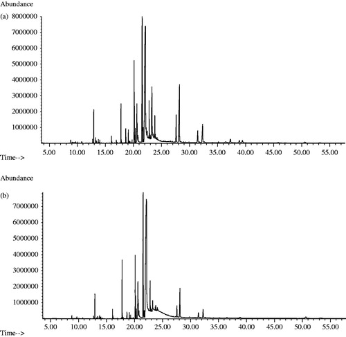
Table 3. Main non-polar constituents tentatively identified by GC-MS in aerial parts and seeds of C. halicacabum.
A total of 33 constituents were tentatively identified in the n-hexane fraction of the seeds obtained by Naviglio while 29 compounds characterized the n-hexane fraction of the seeds obtained by Soxhlet. The dominant constituent of both fractions was eicosanoic acid (39.6 and 41.7% for SN and SS, respectively). Other abundant compounds identified in both fractions are methyl 11-eicosenoate, oleic acid, methyl oleate, and methyl erucate. 1-Octanol, 2-nonenal, caprylic acid methyl ester, capric acid methyl ester, lauric acid and 6,10,14-trimethyl-2-pentadecanone were not identified in SS n-hexane fraction.
Antioxidant properties
The term “oxidative stress” indicates the profound alteration of the dynamic balance between oxidants and antioxidants in favour of seconds with generation of oxidative potential in the cellular districts and in biological fluids. A large body of scientific evidence suggests the direct and/or indirect involvement of oxidative stress in the onset and development of a number of pathologies. The role played by oxidative stress is clearly different depending on the disease, but can be attributed to a series of modifications in biological molecules such as polyunsaturated fatty acids, nucleic acids, proteins and carbohydrates, modifications that may be due to profound alterations of the processes metabolic and functional up to cell death by necrosis or apoptosis. Hence the importance of determining the antioxidant activity using different methodological approaches that can monitor the oxidation process in all its complexity.
The radical scavenging activity of total extracts and fractions of C. halicacabum has been evaluated by using DPPH and ABTS testCitation20. Data are reported in .
Table 4. Antioxidant activity of total extracts and fractions of C. halicacabum.
All samples showed antioxidant activity in a concentration-dependent manner. The best radical scavenging activity assessed by the DPPH test is demonstrated by the methanol fractions of the four samples with IC50 values in the range of 33.1–40.6 µg/mL. By comparing the total extracts obtained by the two different extraction techniques it is possible to highlight a better radical scavenging activity of the extracts obtained by the Soxhlet apparatus with IC50 values of 53.6 and 87.3 µg/mL, respectively, for the aerial parts and seeds. This activity can be in close relation with the highest content of phenols and flavonoids showed in extracts obtained by Soxhlet. In a previous study ethanol and water extracts of C. halicacabum have demonstrated EC50 values of 198.26 and 357.18 mg/mL, respectively in DPPH testCitation6.
The same trend is observed in the other assay that allows you to evaluate the radical scavenging activity, the ABTS test. Indeed, the most active samples are the methanol fractions of the aerial parts with IC50 values of 40.2 and 90.1 µg/mL for SP and NP, respectively. In the FRAP test, the total extract and methanol fraction of NP showed the highest activity with values of 10.1 and 12.8 µM Fe(II)/g, respectively.
The potential of C. halicacabum to inhibit lipid peroxidation was evaluated using the β-carotene bleaching test, which measures the capacity to inhibit the conjugated diene hydroperoxides formation upon linoleic acid oxidation. PS exhibited the highest antioxidant activity with IC50 values of 6.9 and 6.5 µg/mL after 30 and 60 min of incubation, respectively. Interesting results were founded also for the methanol fraction of SS with IC50 values of 7.5 and 8.1 µg/mL after 30 and 60 min of incubation, respectively. Both the total phenols content and the total flavonoids content are greater in samples obtained by extraction with Soxhlet apparatus. Additionally, the HPLC profile demonstrates that rutin, apigenin, apigenin-7-O-glucoside and luteolin contents were highest in PS methanol fraction. Considering that this fraction also demonstrated the highest anti-radical activity, these flavonoids may play an important role. This observation is supported by much evidence that has shown that the antioxidative effects of phenolic compounds could play a critical role, not only in free radical inhibition, but also in the prevention of cell damage. In fact, these compounds have been reported to possess strong antioxidant activitiesCitation27–29. In a previous work, the radical scavenging activity of C. halicacabum var. microcarpum seeds extracts obtained by Soxhlet apparatus with petroleum ether, benzene, chloroform and ethanol, was studied by using different antioxidant modelsCitation30. Free radicals were scavenged by the extracts in a concentration-dependent manner in all used models.
Tyrosinase inhibitory activity
Several scenarios can be envisaged whereby tyrosinase would play a central role in the etiology of PDCitation31. Since dopaminergic neurons synthesize dopamine and specifically sequester the neurotransmitter (via the dopamine transporter), increased levels of the free neurotransmitter could compromise the neuron in the presence of tyrosinase. In addition, PD may be associated with decreased antioxidant levels or result from increased oxidant stress on an individual. Under these conditions, the production of reactive dopamine quinone by autoxidation or enzyme-catalysed conversion would not be counterbalanced by antioxidant scavengers. Finally, although the disease is not believed to have a strong hereditary component, genetic overexpression of tyrosinase or expression of an altered form of the enzyme could contribute to disease progression.
Cardiospermum halicacabum was tested for tyrosinase inhibitory activity using l-tyrosine as the substrate. As reported in , results showed that all extracts and fractions were able to inhibit the activity of tyrosinase. Considering the aerial parts, PN total extract was the most active (IC50 of 10.8 µg/mL) followed by PS total extract (IC50 value of 47.0 µg/mL). The total extract was subjected to partitioning. Among obtained fractions, the most active were the methanol fractions with IC50 values of 64.1 and 100.0 µg/mL for PN and PS, respectively. Several plant extracts and their bioactive constituents have been explored previously for tyrosinase inhibitory activity. In a previous study, the inhibitory effects of some flavonoids on the activity of mushroom tyrosinase were evaluatedCitation32. Flavonoids can lead to reversible inhibition of the enzyme. A kinetic analysis showed that the flavonols are competitive inhibitors, whereas luteolin is an uncompetitive inhibitor. The rank order of inhibition was: quercetin >galangin > morin; fisetin > 3,7,4;-trihydroxyflavone; luteolin > apigenin > chrysin. Luteolin was easily oxidized by tyrosinase due to the presence of catechol moiety at B ring. Moreover, luteolin has been reported as melanin biosynthesis inhibitor on B61 melanoma cells by reducing the signal of cAMP (cyclic adenosine monophosphate) which decreased the melanin contentCitation33,Citation34.
Table 5. Cholinesterases and tyrosinase inhibitory activity of (IC50 µg/mL) of total extracts and fractions of C. halicacabum.
Seeds extracts of C. halicacabum showed significant inhibition of tyrosinase activity with IC50 values of 78.4 and 87.2 µg/mL for SS and SN, respectively.
After partitioning, the less polar fractions (n-hexane) exhibited the highest anti-tyrosinase activity with IC50 values of 20.9 and 21.0 µg/mL for SN and SS, respectively. Eicosanoic acid, methyl 11-eicosenoate, methyl 11,13-eicosadienoate, methyl erucate and methyl oleate were identified by GC-MS analyses as main components of these fractions. In a previous study, Huh et al.Citation35 reported the inhibitory tyrosinase activity of some methyl and ethyl esters of fatty acids isolated from Oxalis triangularis. In literature, it has been reported that unsaturated fatty acids decrease melanin synthesis and tyrosinase activity, and that these inhibitory effects occur in proportion to the number of unsaturated bondsCitation36.
Cholinesterase inhibitory activities
Inhibition of cholinesterase has attracted much attention recently because of its potential for the treatment of AD. Several compounds of natural origin have proved able of inhibiting the cholinesteraseCitation16, and if they are always looking for new to ensure a lower toxicity and allow a better bioavailability, increasing the odds of drug in brain areas of interest. AChE inhibitors are well used for the management of mild to moderate AD. Nevertheless, at the late stage of AD, when AChE declined by up to 85%, BChE takes over for the hydrolysis of acetylcholine. The specificity of BChE is very less towards hydrolyzing acetylcholine but it is generally viewed as a backup enzyme for the homologous AChE, thus plays an important role in aggravating AD, especially at a low concentration of AChE. Thus, the BChE inhibition emerged to be another approach to intervene in the progression of AD. In this work, the anticholinesterase activities of extracts and fractions of C. halicacabum were investigated using Ellman’s colorimetric method. Data, reported in , showed that all samples inhibited AChE and BChE in a concentration-dependent manner. Interestingly, except for the methanol fraction of NP, all samples of C. halicacabum exhibited a major inhibitory activity against BChE respect to AChE.
By comparing the activity of total extracts, the best results were obtained with the seeds with IC50 values of 112.4 and 150.4 µg/mL for SS and SN, respectively. The n-hexane fractions of SN and PN exhibited the best activity with IC50 values of 57.9 and 59.8 µg/mL, respectively. GC and GC-MS analyses revealed the presence of several fatty acids that were implicated in enzymatic and anti-radical activitiesCitation37. However, we can consider that other minor compounds present in the extract may be also responsible for the observed activities.
Conclusions
The history of drug development has its foundation in the study of natural remedies used to treat human disease over centuries. In this context, this study reports for the first time the chemical composition, the cholinesterase and tyrosinase inhibitory activities and the antioxidant properties of C. halicacabum aerial parts and seeds.
Our results demonstrated the potential interest for C. halicacabum as source of agents in the treatment of AD and PD, and lend support to the reported traditional use of C. halicacabum in the treatment and management of neurodegenerative diseases. Thus, this may be an interesting candidate for evaluation in more complex biological assays and eventually in in vivo assays.
Declaration of interest
This work was supported by European Community POR (Programmi Operativi Regionali) Calabria FSE (Fondo Sociale Europeo) 2007/2013. The authors report no conflicts of interest. The authors alone are responsible for the content and writing of this article.
Acknowledgements
The authors thank Dr N. Benanti for technical assistance.
References
- Neuwinger HD. African traditional medicine: a dictionary of plant use and applications. Stuttgart, Germany: Medpharm GmbH Scientific Publishers; 2000
- Venkatesh Babu KC, Krishnakumari S. Cardiospermum halicacabum suppresses the production of TNF-alpha and nitric oxide by human peripheral blood mononuclear cells. Afr J Biomed Res 2006;9:95–9
- Pillai NR, Vijayamma N. Some pharmacological studies on Cardiospermum halicacabum Linn. Anc Sci Life 1985;5:32–6
- Venkat Rao N, Chandra Prakash K, Shanta Kumar SM. Pharmacological investigation of Cardiospermum halicacabum (Linn) in different animal models of diarrhoea. Indian J Pharm 2006;38:346–9
- Kumaran A, Karunakaran RJ. Antioxidant activities of the methanol extract of Cardiospermum halicacabum. Pharm Biol 2006;44:146–51
- Huang MH, Huang SS, Wang BS, et al. Antioxidant and anti-inflammatory properties of Cardiospermum halicacabum and its reference compounds ex vivo and in vivo. J Ethnopharmacol 2011;133:743–50
- Khan MSY, Arya M, Javed K, Khan MH. Chemical examination of Cardiospermum halicacabum. Linn. Indian Drugs 1990;27:257–8
- Subramanyam R, Newmaster SG, Paliyath G, Newmaster CB. Exploring ethnobiologucal classifications for novel alternative medicine: a case study of Cardiospermum halicacabum (Modakathon, Balloon Vine) as a traditional herb for treating rheumatoid arthritis. Ethnobotany 2007;19:1–18
- Deepan T, Alekhya V, Saravanakumar P, Dhanaraju MD. Phytochemical and anti-microbial studies on the leaves extracts of Cardiospermum halicacabum Linn. Adv Biol Res 2012;6:14–8
- Aviram M. Review of human studies on oxidative damage and antioxidant protection related to cardiovascular diseases. Free Rad Res 2000;33:85–97
- Christen Y. Oxidative stress and Alzheimer disease. Am J Clin Nutr 2000;71:621S–9S
- Rao KSJ, Hegde ML, Anitha S, et al. Amyloid β and neuromelanin – toxic or protective molecules? The cellular context makes the difference. Prog Neurobiol 2006;78:364–73
- Tessari I, Bisaglia M, Valle F, et al. The reaction of α-synuclein with tyrosinase: possible implications for Parkinson disease. J Biol Chem 2008;283:16808–17
- Zahid M, Saeed M, Yang L, et al. Formation of dopamine quinone-DNA adducts and their potential role in the aetiology of Parkinson’s disease. IUBMB Life 2011;63:1087–93
- Greggio E, Bergantino E, Carter D, et al. Tyrosinase exacerbates dopamine toxicity but is not genetically associated with Parkinson’s disease. J Neurochem 2005;93:246–56
- Supuran CT. Carbonic anhydrases: novel therapeutic applications for inhibitors and activators. Nat Rev Drug Discov 2008;7:168–81
- Ilies M, Banciu MD, Ilies MA, et al. Carbonic anhydrase activators: design of high affinity isozymes I, II, and IV activators, incorporating tri-/tetrasubstituted-pyridinium-azole moieties. J Med Chem 2002;45:504–10
- Temperini C, Scozzafava A, Supuran CT. Carbonic anhydrase activation and the drug design. Curr Pharm Des 2008;14:708–15
- Loizzo MR, Tundis R, Menichini F, Menichini F. Natural products and their derivatives as cholinesterase inhibitors in the treatment of neurodegenerative disorders: an update. Curr Med Chem 2008;15:1209–28
- Loizzo MR, Tundis R, Bonesi M, et al. Radical scavenging, antioxidant and metal chelating activities of Annona cherimola Mill. (cherimoya) peel and pulp in relation to their total phenolic and total flavonoid contents. J Food Comp Anal 2012;25:179–84
- Bonesi M, Okusa PN, Tundis R, et al. Chemical composition, antioxidant properties and anti-cholinesterase activity of Cordia gilletii (Boraginaceae) leaves essential oil. Nat Prod Commun 2011;6:253–7
- Tundis R, Bonesi M, Menichini F, et al. Antioxidant and anti-cholinesterase activity of Globularia meridionalis extracts and isolated constituents. Nat Prod Commun 2012;7:1015–20
- Tundis R, Loizzo MR, Bonesi M, et al. Comparative study on the antioxidant capacity and cholinesterase inhibitory activity of Citrus aurantifolia Swingle, C. aurantium L., and C. bergamia Risso and Poit. peel essential oils. J Food Sci 2012;77:H40–6
- Tundis R, Nadjafi F, Menichini F. Angiotensin-converting enzyme inhibitory activity and antioxidant properties of Nepeta crassifolia Boiss & Buhse and Nepeta binaludensis Jamzad. Phytother Res 2013;27:572–80
- Bao K, Dai Y, Zhu Z-B, et al. Design and synthesis of biphenyl derivatives as mushroom tyrosinase inhibitors. Bioorg Med Chem 2010;18:6708–14
- Ellman GL, Courtney KD, Andres V Jr., Feath-Erstone RM. A new and rapid colorimetric determination of acetylcholinesterase activity. Biochem Pharmacol 1961;7:88–95
- Shen SC, Lee WR, Lin HY, et al. In vitro and in vivo inhibitory activities of rutin, wogonin, and quercetin on lipopolysaccharide-induced nitric oxide and prostaglandin E(2) production. Eur J Pharmacol 2002;446:187–94
- Shan BE, Wang MX, Li RQ. Quercetin inhibit human SW480 colon cancer growth in association with inhibition of cyclin D1 and survivin expression through Wnt/beta-catenin signaling pathway. Cancer Invest 2009;27:604–12
- Xie T, Wang WP, Mao ZF, et al. Effects of epigallocatechin-3-gallate on pentylenetetrazole-induced kindling, cognitive impairment and oxidative stress in rats. Neurosci Lett 2012;516:237–41
- Jayanthi G, Sathishkumar T, Senthilkumar T, Jegadeesan M. Free radical scavenging potential of Cardiospermum halicacabum L. var. microcarpum (Kunth) Blume seeds. Int Res J Pharm App Sci 2012;2:41–8
- Xu Y, Stokes AH, Roskoski R Jr, Vrana KE. Dopamine, in the presence of tyrosinase, covalently modifies and inactivates tyrosine hydroxylase. J Neurosci Res 1998;54:691–7
- Xie LP, Chen QX, Huang H, et al. Inhibitory effects of some flavonoids on the activity of mushroom tyrosinase. Biochemistry (Mosc) 2003;68:487–91
- Choi MY, Song HS, Hur HS, Sim SS. Whitening activity of luteolin related to the inhibition of cAMP pathway in alpha-MSH-stimulated B16 melanoma cells. Arch Pharm Res 2008;31:1166--71
- An SM, Kim HJ, Kim JE, Boo YC. Flavonoids, taxifolin and luteolin attenuate cellular melanogenesis despite increasing tyrosinase protein levels. Phytother Res 2008;22:1200–7
- Huh S, Kim YS, Jung E, et al. Melanogenesis inhibitory effect of fatty acid alkyl esters isolated from Oxalis triangularis. Biol Pharm Bull 2010;33:1242–5
- Ando H, Ryu A, Hashimoto A, et al. Linoleic acid and alpha-linolenic acid lightens ultraviolet-induced hyperpigmentation of the skin. Arch Dermatol Res 1998;290:375–81
- Ren Y, Houghton P, Hider RC. Relevant activities of extracts and constituents of animals used in traditional Chinese medicine for central nervous system effects associated with Alzheimer’s disease. J Pharm Pharmacol 2006;58:989–96