
Abstract
A series of urea derivatives bearing nitroaryl moiety has been synthesized and assayed for their potential antiproliferative activities. Some of the tested compounds displayed activity in RK33 laryngeal cancer cells and TE671 rhabdomyosarcoma cells while being generally less toxic to healthy HSF human fibroblasts cells. One compound was demonstrated to be a moderate CDK2 inhibitor with IC50 = 14.3 µM. Its structure was solved by an X-ray crystallography and molecular modelling was performed to determine structure-activity relationship. Obtained compounds constitute novel structures and generally demonstrated greater cytotoxicity in comparison to cisplatin. This study offers new structural motifs with potential for further development.
Introduction
Cancer is the leading cause of death worldwide accounting for 8.2 million deaths in 2012 and it is expected that 14.1 million of all cancer cases in 2012 will rise to 22 million in the next two decadesCitation1. Currently available options for treatments of cancer involve surgery, radiotherapy and pharmacotherapy, but the general efficacy is not satisfying. Therefore there is an ongoing need for development of new and more effective drugs.
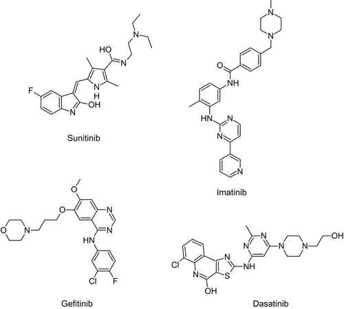
Protein kinases are well-established targets for anticancer drug discoveryCitation2,Citation3. They regulate diverse cellular functions by phosphorylating threonine, serine and tyrosine residues at specific positions in proteinsCitation4. Protein kinases can be altered in many disorders such as cancer and therefore are attractive molecular targets in drug discovery programmesCitation5. These programmes yielded so far several small molecule inhibitors of protein kinases that have been approved for clinical applications in oncology (sunitinibCitation6, imatinibCitation7, gefitinibCitation8, dasatinibCitation9 – Scheme 1).
Scheme 1. Structures of sunitinib, imatinib, gefitinib and dasatinib.
There is an ever increasing body of evidence linking CDK-related malfunction with cancer developmentCitation10,Citation11. CDKs (cyclin dependent kinases) are enzymes that have been identified originally to play a key role in regulating the cell cycle machineryCitation12. They constitute a family of proteins assigned Cdk1–Cdk20Citation13. Similar other protein kinases, CDKs also have two-lobed structure with the active site sandwiched between an amino-terminal lobe consisting of beta-sheets and a carboxy-terminal lobe based on alpha-helicesCitation12. Activities of CDKs are dependent on regulatory subunits -- proteins called cyclins. The function of CDKs lies in regulation of transcription in response to a mitogenic stimulation. This mainly occurs due to phosphorylation and subsequent inactivation of retinoblastoma protein (Rb) resulting in de-repression of multiple genes involved in DNA synthesis and mitosisCitation14. There are many compoundsCitation15 spanning from broad-range inhibitors such as flavopidirolCitation16 and roscovitineCitation17 to selective inhibitors such as palbociclib (PD-033299)Citation18 or PHA-767491Citation19.
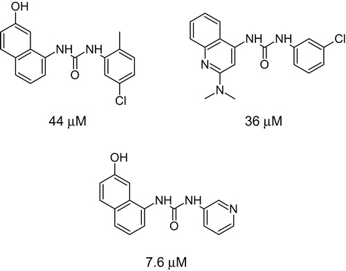
Although 20 CDK inhibitors have entered clinical trials, none has been approved for therapy yet. This prompted us to look for a scaffold which would combine structural features of known CDK inhibitors together with urea moietyCitation20. Our intention was further supported with findings of urea derivatives active towards CDKsCitation21 (Scheme 2). The aim of our work was synthesis, in vitro studies and molecular modelling of novel nitroaryl urea derivatives (NUD) which displayed promising antiproliferative properties. We chose compounds with different substituents to be able to investigate possible structure-activity relationship.
Scheme 2. Urea compounds active in CDK assay with their IC50 valuesCitation21.
Experimental
Synthesis
All reagents were obtained from Sigma-Aldrich (St. Louis, MO) and used without further purification unless stated otherwise. NMR spectra were aqcuired on Bruker AVANCE III 600 MHz and Bruker Fourier 300 MHz spectrometers (Fallanden, Switzerland). Solvents for NMR spectroscopy were purchased from Armar Chemicals (Dottingen, Switzerland). MS spectra were recorded on Bruker micrOTOF-Q II spectrometer using direct probe method. TLC analysis was carried out on Merck HPTLC Silica gel 60 F254 plates (Darmstadt, Germany) prewashed with MeOH. Merck Silica gel 60 15–40 µm particle size was used for DCVC chromatographyCitation22.
N-(4-nitrophenyl)ethane-1,2-diamine (3)
10 g of 1-chloro-4-nitrobenzene (1) (0.06 mol) and 40 g of ethylenediamine (0.6 mol) were refluxed together for 90 minutes. Excess of ethylenediamine was distilled and the remaining residue was dissolved in hot water, filtered while hot and allowed to cool. Crystals formed very quickly. They were filtered, washed with water and air dried. Thus 9.6 g (83%) of orange solid was obtained. All analytical data corresponds to that found in the literatureCitation23 m.p. 148–150 °C. 1H NMR (300 MHz, DMSO-d6) δ 7.89–8.07 (m, 2H), 7.30 (br t, J = 4.66 Hz, 1H), 6.56–6.72 (m, 2H), 3.13 (q, J = 6.08 Hz, 2H), 2.73 (t, J = 6.29 Hz, 2H), 1.60 (br s, 2H); 13C NMR (75 MHz, DMSO-d6) δ 155.2, 135.9, 126.7, 111.1, 46.5, 41.1.
N-(5-nitropyridin-2-yl)ethane-1,2-diamine (4)
4.8 g of 2-chloro-5-nitropyridine (2) (0.03 mol) was carefully added portionwise to a chilled ethylenediamine 40 g (0.6 mol). The mixture was stirred for 30 minutes and excess of ethylenediamine was removed by distillation. The residue was chromatographed over Silica gel with MeOH–DCM–25% NH3aq 10–90–1 to afford 3.8 g of orange solid (69%) with m.p. 124–126 °C in accordance to the literature dataCitation24. 1H NMR (600 MHz, METHANOL-d4) δ 8.93 (d, J = 2.75 Hz, 1H), 8.14 (br d, J = 8.36 Hz, 1H), 6.55 (d, J = 9.35 Hz, 1H), 3.53 (br s, 2H), 2.87 (t, J = 6.33 Hz, 2H); 13C NMR (151 MHz, METHANOL-d4) δ 161.7, 146.2, 135.1, 131.7, 107.9, 43.7, 40.4.
General procedure to obtain compounds 5a–5d and 6a–6f
1 mmol of N-(4-nitrophenyl)ethane-1,2-diamine (3) or N-(5-nitropyridin-2-yl)ethane-1,2-diamine (4) was dissolved in a suitable solvent and solution of 1 mmol of isocyanate in the same solvent was added dropwise at room temperature. The mixture was stirred for 30 minutes at RT. Formed precipitate was collected and purified as indicated for each individual compound.
1-Naphthalen-1-yl-3-{2-[(4-nitrophenyl)amino]ethyl}urea (5a)
Compound 5a was obtained using 1-naphthyl isocyanate and ACN as solvent. Product was recrystallized from AcOH. TLC on Silica gel EtOAc–hexane 8–2 Rf = 0.41. Elemental analysis C19H18N4O3 Calcd. C(65.13%) H(5.18%) N(15.99%) exp. C(64.86%) H(5.11%) N(15.91%). HRMS [M + H]+ Calcd. 351.1452; exp. 351.1453. 1H NMR (600 MHz, DMSO-d6) δ 8.63 (s, 1H), 8.05–8.08 (m, 1H), 8.00–8.04 (m, 2H), 7.96 (dd, J = 0.80, 7.60 Hz, 1H), 7.88–7.92 (m, 1H), 7.58 (d, J = 8.20 Hz, 1H), 7.50–7.56 (m, 2H), 7.42–7.46 (m, 2H), 6.71–6.76 (m, 3H), 3.32–3.40 (m, 4H); 13C NMR (151 MHz, DMSO-d6) δ 156.4, 155.1, 136.2, 135.4, 134.2, 128.8, 126.7, 126.4, 126.3, 126.2, 125.9, 122.8, 122.0, 117.5, 111.2, 43.2, 39.0.
1-(4-Acetylphenyl)-3-{2-[(4-nitrophenyl)amino]ethyl}urea (5b)
Compound 5b was obtained using freshly purified 4-acetylphenyl isocyanate (commercial reagent was heated with carbon tetrachloride, filtered and the solvent evaporated under vacuo leaving pure isocyanate) and THF as solvent. Product was washed with hot AcOH and subsequently with MeOH. TLC on Silica gel EtOAc–hexane 8–2 Rf = 0.24. Elemental analysis C17H18N4O4 Calcd. C(59.64%) H(5.30%) N(16.37%) exp. C(59.54%) H(5.32%) N(16.13%). HRMS [M + H]+ Calcd. 343.1401; exp. 343.1402. 1H NMR (300 MHz, DMSO-d6) δ 9.10 (s, 1H), 8.03 (d, J = 9.22 Hz, 2H), 7.88 (d, J = 8.85 Hz, 2H), 7.56 (d, J = 8.85 Hz, 2H), 7.42 (br s, 1H), 6.73 (d, J = 9.31 Hz, 2H), 6.47 (s, 1H), 3.28–3.36 (m, 4H), 2.52 (s, 3H); 13C NMR (75 MHz, DMSO-d6) δ 196.7, 155.5, 155.1, 145.6, 136.3, 130.4, 130.1, 126.8, 117.2, 111.3, 42.9, 31.2, 26.8.
1-(4-Chlorobenzyl)-3-{2-[(4-nitrophenyl)amino]ethyl}urea (5c)
Compound 5c was obtained using 4-chlorobenzyl isocyanate and THF as solvent. Product was chromatographed over Silica gel with AcOEt–hexane 95–5. TLC on Silica gel EtOAc–hexane 8–2 Rf = 0.19. Elemental analysis C16H17ClN4O3 Calcd. C(55.10%) H(4.91%) Cl(10.16%) N(16.06%) exp. C(55.06%) H(4.92%) Cl(10.18%) N(15.69%). HRMS [M + H]+ Calcd. 349.1062; exp. 349.1065. 1H NMR (600 MHz, DMSO-d6) δ 7.99 (d, J = 9.17 Hz, 2H), 7.34–7.37 (m, 2H), 7.26 (d, J = 8.38 Hz, 2H), 6.67 (d, J = 9.27 Hz, 2H), 6.56 (t, J = 6.06 Hz, 1H), 6.15 (s, 1H), 4.20 (d, J = 6.01 Hz, 2H), 3.20–3.24 (m, 4H); 13C NMR (151 MHz, DMSO-d6) δ 158.7, 155.1, 140.5, 136.1, 131.5, 129.3, 128.6, 126.7, 111.3, 43.4, 42.7, 39.1.
1-(4-Methoxybenzyl)-3-{2-[(4-nitrophenyl)amino]ethyl}urea (5d)
Compound 5d was obtained using 4-methoxybenzyl isocyanate and THF as solvent. Product was washed with THF and subsequently with MeOH. TLC on Silica gel EtOAc–hexane 8–2 Rf = 0.43. Elemental analysis C17H20N4O4 Calcd. C(59.29%) H(5.85%) N(16.27%) exp. C(59.27%) H(5.86%) N(16.22%). HRMS [M + H]+ Calcd. 345.1557; exp. 345.1555. 1H NMR (600 MHz, DMSO-d6) δ 7.97–8.01 (m, 2H), 7.33–7.38 (m, 1H), 7.14–7.19 (m, 2H), 6.84–6.88 (m, 2H), 6.67 (d, J = 9.35 Hz, 2H), 6.42 (t, J = 5.93 Hz, 1H), 6.03–6.10 (m, 1H), 4.14 (d, J = 5.93 Hz, 2H), 3.73 (s, 3H), 3.19–3.24 (m, 4H); 13C NMR (151 MHz, DMSO-d6) δ 158.7, 158.5, 155.2, 136.1, 133.2, 128.8, 126.7, 114.1, 111.2, 55.5, 43.5, 42.9, 39.1.
1-Naphthalen-1-yl-3-{2-[(5-nitropyridin-2-yl)amino]ethyl} urea (6a)
Compound 6a was obtained using 1-naphthyl isocyanate and ACN as solvent. Product was recrystallized from aqueous MeOH. TLC on Silica gel MeOH–CHCl3–NH3 25%aq 10–90–1 Rf = 0.39. Elemental analysis C18H17N5O3 Calcd. C(61.53%) H(4.88%) N(19.93%) exp. C(61.15%) H(4.87%) N(19.62%). HRMS [M + H]+ Calcd. 352.1404; exp. 352.1403. 1H NMR (600 MHz, DMSO-d6) δ 8.92 (d, J = 2.68 Hz, 1H), 8.56 (br. s., 1H), 8.23–8.34 (m, 1H), 8.12 (br. s., 1H), 8.03–8.06 (m, 1H), 7.93–7.97 (m, 1H), 7.87–7.92 (m, 1H), 7.57 (d, J = 8.13 Hz, 1H), 7.49–7.55 (m, 2H), 7.43 (t, J = 7.85 Hz, 1H), 6.68–6.73 (m, 1H), 6.61 (d, J = 8.59 Hz, 1H), 3.57 (br. s., 2H), 3.38 (q, J = 6.07 Hz, 2H); 13C NMR (151 MHz, DMSO-d6) δ 162.1, 156.3, 147.3, 135.5, 134.8, 134.2, 132.2, 128.8, 126.4, 126.2, 125.9, 122.8, 121.9, 117.4, 109.2, 41.6, 39.3.
1-(4-Acetylphenyl)-3-{2-[(5-nitropyridin-2-yl)amino]ethyl} urea (6b)
Compound 6b was obtained using freshly purified 4-acetylphenyl isocyanate and ACN as solvent. Product was recrystallized from acetone/cyclohexane. TLC on Silica gel MeOH–CHCl3–NH3 25%aq 10–90–1 Rf = 0.25. Elemental analysis C16H17N5O4 Calcd. C(55.97%) H(4.99%) N(20.40%) exp. C(55.91%) H(4.93%) N(20.32%). HRMS [M + H]+ Calcd. 344.1353; exp. 344.1354. 1H NMR (600 MHz, DMSO-d6) δ 9.00 (br. s., 1H), 8.92 (d, J = 2.42 Hz, 1H), 8.24 (br. s., 1H), 8.11 (br. s., 1H), 7.83–7.87 (m, 2H), 7.50–7.54 (m, 2H), 6.59 (d, J = 8.56 Hz, 1H), 6.40–6.49 (m, 1H), 3.53 (br. s., 2H), 3.30–3.35 (m, 2H), 2.49 (s, 3H); 13C NMR (151 MHz, DMSO-d6) δ 196.7, 162.0, 155.4, 147.3, 145.6, 134.9, 132.2, 130.3, 130.1, 117.1, 109.2, 41.4, 39.2, 26.8.
1-(4-Chlorobenzyl)-3-{2-[(5-nitropyridin-2-yl)amino]ethyl} urea (6c)
Compound 6c was obtained using 4-chlorobenzyl isocyanate and ACN as solvent. Product was recrystallized from EtOAc. TLC on Silica gel acetone–hexane 1–1 Rf = 0.31. Elemental analysis C15H16ClN5O3 Calcd. C(51.51%) H(4.61%) Cl(10.14%) N(20.02%) exp. C(51.36%) H(4.56%) Cl(10.17%) N(19.99%). HRMS [M + H]+ Calcd. 350.1014; exp. 350.1012. 1H NMR (600 MHz, DMSO-d6) δ 8.91 (d, J = 2.10 Hz, 1H), 8.15–8.24 (m, 1H), 8.06–8.14 (m, 1H), 7.35 (d, J = 7.82 Hz, 2H), 7.25 (d, J = 8.38 Hz, 2H), 6.56 (d, J = 9.21 Hz, 1H), 6.50 (br. s., 1H), 6.14 (br. s., 1H), 4.19 (d, J = 6.08 Hz, 2H), 3.39–3.53 (m, 2H), 3.23 (q, J = 6.20 Hz, 2H); 13C NMR (151 MHz, DMSO-d6) δ 162.0, 158.6, 147.3, 140.5, 134.8, 132.1, 131.5, 129.3, 128.6, 109.1, 42.7, 41.8, 39.4.
1-(4-Methoxybenzyl)-3-{2-[(5-nitropyridin-2-yl)amino]ethyl} urea (6d)
Compound 6d was obtained using 4-methoxybenzyl isocyanate and ACN as solvent. Product was recrystallized from MeOH. TLC on Silica gel acetone–hexane 6–4 Rf = 0.33. Elemental analysis C16H19N5O4 Calcd. C(55.64%) H(5.55%) N(20.28%) exp. C(55.44%) H(5.49%) N(20.32%). HRMS [M + H]+ Calcd. 346.1510; exp. 346.1513. 1H NMR (600 MHz, DMSO-d6) δ 8.90 (d, J = 2.49 Hz, 1H), 8.20 (br. s., 1H), 8.10 (br. s., 1H), 7.15 (d, J = 8.06 Hz, 2H), 6.85 (d, J = 8.02 Hz, 2H), 6.56 (d, J = 9.46 Hz, 1H), 6.35 (br. s., 1H), 6.04 (br. s., 1H), 4.13 (d, J = 5.95 Hz, 2H), 3.72 (s, 3H), 3.45 (br. s., 2H), 3.23 (q, J = 6.25 Hz, 2H); 13C NMR (151 MHz, DMSO-d6) δ 171.1, 167.9, 167.8, 157.0, 144.9, 143.4, 142.4, 139.2, 125.0, 120.3, 68.6, 56.4, 55.5, 53.1.
1-(3,4-Dichlorophenyl)-3-{2-[(5-nitropyridin-2-yl)amino] ethyl}urea (6e)
Compound 6e was obtained using 3,4-dichlorophenyl isocyanate and ACN as solvent. Precipitated product was washed with ACN. TLC on Silica gel acetone–hexane 4–6 Rf = 0.30. Elemental analysis C14H13Cl2N5O3 Calcd. C(45.42%) H(3.54%) Cl(19.15%) N(18.92%) exp. C(45.41%) H(3.50%) Cl(19.17%) N(18.97%). HRMS [M + H]+ Calcd. 370.0468; exp. 370.0469. 1H NMR (600 MHz, DMSO-d6) δ 8.83–8.98 (m, 2H), 8.22 (br. s., 1H), 8.10 (br. s., 1H), 7.84 (d, J = 2.54 Hz, 1H), 7.44 (d, J = 8.91 Hz, 1H), 7.25 (dd, J = 2.49, 8.83 Hz, 1H), 6.58 (d, J = 8.78 Hz, 1H), 6.39–6.46 (m, 1H), 3.42–3.60 (m, 2H), 3.31 (d, J = 5.98 Hz, 2H); 13C NMR (151 MHz, DMSO-d6) δ 162.0, 155.5, 147.3, 141.2, 134.9, 132.2, 131.4, 130.9, 122.7, 119.2, 118.2, 109.2, 41.4, 39.2.
1-(4-Methoxyphenyl)-3-{2-[(5-nitropyridin-2-yl)amino]ethyl} urea (6f)
Compound 6f was obtained using 4-methoxyphenyl isocyanate and ACN as solvent. Product was recrystallized from MeOH/DCM. TLC on Silica gel acetone–hexane 8–2 Rf = 0.48. Elemental analysis C15H17N5O4 Calcd. C(54.38%) H(5.17%) N(21.14%) exp. C(54.25%) H(5.09%) N(21.07%). HRMS [M + H]+ Calcd. 332.1353; exp. 332.1355. 1H NMR (600 MHz, DMSO-d6) δ 8.92 (br. s., 1H), 8.32 (br. s., 1H), 8.23 (br. s., 1H), 8.11 (br. s., 1H), 7.24–7.31 (m, 2H), 6.78-6.85 (m, 2H), 6.58 (d, J = 8.51 Hz, 1H), 6.16 (br. s., 1H), 3.69 (s, 3H), 3.42-3.58 (m, 2H), 3.27–3.32 (m, 2H); 13C NMR (151 MHz, DMSO-d6) δ 162.0, 156.1, 154.5, 147.3, 134.8, 134.0, 132.2, 120.1, 114.3, 109.1, 55.6, 41.6, 39.2.
1-Methyl-3-{2-[(5-nitropyridin-2-yl)amino]ethyl}urea (6g)
182 mg (1 mmol) of N-(5-nitropyridin-2-yl)ethane-1,2-diamine was added to a solution of 125 mg (1 mmol) N-methyl-1H-imidazole-1-carboxamide in 10 ml of DCM. This was followed by addition of 152 µl (1.1 mmol) of TEA. The mixture was stirred under argon for 24 hours. Solvent was removed on a rotavap and the residue was chromatographed over Silica gel with EtOAc/hexane gradient. Elemental analysis C9H13N5O3 Calcd. C(45.18%) H(5.48%) N(29.27%) exp. C(45.13%) H(5.44%) N(29.10%). HRMS [M + H]+ Calcd. 240.1091; exp. 240.1092. 1H NMR (600 MHz, DMSO-d6) δ 8.91 (d, J = 1.16 Hz, 1H), 8.20 (br. s., 1H), 8.03–8.15 (m, 1H), 6.56 (d, J = 8.59 Hz, 1H), 6.04 (br. s., 1H), 5.81 (br. s., 1H), 3.43 (br. s., 2H), 3.19 (q, J = 6.33 Hz, 2H), 2.54 (d, J = 4.62 Hz, 3H); 13C NMR (151 MHz, DMSO-d6) δ 161.9, 159.3, 147.3, 134.8, 132.1, 109.1, 41.9, 39.3, 26.9.
1-(4-Nitrophenyl)imidazolidin-2-imine hydrobromide (7)
3.18g (30 mmol) of cyanogen bromide dissolved in 50 ml of MeOH was added dropwise to 5.44 g (30 mmol) of N-(4-nitrophenyl)ethane-1,2-diamine (3) suspended in 50 ml of MeOH. During addition the amine went into solution and colour turned orange-brown. The reaction was heated under reflux for 90 minutes and then solvent removed on a rotavap. Yellow residue was recrystallized from EtOH to afford 7.3 g (85%) of yellow solid. The hydrobromide salt was converted to a freebase form by dissolving it in warm water, adding to 5% NaOH (2.5 eq) and filtering resulting precipitate. NMR of the freebase 1H NMR (300 MHz, DMSO-d6) δ 8.09–8.18 (m, 2H), 7.96–8.07 (m, 2H), 6.06 (br s, 2H), 3.81–3.94 (m, 2H), 3.35–3.43 (m, 2H); 13C NMR (75 MHz, DMSO-d6) δ 158.5, 148.2, 140.0, 124.9, 117.1, 47.5, 40.0.
2-Imino-3-(4-nitrophenyl)-N-phenylimidazolidine-1-carboxamide (8)
To a stirred mixture of 1.19 g (10 mmol) phenyl isocyanate in 50 ml of THF there was added 2.06 g (10 mmol) 1-(4-nitrophenyl)imidazolidin-2-imine freebase. The mixture was allowed to stir for 75 minutes after which time it was filtered (filtrate was saved) and washed with THF and hot MeOH resulting in 2.35 g (72%) of yellow solid. TLC on Silica gel EtOAc–hexane–AcOH 10–90–1 Rf = 0.27. Elemental analysis C16H15N5O3 Calcd. C(59.07%) H(4.65%) N(21.53%) exp. C(58.93%) H(4.71%) N(21.71%). HRMS [M + H]+ Calcd. 326.1248; exp. 326.1246. 1H NMR (600 MHz, CHLOROFORM-d) δ 12.11 (br. s., 1H), 8.33–8.42 (m, 2H), 7.53–7.61 (m, 2H), 7.36–7.41 (m, 2H), 7.30–7.36 (m, 2H), 7.09 (tt, J = 1.06, 7.41 Hz, 1H), 6.45 (br. s., 1H), 4.10–4.17 (m, 2H), 3.90–3.97 (m, 2H); 13C NMR (151 MHz, CHLOROFORM-d) δ 152.5, 151.4, 145.1, 144.1, 138.4, 129.1, 129.0, 125.8, 123.5, 120.7, 119.7, 44.8, 40.7.
1-[1-(4-Nitrophenyl)-4,5-dihydro-1H-imidazol-2-yl]-3- phenylurea (9)
The filtrate from the previous experiment was allowed to evaporate overnight. The residue was repeatedly recrystallized from isopropanol/NH3. 127 mg (4 %) of yellow solid was obtained. TLC on Silica gel EtOAc – hexane – AcOH 10–90–1 Rf = 0.44. Elemental analysis C16H15N5O3 Calcd. C(59.07%) H(4.65%) N(21.53%) exp. C(58.43%) H(4.72%) N(21.07%). HRMS [M + H]+ Calcd. 326.1248; exp. 326.1243. 1H NMR (600 MHz, CHLOROFORM-d) δ 9.03 (br s, 1H), 8.28 (br d, J = 7.91 Hz, 2H), 7.90 (br d, J = 8.47 Hz, 2H), 7.52 (br d, J = 6.78 Hz, 2H), 7.33 (t, J = 7.91 Hz, 2H), 7.13 (br s, 1H), 7.07 (br t, J = 6.40 Hz, 1H), 4.01–4.07 (m, 2H), 3.80–3.86 (m, 2H); 13C NMR (151 MHz, CDCl3) δ 159.7, 145.5, 142.6, 139.1, 128.9, 124.8, 124.6, 123.0, 119.1, 118.9, 45.7, 39.9.
Cell lines and cell culture
RK33 laryngeal cancer cells were derived from a patient with diagnosed laryngeal squamous cell carcinoma, as previously describedCitation25. Human rhabdomyosarcoma TE671 cell line (originally identified as medulloblastoma) was obtained from ATCC. HSF (human skin fibroblasts) were obtained as a laboratory strain from a healthy volunteer by using standard outgrowth technique. Cell lines were maintained in Dulbecco's Modified Eagle Medium (DMEM) culture medium (Sigma), and 1:1 mixture of DMEM and Nutrient mixture F-12 Ham (Sigma), for RK33 and TE671 cells respectively. Human skin fibroblasts were cultured in DMEM. Culture media were supplemented with 10% Fetal Bovine Serum (FBS) (Sigma) and antibiotics: penicillin (100 U/ml) and streptomycin (100 µg/ml) (both from Sigma). Cultures were kept at 37 °C in a humidified atmosphere of 95% air and 5% CO2.
Cell viability assessment
Cancer cells were seeded on 96-well microplates (Nunc, Langenselbold, Germany) at a density of 1 × 10⁴ cells/ml. The following day, the culture medium was replaced by serial dilutions of examined compounds in fresh culture medium with 10% FBS. Analyzed compounds were dissolved in DMSO as 25 µM stock solution, the final concentration of solvent did not exceed 0.1% v/v. Cell viability was assessed after 72 h by means of MTT method in which the yellow tetrazolium salt (MTT, Methylthiazolyldiphenyl-tetrazolium bromide) is metabolized by viable cells to purple formazan crystals. The cells were incubated for 3 h in culture medium with MTT solution (5 mg/ml; Sigma). Formazan crystals were solubilized overnight in sodium dodecyl sulfate (SDS) buffer (10% SDS in 0.01 N HCl) and the product was quantified spectrophotometrically by measuring the optical density (OD) at 570 nm wavelength using an Infinite M200 Pro microplate reader (Tecan, Männedorf, Switzerland). The viability of the cells treated with solvent only (0.1% v/v DMSO) was set as 100%, and the viability in the other groups was calculated by comparing the OD reading with the control. The GI50 values were calculated using nonlinear regression analysis on GraphPad Prism 5.04 platform. The selectivity index (SI) was calculated as GI50 for normal cell line/GI50 for cancer cell line ratio. The data were collected from three independent experiments, each in 8 wells (n = 24).
Kinase inhibition assays
CDK2/Cyclin E and c-Abl kinases were produced in Sf9 insect cells via baculoviral infection and purified on a NiNTA column (Qiagen, Valencia, CA). The kinases were assayed with 1 mg/mL histone H1 (CDK2) or with 500 µM GGEAIYAAPFKK peptide (c-Abl) in the presence of 15 µM ATP, 0.05 µCi [γ-33P]ATP and of the test compound in a final volume of 10 µL, all in a reaction buffer (60 mM HEPES-NaOH, pH 7.5, 3 mM MgCl2, 3 mM MnCl2, 3 μM Na-orthovanadate, 1.2 mM DTT, 2.5 μg/50 μl PEG20.000). The reactions were stopped by adding 5 µL of 3 % aq. H3PO4. Aliquots were spotted onto P-81 phosphocellulose (Whatman, Little Chalfont, UK), washed 3× with 0.5 % aq. H3PO4 and finally air-dried. Kinase inhibition was quantified using a FLA-7000 digital image analyzer (Fujifilm, Tokyo, Japan). The concentration of the test compounds required to decrease the CDK activity by 50% was determined from dose-response curves and designated as IC50(37).
Crystallography
X-ray data of 9 were collected on the Bruker SMART APEX II CCD diffractometer; crystal sizes 0.50 × 0.50 × 0.01 mm, CuKα (λ = 1.54178 Å) radiation, ω scans, absorption correction: multi-scan SADABSCitation26, Tmin/Tmax = 0.8308/1.0000. The structure was solved by direct methods using SHELXS97 and refined by full–matrix least–squares with SHELXL97Citation27. The N-bound H atoms were located by difference Fourier synthesis and refined freely. The remaining H atoms were positioned geometrically and treated as riding on their parent C atoms with C–H distances of 0.93 Å (aromatic) and 0.97 Å (CH2). All H atoms were refined with isotropic displacement parameters taken as 1.5 times those of the respective parent atoms. All calculations were performed using WINGX version 1.64.05 packageCitation28 CCDC-1036098 contains the supplementary crystallographic data for this paper. These data can be obtained free of charge at www.ccdc.cam.ac.uk/conts/retrieving.html [or from the Cambridge Crystallographic Data Centre (CCDC), 12 Union Road, Cambridge CB2 1EZ, UK; fax: +44(0) 1223 336 033; email: [email protected]].
Crystal data of 9: C16H15N5O3, M = 325.33, monoclinic, space group P21, a = 5.2582(1), b = 9.7217(1), c = 14.7605(1) Å, β = 91.229(1)°, V = 754.363(17) Å3, Z = 2, dcalc = 1.432 Mg m−3, F(000) = 340, μ(Cu Kα) = 0.855 mm−1, T = 293 K, 8874 measured reflections (θ range 2.99–69.62°), 2649 unique reflections (Rint = 0.028), extinction coefficient: 0.0139(16), final R = 0.029, wR = 0.090, S = 1.115 for 2626 reflections with I > 2σ(I).
Molecular modelling
The investigated compounds were modeled using the LigPrep protocol from the Schrödinger SuiteCitation29. In order to sample different protonation states of ligands in physiological pH, Epik module was usedCitation30. The molecular structures of 5a–5d, 6a–6g, 8 and 9 in the ground state (in vacuo) were further optimized with the B3LYP DFT (the variant of DFT method using Becke’s three-parameter hybrid functional (B3)Citation31 with correlation functional such as the one proposed by Lee, Yang, and Parr (LYP)Citation32 using 6–31 G(d,p) basis set as included in Gaussian09Citation33. Furthermore, electron density and frontal molecular orbital (FMO) analysis was performed with Gaussian09 on the B3LYP/6–31 G(d,p) level of theory. Molecular descriptors were calculated with VegaZZCitation34 (molecular surface area, polar surface area, molecular volume and ovality), Discovery Studio v.3.5Citation35 (lipophilicity, molecular weight, polarizability) or ACDLabsCitation36 software (number of hydrogen bond donors and acceptors).
In order to determine the binding mode of 9 with the active site of CDK2, molecular docking followed by molecular dynamics simulations were performed. Crystallographic structure of 9 was used as a starting conformation for molecular docking to a selected crystal structure of CDK2 (PDB ID: 3QXP)Citation20. Molecular docking was performed using the induced-fit approach of Schrödinger software. The binding pose of 9 was selected from most frequent binding poses obtained from docking which corresponded to the binding pose of the reference inhibitor from the X-ray structure. The inhibitor-enzyme complex was subjected to molecular dynamics with DesmondCitation37. The complex was first hydrated and NaCl was added to the concentration of 0.15 M. The inhibitor-enzyme complex was minimized and subjected to MD first in the NVT ensemble for 1 ns and then in NPT ensemble for 100 ns. Visualization of results was carried out with ArgusLabCitation38 and PyMol v. 0.99Citation39.
Results and discussion
Synthesis
The synthesis of novel nitroaryl urea derivatives was performed as outlined in Scheme 3. The starting 1-chloro-4-nitrobenzene 1 or 2-chloro-5-nitropyridine 2 was reacted with ethylenediamine to afford N-nitroarylethylenediamines 3 and 4. These were then subjected to reaction with variety of isocyanates a–f or with N-methyl carbamoylimidazole g due to toxicity of methyl isocyanateCitation40. Thus, 1-substituted-3-{2-[(4-nitrophenyl)amino]ethyl}urea 5a–d and 1-substituted-3-{2-[(5-nitropyridin-2-yl)amino]ethyl}urea 6a–g were obtained. 2-Imino-3-(4-nitrophenyl)-N-phenylimidazolidine-1-carboxamide 8 and 1-[1-(4-nitrophenyl)-4,5-dihydro-1H-imidazol-2-yl]-3-phenylurea 9 were synthesized in one-step reaction from 1-(4-nitrophenyl)-4,5-dihydro-1H-imidazol-2-amine hydrobromide 7 which had been freebased and then reacted with phenyl isocyanate. Formation of two regioisomers is possible due to presence of guanidine moiety which renders bonds between nitrogens equivalent as demonstrated by equal bond distancesCitation41 in compound 5. However, there is a preference for addition to a nitrogen contained within the ring probably because of a lesser steric hindrance resulting in compound 8 being the major product. Compound 7 was obtained from cyclisation of 3 with cyanogen bromide. Compounds 5, 6, 8 and 9 have been obtained in good to excellent yields in a general reaction of amine and isocyanate and constitute novel compounds. This approach allows for a rapid generation of a library of urea moiety compounds with simple chemistry.
Scheme 3. Synthesis of 1-substituted-3-{2-[(4-nitrophenyl)amino]ethyl}urea (5a–d), 1-substituted-3-{2-[(5-nitropyridin-2-yl)amino]ethyl}urea (6a–g), 2-imino-3-(4-nitrophenyl)-N-phenylimidazolidine-1-carboxamide (8) and 1-[1-(4-nitrophenyl)-4,5-dihydro-1H-imidazol-2-yl]-3-phenylurea (9). (i) NH2CH2CH2NH2, reflux. (ii) RNCO or RNHCOIm, room temperature. (iii) BrCN, MeOH, reflux. (iv) 1. NaOH 2.PhNCO, DCM.
![Scheme 3. Synthesis of 1-substituted-3-{2-[(4-nitrophenyl)amino]ethyl}urea (5a–d), 1-substituted-3-{2-[(5-nitropyridin-2-yl)amino]ethyl}urea (6a–g), 2-imino-3-(4-nitrophenyl)-N-phenylimidazolidine-1-carboxamide (8) and 1-[1-(4-nitrophenyl)-4,5-dihydro-1H-imidazol-2-yl]-3-phenylurea (9). (i) NH2CH2CH2NH2, reflux. (ii) RNCO or RNHCOIm, room temperature. (iii) BrCN, MeOH, reflux. (iv) 1. NaOH 2.PhNCO, DCM.](/cms/asset/97836b71-3100-4e2e-b567-1959fcbf6c8f/ienz_a_1057716_sch0003_c.jpg)
Crystallography
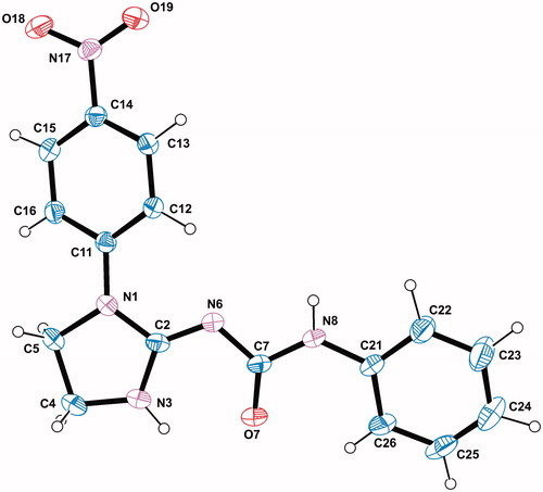
The X-ray analysis of compound 9 was performed in order to confirm the synthesis pathway, identify tautomeric form in the solid state and provide structural information for docking studies. The structure and conformation of the molecule 9 in the crystal is shown in . In the crystalline state, the molecule exists in N3-amino/N6-imino/O7-keto/N8-amino tautomeric form, as evidenced by the C7–O7 bond length of 1.2275(17) Å typical for the carbonyl group and the positions of the H atoms in the vicinity of N3 and N8 in the difference electron-density map. The bond distances and angles are in normal rangesCitation42 and are comparable to the corresponding values observed in closely related structure of 1-(3-chlorophenyl)-3-(1-p-tolylimidazolidin-2-ylidene)ureaCitation43. The tautomeric form and the nearly planar conformation of the molecule as a whole with the torsion angles C12–C11–N1–C2, N1–C2–N6–C7 and C22–C21–N8–C7 of −23.21(19), −178.06(12) and −175.04(15)°, respectively, are stabilized by the strong N3–H3 … O7 [N3–H3 = 0.84(3), H3 ⋯ O7 = 2.03(3), N3 ⋯ O7 = 2.608(2) Å and N3–H3 ⋯ O7 = 125(2)°], C12–H12 ⋯ N6 [C12–H12 = 0.93, H23 ⋯ N6 = 2.31, C12 ⋯ N6 = 2.8863(18) Å and C12–H12 ⋯ N6 = 120°] and C26–H26 ⋯ O7 [C26–H26 = 0.93, H26 ⋯ O7 = 2.25, C26 ⋯ O7 = 2.848(2) Å and C26–H26 ⋯ O7 = 122°] intramolecular hydrogen bonds.
Figure 1. A view of the X-ray molecular structure of 9 with the atomic labelling scheme. Non H-atoms are represented by displacement ellipsoids of 30% probability.
Biological activity
Cell viability assessment
Newly synthesized compounds were subjected to MTT bioassay to determine whether cancer and normal cells responded with growth inhibition. Cancer and normal cells were treated with increasing concentrations of indicated nitroaryl urea derivatives over a period of 72 h. In general, all of the synthesized compounds significantly reduced cell viability of cancer as well as normal cells compared with non-exposed cells in a concentration-dependent manner (). DMSO used in 0.1% (v/v) concentration as a solvent did not influence viability of analysed cancer cells, notwithstanding the inhibitory effect of the solvent was observed in the human skin fibroblasts HSF culture. The mean HSF cell viability for the solvent only was 94.26% ± 1.42 (standard error) in comparison to the viability of cells cultured with the lack of DMSO, and that difference is statistically significant (p = 0.006, Mann–Whitney test). For that reason the cells treated with DMSO in 0.1% (v/v) were used as a control group. Novel derivatives 5a, 5d, 6d, 6e and 6f showed the strongest inhibitory influence on the cell viability of laryngeal cancer cells RK33. Compounds 5a, 5b, 5d, 6d and 6e were the most potent against rhabdomyosarcoma cells TE671. The highest inhibitory effect on the cell viability was observed for compounds 5a, 5d and 6e against both RK33 and TE671 cells. Compounds 5a, 5d, 6d and also 6f produced the highest inhibitory effect on the HSF cells viability.
Table 1. Effects of the synthesized compounds on cancer and normal cells in vitro.
Next, the selectivity index (SI) was calculated (as was described in Experimental section) to determine the selective activity of investigated compounds against cancer cells over normal HSF. Two compounds, 5c and 6e, showed the highest selectivity against both cancer cell lines in comparison to antiproliferative effects exerted on normal cells. The GI50 value of the compound 6e for HSF cells was 4 and at least 8 times higher than observed for laryngeal cancer cells and rhabdomyosarcoma, respectively. Therefore compound 6e shows the highest SI value against both cancer cell types.
The considerable anticancer properties were shown for compounds 5c, 5d, 6e and 5c, 6c, 6e against RK33 and TE671 cancer cells respectively. Four urea derivatives 6b, 6c, 8 and 9 were active against TE671 cell line, nonetheless not affected on RK33 cells viability. The less active against both cancer cell lines used in this study were compounds 6g, anywise no significant influence on HSF cells viability was observed. Anti-cancer drug cisplatin (CDDP) was used as a positive control of the cell viability assessment. TE671 cancer cell line was the most sensitive against CDDP. Calculated GI50 ± SD values for TE671 and RK33 cells were 56.66 ± 1.2 µM and 83.33 ± 1.1 µM respectively demonstrating greater potency of some newly synthesized compounds on tested cell lines. Healthy human cells showed the greatest resistance against CDDP in MTT assay, the calculated GI50 value was 146.66 ± 1.1 µM. Estimated SI values for both cancer cell lines (TE671 and RK33) were 2.6 and 1.8 respectively.
CDK inhibition
Because of the antiproliferative properties of newly prepared compounds, we sought to find the molecular mechanism of this activity. Aryl substituted urea derivatives have been discovered as potent inhibitors of various kinasesCitation44–47 and sorafenib is a well-known example of a drug used for treatment of some cancersCitation48. We decided to pursue CDK inhibition because of the similarity of our compounds and those described in the literature taking urea moiety into accountCitation21,Citation49,Citation50. Moreover a link between inhibition of CDKs and antiproliferative effect has been well documentedCitation51–54. CDK2 is often deregulated in cancer cells usually by cyclin overexpression or loss of natural inhibitors and provides cells with growth advantage. We therefore assayed all prepared compounds with purified recombinant protein kinases CDK2/cyclin E and c-Abl as described previouslyCitation55. The compounds were tested in a dose-dependent manner starting from 0.1 µM up to their respective solubility limits. Results summarized in illustrate that the majority of tested compounds did not display significant activity against the two kinases, with the exception of 9 that showed modest inhibitory activity towards CDK2 with IC50 of 14.3 µM, indicating some sort of selectivity over tyrosine kinase c-Abl. This finding however suggests a different mechanism of action and prompts for investigation of another targets.
Table 2. Effect of the synthesized compounds on inhibition of protein kinases CDK2/cyclin E and c-Abl.
Molecular modelling
In order to determine the structure-activity relationship regarding the effect of the synthesized compounds on inhibition of protein kinase CDK2/cyclin E, selected molecular descriptors were calculated (). It can be concluded that the active compound 9 has smaller molecular surface area, polar surface area, volume, ovality, polarizability, molecular weight, LUMO energy and number of hydrogen bond donors than most of the inactive compounds. No trend was found for the number of hydrogen bond acceptors, HOMO energy and lipophilicity. presents the HOMO and LUMO orbitals as well as the map of the electrostatic potential onto a surface of the electron density for 9.
Figure 2. HOMO (A) and LUMO (B) orbitals for 9. (C) The map of the electrostatic potential onto a surface of the electron density for 9.
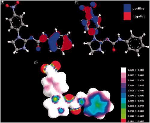
Table 3. Molecular descriptors of the investigated compounds.
Next we determined the binding mode of 9 in the active site of CDK2 using induced-fit molecular docking followed by molecular dynamics. From many available X-ray structures of CDK2 in complex with inhibitors, 3QXPCitation20 was selected due to structural similarity of 9 to the complexed inhibitor. depicts the final binding pose of 9 (A) and this pose aligned to the reference ligand (B). The binding pose of 9 was selected from the most frequent binding poses which corresponded to the binding pose of the reference inhibitor from the X-ray structure.
Figure 3. Compound 9 in stick representation with magenta carbon atoms in the binding pocket of CDK2 (protein colored with green carbon atoms) (A), 2D interaction map (B) and 9 superimposed to the reference ligand (stick representation, cyan carbon atoms, hydrogen atoms not shown in ligands for clarity; protein shown as green cartoon) (C). Hydrogen bonds shown as red dashed lines (color version available online).
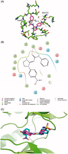
It was found that the nitro group of 9 forms water molecule mediated hydrogen bond with the side chain of Gln131. Moreover, the urea moiety of 9 forms two hydrogen bond with the main chain of Leu83. The compound 9 fits well to the CDK2 binding pocket and is additionally involved in a set of non-polar interactions with the hydrophobic residues of the active site (Ile10, Val18, Ala31, Val64, Phe80, Phe82, Leu134, Ala144). The urea moiety seems to be a key element of the system. The docking shows that a polar group substituted at N1 phenyl ring is favorable for activity. The nitro group may be exchanged into carboxylic, amide or hydroxyl group. Moreover, as there is some space near phenyl moiety substituted at urea system, some non-polar substituents, such as 2-fluoro, 2-chloro or 2-methyl groups could be favorable. These modifications will be a subject of our future studies. This inhibitor-enzyme complex was found to be stable in molecular dynamics simulations which confirm the probability of rationally selected binding mode.
Conclusion
The novel series of nitroaryl urea derivatives have been synthesized and tested for biological activity. They displayed an antiproliferative effect in the lower micromolar range. Two compounds 5d nitrophenyl (6.7 µM in RK33) and 6e nitropyridyl (6.5 µM in TE671) were the most active from the series. They contain 4-methoxybenzyl and 3,4-dichlorophenyl substituents respectively. Compound 6e proved to be the most selective towards cancer cells. All compounds have also been tested towards CDK. However, all compounds except 9 have not displayed significant activity, suggesting a different mechanism of action which will be further investigated. Compound 9 being active in a CDK2 assay has a rigid structure with urea moiety closer to nitroaryl fragment. The rest of the compounds have greater conformational flexibility together with a more distanced urea fragment. In the case of compound 8 which is similarly rigid as active compound 9, the urea feature is also moved further away from the nitroaryl fragment. This feature renders the compounds significantly less active towards CDK2. Moreover, compound 9 is characterized by decreased values of molecular descriptors which make this molecule more compact. This corresponds well with docking studies which also revealed the importance the nitro group. From the above results, it can be concluded that this scaffold may be developed into more active CDK inhibitors the future.
Declaration of interest
The authors report no declarations of interest.
This paper was developed using the equipment purchased within the Project “The equipment of innovative laboratories doing research on new medicines used in the therapy of civilization and neoplastic diseases” within the Operational Program Development of Eastern Poland 2007–2013. Priority Axis I Modern Economy, Operations I.3 Innovation Promotion. Support of Grant for Young Scientists MNmb 44 is also greatly appreciated. V.K. acknowledges financial support from the Czech Science Foundation (project 15-15264S).
References
- WHO. 2014. Available from: http://globocan.iarc.fr/Pages/fact_sheets_cancer.aspx [last accessed Nov 2014]
- Cohen P. Protein kinases – the major drug targets of the twenty-first century? Nat Rev Drug Discov 2002;1:309–15
- Melnikova I, Golden J. Targeting protein kinases. Nat Rev Drug Discov 2004;3:993–4
- Adams JA. Kinetic and catalytic mechanisms of protein kinases. Chem Rev 2001;101:2271–90
- Giamas G, Stebbing J, Vorgias CE, Knippschild U. Protein kinases as targets for cancer treatment. Pharmacogenomics 2007;8:1005–16
- Mendel DB, Laird AD, Xin X, et al. In vivo antitumor activity of SU11248, a novel tyrosine kinase inhibitor targeting vascular endothelial growth factor and platelet-derived growth factor receptors: determination of a pharmacokinetic/pharmacodynamic relationship. Clin Cancer Res 2003;9:327–37
- Druker BJ, Tamura S, Buchdunger E, et al. Effects of a selective inhibitor of the Abl tyrosine kinase on the growth of Bcr–Abl positive cells. Nat Med 1996;2:561–6
- Barker AJ, Gibson KH, Grundy W, et al. Studies leading to the identification of ZD1839 (iressa™): an orally active, selective epidermal growth factor receptor tyrosine kinase inhibitor targeted to the treatment of cancer. Bioorg Med Chem Lett 2001;11:1911–14
- Das J, Chen P, Norris D, et al. 2-Aminothiazole as a novel kinase inhibitor template. Structure–activity relationship studies toward the discovery of N-(2-Chloro-6-methylphenyl)-2-[[6-[4-(2-hydroxyethyl)-1-piperazinyl)]-2-methyl-4-pyrimidinyl]amino)]-1,3-thiazole-5-carboxamide (Dasatinib, BMS-354825) as a potent pan-Src kinase inhibitor. J Med Chem 2006;49:6819–32
- Hunter T, Pines J. Cyclins and cancer II: cyclin D and CDK inhibitors come of age. Cell 1994;79:573–82
- Krystof V, Uldrijan S. Cyclin-dependent kinase inhibitors as anticancer drugs. Curr Drug Targets 2010;11:291–302
- Malumbres M. Cyclin-dependent kinases. Genome Biol 2014;15:122
- Malumbres M, Harlow E, Hunt T, et al. Cyclin-dependent kinases: a family portrait. Nat Cell Biol 2009;11:1275–6
- Malumbres M, Barbacid M. Milestones in cell division: to cycle or not to cycle: a critical decision in cancer. Nat Rev Cancer 2001;1:222–31
- Cicenas J, Valius M. The CDK inhibitors in cancer research and therapy. J Cancer Res Clin Oncol 2011;137:1409–18
- Blachly JS, Byrd JC. Emerging drug profile: cyclin-dependent kinase inhibitors. Leukemia Lymphoma 2013;54:2133–43
- Havlíček L, Hanuš J, Veselý J, et al. Cytokinin-derived cyclin-dependent kinase inhibitors: synthesis and cdc2 inhibitory activity of olomoucine and related compounds. J Med Chem 1997;40:408–12
- Fry DW, Harvey PJ, Keller PR, et al. Specific inhibition of cyclin-dependent kinase 4/6 by PD 0332991 and associated antitumor activity in human tumor xenografts. Mol Cancer Therap 2004;3:1427–38
- Montagnoli A, Valsasina B, Croci V, et al. A Cdc7 kinase inhibitor restricts initiation of DNA replication and has antitumor activity. Nat Chem Biol 2008;4:357–65
- Schonbrunn E, Betzi S, Alam R, et al. Development of highly potent and selective diaminothiazole inhibitors of cyclin-dependent kinases. J Med Chem 2013;56:3768–82
- Honma T, Hayashi K, Aoyama T, et al. Structure-based generation of a new class of potent Cdk4 inhibitors: new de novo design strategy and library design. J Med Chem 2001;44:4615–27
- Pedersen DS, Rosenbohm C. Dry column vacuum chromatography. Synthesis 2001;16:2431–4
- Allali M, Benoist E, Habbadi N, et al. Design and synthesis of new ethylenediamine or propylenediamine diacetic acid derivatives for Re(I) organometallic chemistry. Tetrahedron 2004;60:1167–74
- Bremer O. Über die Synthese nitrierter 2,3-dihydro-pyrimidazole. Justus Liebigs Annalen der Chemie 1936;521:286–97
- Rzeski W, Paduch R, Klatka J, et al. Establishment and preliminary characterization of two cell lines derived from larynx carcinoma. Folia Histochem Cytobiol 2002;40:195–6
- SADABS. 2004/1 ed. Madison (WI): Bruker AXS Inc.; 2005
- Sheldrick G. A short history of SHELX. Acta Crystallogr Sect A 2008;64:112–22
- Farrugia L. WinGX and ORTEP for Windows: an update. J Appl Crystallogr 2012;45:849–54
- LigPrep. Schrödinger. 2.4 ed. New York: LLC; 2010
- Epik. Schrödinger. 2.1 ed. New York: LLC; 2010
- Becke AD. Density–functional thermochemistry. III: the role of exact exchange. J Chem Phys 1993;98:5648–52
- Lee C, Yang W, Parr R. Development of the Colle–Salvetti correlation-energy formula into a functional of the electron density. Phys Rev B 1988;37:785–9
- Frisch MJ, Trucks GW, Schlegel HB, et al. Gaussian 09. Wallingford (CT): Gaussian, Inc.; 2009
- Pedretti A, Villa L, Vistoli G. VEGA – an open platform to develop chemo-bio-informatics applications, using plug-in architecture and script programming. J Comput-Aided Mol Des 2004;18:167–73
- Discovery Studio. 3.5 ed: Biovia. Available from: http://accelrys.com/products/discovery-studio/ [last accessed Nov 2014]
- ACDLabs. Available from: http://www.acdlabs.com/ [last accessed Nov 2014]
- Bowers KJ, Chow E, Xu H, et al. Scalable algorithms for molecular dynamics simulations on commodity clusters. Proceedings of the 2006 ACM/IEEE conference on Supercomputing. Tampa (FL): ACM; 2006. p. 84
- ArgusLab. Available from: http://www.arguslab.com/arguslab.com/ArgusLab.html [last accessed Nov 2014]
- The PyMOL Molecular Graphics System. Schrödinger. 0.99 ed: LLC. Available from: https://www.pymol.org/pymol [last accessed Nov 2014]
- Duspara PA, Islam MS, Lough AJ, Batey RA. Synthesis and reactivity of N-alkyl carbamoylimidazoles: development of N-methyl carbamoylimidazole as a methyl isocyanate equivalent. J Org Chem 2012;77:10362–8
- Dobrowolski MA, Cyrański MK, Pisklak M, Wawer I, Matosiuk D. Structural studies of 1-aryl-2-aminoimidazolinium bromides: focus on tautomer preference of the 2-aminoimidazoline moiety in the solid state. Pol J Chem 2007;81:1037–48
- Allen FH, Kennard O, Watson DG, et al. Tables of bond lengths determined by X-ray and neutron diffraction. Part 1. Bond lengths in organic compounds. J Chem Soc Perkin Trans 2 1987;12:S1–19
- Wysocki W, Matosiuk D, Karczmarzyk Z, et al. 1-(3-Chlorophenyl)-3-(1-p-tolylimidazolidin-2-ylidene)urea. Acta Crystallogr Sect E 2009;65:o40
- Wang GT, Li G, Mantei RA, et al. 1-(5-Chloro-2-alkoxyphenyl)-3-(5-cyano-pyrazi-2-yl)ureas as potent and selective inhibitors of Chk1 kinase: synthesis, preliminary SAR, and biological activities. J Med Chem 2005;48:3118–21
- Mitchell SA, Danca MD, Blomgren PA, et al. Imidazo[1,2-a]pyrazine diaryl ureas: inhibitors of the receptor tyrosine kinase EphB4. Bioorg Med Chem Lett 2009;19:6991–5
- Chen Z, Venkatesan AM, Dos Santos O, et al. Stereoselective synthesis of an active metabolite of the potent PI3 kinase inhibitor PKI-179. J Org Chem 2010;75:1643–51
- Yin Y, Lin L, Ruiz C, et al. Synthesis and biological evaluation of urea derivatives as highly potent and selective rho kinase inhibitors. J Med Chem 2013;56:3568–81
- Hotte SJ, Hirte HW. BAY 43-9006: early clinical data in patients with advanced solid malignancies. Curr Pharm Design 2002;8:2249–53
- Yue EW, DiMeo SV, Higley CA, et al. Synthesis and evaluation of indenopyrazoles as cyclin-dependent kinase inhibitors. Part 4: heterocycles at C3. Bioorg Med Chem Lett 2004;14:343–6
- Honma T, Yoshizumi T, Hashimoto N, et al. A novel approach for the development of selective Cdk4 inhibitors: Library design based on locations of Cdk4 specific amino acid residues. J Med Chem 2001;44:4628–40
- Vermeulen K, Strnad M, Kryštof V, et al. Antiproliferative effect of plant cytokinin analogues with an inhibitory activity on cyclin-dependent kinases. Leukemia 2002;16:299–305
- Gray NS, Wodicka L, Thunnissen A-MWH, et al. Exploiting chemical libraries, structure, and genomics in the search for kinase inhibitors. Science 1998;281:533–8
- Wang S, Meades C, Wood G, et al. 2-Anilino-4-(thiazol-5-yl)pyrimidine CDK inhibitors: synthesis, SAR analysis, X-ray crystallography, and biological activity. J Med Chem 2004;47:1662–75
- Chu X-J, DePinto W, Bartkovitz D, et al. Discovery of [4-Amino-2-(1-methanesulfonylpiperidin-4-ylamino)pyrimidin-5-yl](2,3-difluoro-6-methoxyphenyl)methanone (R547), a potent and selective cyclin-dependent kinase inhibitor with significant in vivo antitumor activity. J Med Chem 2006;49:6549–60
- Kryštof V, Cankař P, Fryšová I, et al. 4-Arylazo-3,5-diamino-1H-pyrazole CDK inhibitors: SAR study, crystal structure in complex with CDK2, selectivity, and cellular effects. J Med Chem 2006;49:6500–9