
Abstract
A number of immunomodulatory therapeutics increase the risk of disease associated with latent herpesviruses such as cytomegalovirus (CMV) and Epstein-Barr virus (EBV), a member of the lymphocryptovirus (LCV) family that infects humans. The diseases associated with loss of immunity to these viruses can have major impacts on patients as well as on the commercial viability of the immunomodulatory therapeutics. In an effort to develop non-clinical methods for measuring effects on anti-viral immunity, we have developed an interferon (IFN)-γ enzyme-linked immunosorbent spot (ELISPOT) assay to quantify the number of CMV or LCV-reactive T-cells in peripheral blood of cynomolgus macaques. After optimization of various parameters, the IFN-γ ELISPOT assay was characterized for specificity, intra-assay, monkey-to-monkey, and longitudinal variability and sensitivity to immunosuppression. The results show that nearly all animals have detectable responses against both CMV and LCV and responses were derived from T-cells specific to the virus of interest. Analyses of variability show assay reproducibility (≤23% CV), and that variability over time in anti-viral responses in individual animals (larger for LCV than for CMV) was ∼2-fold in most animals over a 3-month time period, which is predicted to allow for detection of drug-induced changes when using group sizes typical of non-clinical studies. In addition, the IFN-γ ELISPOT assay was capable of detecting decreases in the numbers of CMV and LCV reactive T-cells induced by immunosuppressive drugs in vitro. This assay may allow for non-clinical assessment of the effects of immunomodulatory therapeutics on anti-viral T-cell immunity in monkeys, and may help determine if therapeutics increase the risk of reactivating latent viral infections.
Introduction
A number of immunosuppressive therapeutics are associated with increased rates of infections (Brennan et al., Citation2010; Major, Citation2010; Rychly & DiPiro, Citation2005), which can limit or prevent the use of these therapeutics and, therefore, limit the possible benefit to patients. A major challenge to the development of immunomodulatory therapeutics is that their liability to increase infection risk is often not known until large numbers of patients have been exposed in the clinic. Non-clinical methods for detecting increased risk of infection may facilitate the development of immunomodulatory therapeutics.
When immune responses are compromised, latent or persistent viruses may replicate and cause disease. Herpesviruses are among the most common latent infections and produce diseases with immunosuppression as observed in immunosuppressed transplant patients or HIV+ patients. Examples of well-studied herpesviruses in humans are the β herpesvirus cytomegalovirus (CMV) and the γ herpesvirus Epstein-Barr virus (EBV), a member of the lymphocryptovirus (LCV) genus. Following infection of normal immunocompetent hosts, replication and spread of the viruses is controlled by the immune response of the host, after which the viruses exist in a latent form that can periodically reactivate and undergo viral replication leading to viral shedding. The immune system must actively control CMV and EBV during primary infection as well as throughout latency, and T-cells appear to be the most critical cell types exerting this immune control (Hislop et al., Citation2007; Waller et al., Citation2008).
In immunocompetent hosts, infection with CMV or EBV/LCV typically causes no overt disease. However, CMV and LCV (EBV in humans) both cause serious disease under conditions of strong immunosuppression, including HIV/AIDS, genetic deficiencies affecting T-cells and immunosuppression with T-cell targeting therapeutics. In the case of EBV, patients receiving immunosuppression can develop post-transplant lymphoproliferative disease (PTLD) or lymphoma because the loss of immune control allows for viral transformation and outgrowth of latently infected B-cells (Gottschalk et al., Citation2005; Haque et al., Citation2002; Smets & Sokal, Citation2002). CMV also commonly causes disease in transplant patients that can affect multiple organs and include colitis, pneumonitis, hepatitis, and retinitis (Fishman, Citation2007; Kotton, Citation2010; Mori & Kato, Citation2010; Nashan et al., Citation2012; Sagedal et al., Citation2004; Steininger, Citation2007; Vallejo et al., Citation2011). Both viruses can cause disease in other patient populations with immunosuppression, such as those with HIV/AIDS (Steininger, Citation2007; van Baarle et al., Citation2001) or those receiving immunosuppressive therapies for autoimmune conditions like lupus, rheumatoid arthritis, Crohn’s disease, and psoriasis (Afif & Loftus, Citation2009; Gentile & Foa, Citation2011; Woodrick & Ruderman, Citation2011). As a result, both CMV and EBV are a major concern in clinical situations involving immunosuppression.
CMV and LCV are present not only in humans but in other non-human primates (NHP). Macaques harbour CMV and LCV that are similar to their human CMV and EBV counterparts in terms of overall genetic organization, tropism, disease caused during immunosuppression and the immune cells important for control of the viruses (Carville & Mansfield, Citation2008; Moghaddam et al., Citation1997; Rivailler et al., Citation2004; Yue & Barry, Citation2008). In particular, T-cells are critical for control of both CMV and LCV, and depletion of T-cells due to SIV infection or by T-cell targeting therapeutics leads to loss of immune control and overt disease in macaques (Haustein et al., Citation2008; Kaur et al., Citation2002, Citation2003; Rivailler et al., Citation2004; Schmidtko et al., Citation2002). Because T-cells are critical for control of CMV and LCV in both humans and macaques, the macaque may serve as a useful animal model for determining the ability of a therapeutic to impair anti-viral T-cell immunity and, in turn, to increase the risk of virus-associated disease.
The overall objective of these studies was to generate methods for predicting viral disease liability of therapeutics in the non-clinical stage of drug development. It is important to have such methods available for NHP because many immunomodulatory therapeutics are antibodies or antibody-derived molecules that do not cross-react with species other than NHP and because viruses similar to human CMV and EBV exist in NHP. With this in mind, we have developed an interferon (IFN)-γ ELISPOT assay for enumerating memory T-cells responsive to CMV and LCV in cynomolgus macaques. The IFN-γ ELISPOT was chosen as it can detect low frequencies of antigen-specific T-cells; is relatively simple to perform; and does not have to be performed at the time/place of blood collection (Kamperschroer et al., Citation2012; Kaur et al., Citation2002; McCutcheon et al., Citation1997). This assay should provide a non-clinical tool for determining the effects of therapeutics on herpesvirus-specific T-cells. The ability to monitor anti-viral T-cell responses and, by inference, immune control over herpesviruses, may aid in our ability to determine infection risk.
Materials and methods
Animals
Mauritius-origin adult cynomolgus macaques (Macaca fascicularis), obtained from Charles River Laboratories (Houston, TX) and housed at Pfizer’s facilities located in Groton, were used for all experiments unless indicated otherwise. For specificity testing, control rhesus macaques (Macaca mulatta) blood samples were used. Samples were obtained from CMV- and LCV-seronegative rhesus monkeys housed in a specific pathogen-free colony or from CMV- and LCV-seropositive rhesus monkeys housed in a conventional colony at the New England Regional Primate Research Center (Southborough, MA). Viral status of rhesus monkeys was routinely determined by ELISA and in some cases was confirmed by polymerase chain reaction (PCR). All procedures were conducted in accordance with the guidelines of the National Institutes of Health Guide for the Care and Use of Laboratory Animals.
Antigens
CMV antigen consisted of a freeze-thaw/sonicated lysate prepared from MRC-5 cells infected with rhesus monkey CMV strain 68-1 (Oregon Health Sciences University Virology Core Facility, Beaverton, OR). Dose response curves were performed with each lysate preparation to determine the amount to use in the IFN-γ ELISPOT assay.
LCV antigen consisted of a freeze-thaw/sonicated lysate prepared from cynomolgus monkey LCV-transformed lymphoblastic cell lines (LCL). The LCV-transformed LCL were expanded from long-term cultures of normal healthy cynomolgus peripheral blood mononuclear cells (PBMC) grown in the presence of cyclosporine. The presence of LCV genome and protein expression of LCV latent and lytic phase proteins were confirmed (refer to supplemental data section for additional details). Lysates were prepared from non-treated LCL or LCL treated with 20 ng phorbol-12-myristate 13-acetate (PMA)/ml (Sigma, St. Louis, MO) + 3 mM sodium butyrate (NaB) (Sigma) for 72 h to enhance LCV gene expression (Gradoville et al., Citation2002). Lot-specific titers for use in the IFN-γ ELISPOT assay were determined from dose-response curves; both LCV seronegative and seropositive PBMC were routinely tested to ensure specificity of responses.
Preparation of PBMC
Blood was collected directly into CPT Vacutainer tubes (BD, Franklin Lakes, NJ) and isolated by density gradient sedimentation according to manufacturer recommendations, with minor modification. Briefly, blood samples were centrifuged at 1600 × g for 40 min. The resulting mononuclear cell fraction was washed once with phosphate-buffered saline (PBS, pH 7.4), followed by lysis of red blood cells using ACK Lysing Buffer (Invitrogen, Grand Island, NY) for 5 min at room temperature (RT). Resulting PBMC were cryopreserved at 107 cells/ml in Cell Recovery Medium (Invitrogen), placed in a Nalgene Cryo 1 °C Freezing Container (Nalge Co., Rochester, NY) at −80 °C for ≥24 h prior to transfer to liquid nitrogen for long-term storage.
IFN-γ ELISPOT
The frequencies of IFN-γ-producing cells were measured using a ELISpotPLUS kit for monkey IFN-γ from MABTECH (Nacka Strand, Sweden). Briefly, polyvinylidene difluoride (PVDF) membrane plates pre-coated with anti-human IFN-γ monoclonal antibody (mAb) GZ-4 were washed with PBS and blocked with the same medium used for cell suspensions for ≥30 min at RT. Thawed PBMC were suspended in RPMI medium containing 5% fetal bovine serum (FBS), 2 mM L-glutamine, 0.1 mM MEM-non-essential amino acids, 1 mM MEM-sodium pyruvate, and 10 mM HEPES (R-5 medium). FBS was purchased from Hyclone (Logan, UT); all other media and supplements were purchased from Invitrogen. Serial dilutions of PBMC were stimulated with R-5 medium alone, predetermined amounts of CMV and LCV lysate or 1:2000 dilution of the anti-CD3 (αCD3) antibody provided in the IFN-γ ELISPOT kit. Following a 48 h incubation at 37 °C, 5% CO2, the plates were washed 5-times with PBS using a plate washer (Biotek Instruments, Winooski, VT) and incubated with biotinylated anti-human IFN-γ mAb 7-B6-1 at 1 µg/ml in PBS containing 0.5% FBS (blocking buffer) for 2 h at RT. Plates were washed 5-times with PBS and incubated with streptavidin-horseradish peroxidase solution diluted 1:1000 in blocking buffer for 1 h at RT. Plates were washed as before, followed by the addition of kit-provided tetramethylbenzidine (TMB) substrate solution until distinct spots emerged. Color development was stopped with extensive washing with tap water and plates were allowed to completely dry before analysis. Resulting spots were counted and analyzed with an automated Immunospot Reader from Cellular Technology Ltd (CTL, Shaker Heights, OH) using Image Acquisition software (version 4.5) and Immunospot software (version 5.0) (CTL). Data were expressed as mean number of spot-forming cells (SFC)/106 PBMC, calculated by subtracting the mean of SFC obtained in unstimulated media control wells from the mean SFC of the antigen-stimulated wells at each cell concentration. The mean SFC responses at each dilution of cells were normalized to SFC/106 PBMC and averaged.
T-cell depletion experiments
T-cells (CD3+) were depleted from PBMC using sheep anti-mouse IgG Dynabeads (Invitrogen) according to manufacturer recommendations. Briefly, thawed PBMC were suspended at 107/ml in cold PBS/0.1% BSA (Buffer) and incubated with 1 µg purified αCD3 antibody (clone SP34-2, BD) or isotype matched control antibody (clone MOPC-21, BD) per 106 PBMC in cold buffer for 1 h at 4 °C. Antibody-coated cells were washed once with cold buffer and combined with pre-washed Dynabeads at a final concentration of 4 beads/target cell. After 1 h incubation at 4 °C, antibody-bead coated cells were depleted using a magnet. Proportions of CD3+ cells in the unfractionated and CD3-depleted PBMC were determined by flow cytometry after staining with a non-competing αCD3 antibody (clone 10D12 Miltenyi Biotech, Bergish Gladbach, Germany). The CD3-depleted samples were tested in the IFN-γ ELISPOT assay at a cell concentration adjusted to account for loss of CD3+ T-cells, such that equal numbers of non-CD3+ cells were compared in the assay.
Assay variability in the IFN-γ ELISPOT
Intra-assay precision was determined from two independent assays. Forty-eight replicates of media control and αCD3 antibody stimulations were assayed on each plate. Precision was calculated by averaging the daily coefficient of variance (CV) and was expressed as % CV.
Inter-assay precision was calculated from independent runs of IFN-γ ELISPOT results generated using the same stock of frozen PBMC tested on separate days using the same batch of CMV and LCV antigen for stimulations. The average SFC/106 PBMC from each day was used to calculate the mean, standard deviation, and coefficient of variation.
Longitudinal variability of monkey T-cell responses
PBMC from five male and five female untreated monkeys were bled every 2 weeks for a period spanning 3 months and PBMC from each time point were frozen. All of the samples collected at the different time points were analyzed on the same day for IFN-γ ELISPOT responses to CMV, LCV, or αCD3 antibody. Using longitudinal ELISPOT data, an analysis of statistical power was performed according to the formula , where
and
are variability estimates for day and technical replicate, respectively, and
is the number of technical replicates (
= 2 for duplicates and 3 for triplicates, etc.). This analysis assumes a paradigm in which individual animals will be assessed relative to their own baseline values prior to administration of a given test article. A power analysis based upon paired means and the above variability estimate was carried out for each antigen separately.
Sensitivity to immune suppression
To determine the ability of the IFN-γ ELISPOT assay to detect immunosuppression, known immunosuppressive compounds were added in vitro during the incubation period for the assay. Stock solutions of compounds were prepared in manufacturer recommended solvent. Tacrolimus (Sigma) was prepared in DMSO, dexamethasone (Sigma) and mycophenolic acid (Sigma) were prepared in methanol, and Cyclosporine A (Novartis, Basel, Switzerland) was purchased in solution from Northeast Medical products (Old Saybrook, CT). Subsequent dilutions of each compound were made in R-5 medium and added to PBMC for 1 h at 37 °C, 5% CO2 prior to stimulating the PBMC with media alone, CMV, LCV, or αCD3 antibody for 48 h in the IFN-γ ELISPOT assay.
Results
Specificity of cellular responses to CMV and LCV as measured in the IFN-γ ELISPOT
To allow for monitoring of memory T-cell responses specific for CMV and LCV, an IFN-γ ELISPOT assay was developed for use with cynomolgus macaque PBMC. To determine whether responses measured in the IFN-γ ELISPOT assay are indeed antigen-specific, PBMC from seropositive and seronegative control rhesus monkeys were evaluated (seronegative and seropositive cynomolgus monkeys are not available). As shown in , in the absence of antigen, few if any IFN-γ spot-forming cells (SFC) were observed; however, stimulation with relevant viral antigen resulted in a clear induction of IFN-γ SFC in seropositive animals but not naïve animals. αCD3 antibody-induced responses were present in all animals (data not shown). Therefore, the anti-viral responses measured in the ELISPOT assay were antigen-specific.
Figure 1. Specificity of immune responses to CMV and LCV antigens as measured by IFN-γ ELISPOT. PBMC were stimulated with media alone, CMV antigen, LCV antigen, or stimulatory anti-CD3 (αCD3) antibody and IFN-γ producing cells were enumerated by ELISPOT assay. (a) PBMC from CMV and LCV seropositive (+) versus seronegative (−) rhesus monkeys were used as specificity controls for responses following stimulation with the relevant antigen. Images showing representative ELISPOT wells are shown. (b) T-cell contribution to IFN-γ production was determined using PBMC from cynomolgus monkeys that were T-cell depleted with αCD3 antibody (αCD3 Depletion) or mock depleted with an isotype matched control antibody (Mock Depletion) prior to seeding in the ELISPOT assay. PBMC seeding density was adjusted to account for CD3+ T-cell loss, so that equivalent numbers of non-T-cells were compared. The cells were stimulated with CMV, LCV, or αCD3 antibody. The numbers of spot-forming cells (SFC)/106 PBMC are shown. Data shown are representative of two independent experiments.
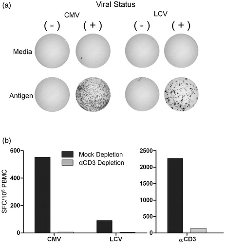
To determine whether antigen-induced IFN-γ SFC are T-cells and not other cell types (such as NK cells), PBMC from cynomolgus monkeys were depleted of T-cells prior to measuring anti-viral responses in the ELISPOT assay. As shown in , mock-depleted samples to which an isotype-matched control antibody was added demonstrate the presence of IFN-γ-secreting cells in response to stimulation with both CMV and LCV lysate antigens, as well as to αCD3 antibody. Following the removal of T-cells from total PBMC, CMV-specific responses decreased by 99%, LCV-specific responses decreased by 96%, and αCD3 antibody stimulated responses decreased by 94%, indicating that T-cells are responsible for the IFN-γ production detected in the ELISPOT assay. A small number of CD3+ T-cells remained after the depletion process (<1%; data not shown), which may at least partially account for the residual responses.
Optimization of the LCV antigen used in the IFN-γ ELISPOT assay
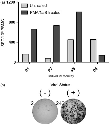
Preliminary studies demonstrated that LCV-specific responses were relatively weak, with SFC numbers often near those detected in media control samples. Therefore, additional experiments focused on increasing the immunogenicity of the LCV antigen preparation to increase LCV-specific T-cell responses.
Although the LCL used to generate LCV antigen preparation expresses both latent and lytic LCV proteins (supplemental data Figure 1), experiments were conducted to increase the expression of lytic phase proteins by LCL to enhance the T-cell responses. It has been reported that latently infected LCL can be driven to enter into the lytic phase of the EBV life cycle by exposure to PMA, a protein kinase C agonist, and sodium butyrate (NaB), a histone deacetylase inhibitor (Bhaduri-McIntosh & Miller, Citation2006; Gradoville et al., Citation2002), so protein lysates were prepared from LCL that were either treated with PMA + NaB or left untreated. In three out of four animals tested, a higher frequency of LCV-specific cells was detected when lysate from PMA/NaB-treated LCL was used as the source of LCV antigen (). Consequently, experiments performed subsequent to this finding used LCV lysate from PMA and NaB-treated LCL. To ensure that the PMA/NaB treatment did not lead to non-specific responses, each batch of LCV antigen was tested to determine the concentration of lysate which yielded no SFC response above unstimulated controls in samples from LCV seronegative animals while retaining positive responses in samples from LCV seropositive animals ().
Figure 2. LCV-specific responses using LCV antigen generated from LCL pre-treated with PMA and sodium butyrate. PBMC were stimulated with media alone or LCV antigen generated from either non-treated LCL (Untreated) or LCL treated with 20 ng PMA/ml and 3 mM sodium butyrate for 72 h (PMA/NaB treated), and then IFN-γ-secreting cells were assessed by ELISPOT assay. (a) T-cell responses of four individual animals stimulated with untreated or PMA/NaB-treated LCL are shown. (b) PBMC from LCV seronegative (−) and seropositive (+) rhesus monkeys were routinely tested to ensure specificity of LCV antigen responses. Data shown are representative of 11 independent experiments encompassing 12 seronegative and 15 seropositive rhesus monkeys. Numbers indicate SFC for each image.
Optimization of assay parameters in the IFN-γ ELISPOT
A number of experiments were performed to improve the signal-to-noise ratio of the IFN-γ ELISPOT assay by reducing the spontaneous IFN-γ production frequently observed in the media control wells while optimizing the signal obtained following antigenic stimulation. These results are summarized in . The findings from these experiments indicate that both fresh and frozen PBMC can be used. The choice of serum source had no significant impact, nor did resting cells prior to seeding, and a 48 h stimulation time was optimal. The assay parameter that reduced background spot formation the most was seeding density; plating PBMC at ≤300 000 cells/well yielded a low background while retaining strong antigen-specific responses in both CMV and LCV-stimulated samples (refer to supplemental Figure 2 for supporting data).
Table 1. Parameters evaluated to assess impact on assay background.
Statistical analysis of precision in the IFN-γ ELISPOT
To evaluate overall reproducibility of the IFN-γ ELISPOT assay, both intra- and inter-assay precision were measured. Intra-assay precision of 48 replicates was tested on multiple days and the average CV was 5.1% for αCD3 antibody-stimulated samples (data not shown). Inter-assay precision was determined for seven monkeys by comparing IFN-γ ELISPOT results conducted using the same frozen stock of PBMC stimulated with the same batch of CMV and LCV antigen on different days. The inter-assay precision measurements () ranged from 2.0–19.7% for CMV-specific responses and from 10.2–22.9% for LCV-specific responses.
Table 2. Inter-assay precision analysis.
Assessment of longitudinal variability in T-cell responses as measured by IFN-γ ELISPOT
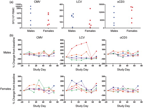
Given the small number of animals assigned per group in monkey studies, pre-study values are typically used as controls to determine whether administration of a test article alters a given end-point. It is therefore important to understand the normal degree of fluctuation in antigen-specific T-cell responses over time. To determine the longitudinal variability, PBMC from a cohort of normal cynomolgus monkeys were isolated bi-weekly for 3 months and tested in the IFN-γ ELISPOT. Results from the longitudinal sampling are depicted in . All animals responded to each of the antigens on each study day with notable differences in the starting frequency of IFN-γ-secreting cells detected at the first blood collection timepoint of the study (). Comparing each individual monkey’s T-cell response to its pre-study value, there was more variability in LCV-induced responses than there was in the CMV- and αCD3 antibody-induced responses. The percent change in the frequencies of CMV-specific IFN-γ-secreting cells ranged from −49.68% to +79.56% in males and −66.93% to +224.74% in females. Longitudinal LCV-specific responses were more variable, ranging from −95.35% to +430.77% in males and from −54.71% to +213.43% in females; αCD3 responses ranged from −67.50% to +81.97% in males and from −38.23% to +90.32% in females (). Based on these longitudinal data, a statistical sample size and power analysis of the monkey IFN-γ ELISPOT assay was performed.
Figure 3. Biological variability of monkey T-cell responses. Five male and five female untreated monkeys were bled every 2 weeks for 3 months and PBMC from each timepoint were frozen. PBMC were then stimulated with media alone, CMV antigen, LCV antigen, or αCD3 antibody and IFN-γ-producing cells were then enumerated by ELISPOT assay. (a) SFC/106 PBMC in response to CMV antigen, LCV antigen, and αCD3 antibody stimulation at outset of study. Data points represent individual animals. (b) Percent change in SFC/106 PBMC, relative to pre-treatment starting values, for T-cell responses to CMV, LCV, and αCD3. Each line represents an individual animal.
Given the underlying assumption that the responses in a given test article-treated animal will be compared to its own pre-study values, a power analysis was performed to predict the number of animals required to detect a given percent change in T-cell responses. For robust responses, such as those resulting from CMV and αCD3 antibody stimulation, an estimated sample size of n = 4 would be sufficient to detect a 50% change, whereas the sample size requirement for detecting a similar degree of change in weaker LCV responses would be n = 14 (). Given that typical non-human primate safety studies are limited in sample size for each dose group, these results suggest that the IFN-γ ELISPOT would be able to detect modest (≥50%) changes in T-cell responses against CMV or αCD3 antibody, but would detect only larger changes (∼80%) in responses against LCV, unless unusually large group sizes were used. It is worth noting that improvements in the immunogenicity of the LCV antigen source () have been made since this data set was generated, and that these improvements are expected to increase the ability to detect changes in LCV-specific responses.
Table 3. Sample size and power analysis.
Ability of the IFN-γ ELISPOT to detect in vitro immune suppression
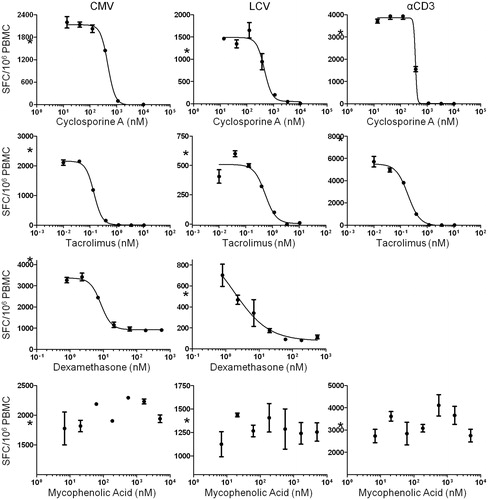
A panel of immunosuppressive drugs with different mechanisms of action was tested to determine whether the IFN-γ ELISPOT assay was capable of detecting the suppression of T-cell responses. Cyclosporine A and tacrolimus are both calcineurin inhibitors, dexamethasone (DEX) is a glucocorticoid agonist, and mycophenolic acid (MPA; active moiety of mycophenolate mofetil) is an anti-proliferative agent which inhibits de novo synthesis of purines (Lee et al., Citation2007; Lowenberg et al., Citation2007, Citation2008; Ransom, Citation1995).
Dose-dependent immune suppression was evident in the cyclosporine A-, tacrolimus-, and DEX-treated PBMC. In each case, suppression of CMV-, LCV-, and αCD3 antibody-mediated responses was apparent (). The effect of DEX on αCD3 antibody-induced responses is not represented because the number of SFC was too numerous to measure accurately. In contrast, MPA, at all concentrations tested, demonstrated no apparent suppression of IFN-γ secretion. This result was anticipated since proliferation is not required for cytokine secretion by T-cells following ex vivo stimulation (Murali-Krishna et al., Citation1998). The concentration of relevant vehicle contained in the highest concentration of drug tested, ranging from 0.02–0.06%, was always used as a control and showed no effect on cell viability. In addition, no effect on cell viability was seen at any concentration of any drug tested (data not shown). These results show that the IFN-γ ELISPOT assay is capable of detecting suppression of T-cell responses to CMV, LCV, and αCD3 antibody, with in vitro exposure to immunosuppressive drugs with different mechanisms of action.
Figure 4. Sensitivity of antigen-specific T-cell responses to in vitro immune suppression as measured by IFN-γ ELISPOT. PBMC were stimulated with media alone, CMV antigen, LCV antigen, and αCD3 antibody in the presence of serial dilutions of cyclosporine A (13.7–10 000 nM), tacrolimus (0.01–10.5 nM), dexamethasone (0.8–556 nM), and mycophenolic acid (68.6–50 000 nM), and IFN-γ-producing cells were then quantified. Each data point represents the mean SFC/106 PBMC (±SD) of duplicate wells. Each plot shows data from an individual animal. Asterisks (*) on y-axes indicate SFC obtained in vehicle control wells. Data is representative of multiple independent experiments with multiple animals (cyclosporine A, n = 5; tacrolimus, n = 5; dexamethasone, n = 5; mycophenolic acid, n = 3).
Discussion
We have developed an interferon (IFN)-γ ELISPOT assay capable of quantifying T-cells responsive to the persistent herpesviruses CMV and LCV in cynomolgus macaques. Such an assay was not previously available for cynomolgus macaques. Using the IFN-γ ELISPOT, CMV-reactive T-cells were detected at approximate frequencies in the range of ≈500–3000 SFC/106 PBMC (1/2000 to 1/300, respectively), and LCV-reactive T-cell frequencies in the range of ≈50–500 SFC/106 PBMC (1/20 000 to 1/2000, respectively). The frequencies we detect are similar to those reported in rhesus macaques for CMV (Ambagala et al., Citation2011; Kaur et al., Citation2002; Pitcher et al., Citation2002) and LCV (Fogg et al., Citation2005, Citation2006), as well as those reported in humans for CMV and EBV (Hislop et al., Citation2007; Tan et al., Citation1999; Waldrop et al., Citation1997), indicating that cynomolgus macaques are similar to rhesus macaques and humans with regards to the size of the memory T-cell pool specific for CMV and LCV.
There are both pros and cons to using the IFN-γ ELISPOT assay. The IFN-γ ELISPOT is very sensitive and able to detect antigen-specific T-cells at low frequencies (McCutcheon et al., Citation1997; Kaur et al., Citation2002). Another advantage of the assay is that it can be done using frozen PBMC (), providing more flexibility in terms of when and where the samples are analyzed, and allowing for samples collected on multiple days to be run at the same time to prevent day-to-day variability. One consideration with the ELISPOT, like any ex vivo assay, is that it can only assess T-cell responses from the tissue that is collected. For some T-cell targeting treatments, effects in the peripheral blood have been shown to reflect effects in lymphoid organs (Aarntzen et al., Citation2011), but this may not always be the case. Another potential limitation of the ELISPOT is that it does not directly provide phenotypic information about the responding cells. A bias towards CD4 T-cells would be expected using protein lysates as antigen, and this has previously been reported for responses against CMV lysate in macaques (Kamperschroer et al., Citation2012; Kaur et al., Citation2002). Depletion of CD4 or CD8 T-cells prior to conducting the IFN-γ ELISPOT confirmed that responses to CMV come almost exclusively from CD4 T-cells, and suggested that responses to LCV and αCD3 antibody stimulation involve both CD4 T-cells and CD8 T-cells (data not shown). Other T-cell subsets (e.g. effector or central memory T-cells) are difficult to evaluate using ELISPOT. As a result, we are pursuing flow cytometric analysis of intracellular cytokine staining as a complementary method for situations where detailed phenotypic information about the responding cells is important.
Another important consideration is that, during the preparation of PBMC, the test article may be washed away and will not be present during the antigen stimulation step of the IFN-γ ELISPOT. For therapeutics in which the immunomodulatory effects are rapidly reversible and removed during PBMC preparation, the IFN-γ ELISPOT assay only measures the number of memory T-cells in the PBMC with the capacity to respond to antigen. In contrast, ex vivo T-cell assays that use whole blood will detect test article effects not only on the number of memory T-cells that are capable of responding, but also will detect effects on the ability of those cells to respond to ex vivo antigen-stimulation in the presence of test article in the blood. When used in combination with assays employing whole blood, the IFN-γ ELISPOT could be useful for determining whether suppression of T-cell responses is due to a decrease in the numbers of functional memory T-cells or to ongoing suppression of their function by test article in the blood.
To date, we have used protein lysates of virus-infected cells as antigen. Although the numbers of T-cells that respond to these lysates can vary considerably among individual animals, non-responders are rare. Also, cynomolgus monkey cells responded against rhesus CMV antigen and rhesus monkey cells responded against cynomolgus LCV antigen, indicating that there is sufficient similarity in the viruses to allow for cross-species reactivity to both. These findings suggest that the CMV and LCV antigens used for this study will induce detectable responses from the vast majority of animals, such that pre-screening for responsiveness should not be required. They also suggest that both antigens could be used for studies with cynomolgus monkeys from various geographical areas, and even with rhesus macaques.
For the LCL lysate preparations, batch-to-batch differences in IFN-γ ELISPOT response have been observed. Thus, each batch must be titrated to determine the concentration that gives the greatest response without increased background. For LCV, the lysates were prepared from cynomolgus lymphoblastic cell lines (LCL) generated in our lab, which to our knowledge are not available elsewhere. In the future, LCV peptides should be considered. By using Mauritian cynomolgus macaques, a relatively small number of defined immunogenic peptides would likely induce responses in all animals because there are only six-to-seven major histocompatibility complex (MHC) haplotypes in the Mauritian population (Budde et al., Citation2010; Burwitz et al., Citation2009; O’Conor et al., Citation2007). This lack of MHC diversity would also make tools like MHC-peptide multimers a viable option, and we are currently working to identify peptide epitopes.
The assay described here was developed with the goal in mind of testing whether therapeutics under development affect T-cell control over herpesviruses like CMV and LCV. When used on in vivo studies, the intent is to test for effects by comparing T-cell responses at baseline to responses following administration of the test article. In this testing paradigm, the monkey-to-monkey variability in the IFN-γ ELISPOT responses observed is not a concern since the change from baseline SFC (not the absolute SFC number) will be used to determine if test article-related changes are observed. The extent of longitudinal variability we observed allows us to predict that moderate changes in T-cell responses against CMV and αCD3 antibody (≥50%) and larger changes in responses against LCV (≥80%) could be detected with group sizes typically used for studies with non-human primates.
The anti-viral T-cell responses measured in the IFN-γ ELISPOT were decreased with in vitro exposure to tacrolimus, cyclosporine, and dexamethasone, drugs known to suppress the functions of T-cells. Cyclosporine and tacrolimus prevent calcium-dependent signaling through the T-cell receptor by inhibiting the ability of calcineurin to activate nuclear factor of activated T-cells (NF-AT), which prevents NF-AT from activating transcription of genes (e.g., cytokines like interleukin [IL]-2) associated with T-cell activation (Lee et al., Citation2007; Malek, Citation2008). Dexamethasone is a glucocorticoid agonist that acts primarily to suppress factors that promote cytokine and chemokine production, but can also inhibit T-cell receptor signaling (Lowenberg et al., Citation2007, Citation2008). Mycophenolate mofetil (MMF) did not suppress T-cell responses in vitro when added to the ELISPOT (Ransom, Citation1995), and this result was expected because MMF has been shown to inhibit T-cell proliferation but not cytokine secretion (Kamar et al., Citation2006). However, monkeys treated with MMF may show effects in the ELISPOT ex vivo if the documented suppression of T-cell proliferation (Klupp et al., Citation2003) led to reduced numbers of antigen-specific T-cells. Effects on anti-viral T-cells in the ELISPOT assay following in vivo immunosuppressive treatment is currently being evaluated.
The ELISPOT assay described in this report allows for the monitoring of T-cell responses against CMV and LCV and may be useful for testing the effects of test articles on herpesvirus-specific T-cells in this important animal model. In vivo studies with known immunosuppressive compounds are needed to define its utility in predicting the risk of CMV or LCV reactivation.
Declaration of interest
All authors are employees of Pfizer, Inc.
Supplementary Material
Download PDF (270.6 KB)Acknowledgments
The authors would like to acknowledge Angela Carville and Keith Mansfield at the New England Primate Research Center for providing PBMC from CMV/LCV positive and negative rhesus macaques for specificity controls, Donald Siess, Andrew Sylwester, and Louis Picker for providing CMV antigen, Ellen Evans for critical review of the manuscript, and Harshan Pisharath for helpful discussion.
References
- Aarntzen, E. H., Srinivas, M., de Wilt, J. H., et al. (2011). Early identification of antigen-specific immune responses in vivo by [18F]-labeled 3′-fluoro-3′-deoxy-thymidine ([18F]FLT) PET imaging. Proc. Natl. Acad. Sci. USA 108:18396–18399
- Afif, W., and Loftus, E. V. Jr (2009). Safety profile of IBD therapeutics: infectious risks. Gastroenterol. Clin. North Am. 38:691–709
- Ambagala, A. P., Marsh, A., Chan, J., et al. (2011). Isolation and characterization of cynomolgus macaque (Macaca fascicularis) cytomegalovirus (CyCMV). Virology 412:125–135
- Bhaduri-McIntosh, S., and Miller, G. (2006). Cells lytically infected with Epstein-Barr virus are detected and separable by immunoglobulins from EBV-seropositive individuals. J. Virol. Meth. 137:103–114
- Brennan, F. R., Mroton, L. D., Spindeldreher, S., et al. (2010). Safety and immunotoxicity assessment of immunomodulatory monoclonal antibodies. MAbs 2:233–255
- Budde, M. L., Wiseman, R. W., Karl, J. A., et al. (2010). Characterization of Mauritian cynomolgus macaque major histocompatibility complex Class I haplotypes by high-resolution pyrosequencing. Immunogenetics 62:773–780
- Burwitz, B. J., Pendley, C. J., Greene, J. M., et al. (2009). Mauritian cynomolgus macaques share two exceptionally common major histocompatibility complex Class I alleles that restrict simian immunodeficiency virus-specific CD8+ T-cells. J. Virol. 83:6011–6019
- Carville, A., and Mansfield, K. G. (2008). Comparative pathobiology of macaque lymphocrypto-viruses. Comp. Med. 58:57–67
- Fishman, J. A. (2007). Infection in solid-organ transplant recipients. New Engl. J. Med. 357:2601–2614
- Fogg, M. H., Garry, D., Awad, A., et al. (2006). The BZLF1 homolog of an Epstein-Barr-related γ herpesvirus is a frequent target of the CTL response in persistently infected rhesus macaques. J. Immunol. 176:3391–3401
- Fogg, M. H., Kaur, A., Cho, Y. G., and Wang, F. (2005). The CD8+ T-cell response to an Epstein-Barr virus-related γ herpesvirus infecting rhesus macaques provides evidence for immune evasion by the EBNA-1 homologue. J. Virol. 79:12681–12691
- Gentile, G., and Foa, R. (2011). Viral infections associated with the clinical use of monoclonal antibodies. Clin. Microbiol. Infect. 17:1769–1775
- Gottschalk, S., Rooney, C. M., and Heslop, H. E. (2005). Post-transplant lymphoproliferative disorders. Annu. Rev. Med. 56:29–44
- Gradoville, L., Kwa, D., El-Guindy, A., and Miller, G. (2002). Protein kinase C-independent activation of the Epstein-Barr virus lytic cycle. J. Virol. 76:5612–5626
- Haque, T., Wilkie, G. M., Taylor, C., et al. (2002). Treatment of Epstein-Barr-virus-positive post-transplantation lymphoproliferative disease with partly HLA-matched allogeneic cytotoxic T-cells. Lancet. 360:436–442
- Haustein, S. V., Kolterman, A. J., Sunblad, J. J., et al. (2008). Non-human primate infections after organ transplantation. ILAR J. 49:209–219
- Hislop, A. D., Taylor, G. S., Sauce, D., and Rickinson, A. B. (2007). Cellular responses to viral infection in humans: lessons from Epstein-Barr virus. Annu. Rev. Immunol. 25:587–617
- Kamar, N., Glander, P., Nolting, J., et al. (2006). Effect of mycophenolate mofetil monotherapy on T-cell functions and inosine monophosphate dehydrogenase activity in patients undergoing a kidney transplantation. Transplant. Proc. 38:2292–2294
- Kamperschroer, C., Kaur, A., and Lebrec, H. (2012). A summary of meeting proceedings for ‘Measuring immune responses in non-human primates for drug development opportunities and challenges for predicting human efficacy and immunotoxicity’. J. Immunotoxicol. 9:108–120
- Kaur, A., Hale, C. L., Noren, B., et al. (2002). Decreased frequency of cytomegalovirus (CMV)-specific CD4+ T lymphocytes in simian immunodeficiency virus-infected rhesus macaques: inverse relationship with CMV viremia. J. Virol. 76:3646–3658
- Kaur, A., Kassis, N., Hale, C. L., et al. (2003). Direct relationship between suppression of virus-specific immunity and emergence of cytomegalovirus disease in simian AIDS. J. Virol. 77:5749–5758
- Klupp, J., Dambrin, C., Hibi, K., et al. (2003). Treatment by mycophenolate mofetil of advanced graft vascular disease in non-human primate recipients of orthotopic aortic allografts. Am. J. Transplant. 3:817–829
- Kotton, C. N. (2010). Management of cytomegalovirus infection in solid organ transplantation. Nat. Rev. Nephrol. 6:711–721
- Lee, C. L., Jiang, P. P., Sit, W. H., and Wan, J. M. (2007). Proteome of human T-lymphocytes with treatment of cyclosporine and polysaccharopeptide: analysis of significant proteins that manipulate T-cells proliferation and immunosuppression. Int. Immunopharmacol. 7:1311–1324
- Lowenberg, M., Stahn, C., Hommes, D. W., and Buttgereit, F. (2008). Novel insights into mechanisms of glucocorticoid action and the development of new glucocorticoid receptor ligands. Steroids 73:1025–1029
- Lowenberg, M., Verhaar, A. P., van den Brink, G. R., and Hommes, D. W. (2007). Glucocorticoid signaling: a non-genomic mechanism for T-cell immunosuppression. Trends Mol. Med. 13:158–163
- Major, E. O. (2010). Progressive multifocal leukoencephalopathy in patients on immunomodulatory therapies. Annu. Rev. Med. 61:35–47
- Malek, T. R. (2008). The biology of IL-2. Annu. Rev. Immunol. 26:453–479
- McCutcheon, M., Wehner, N., Wensky, A., et al. (1997). A sensitive ELISPOT assay to detect low-frequency human T-lymphocytes. J. Immunol. Meth. 210:149–166
- Moghaddam, A., Rosenzweig, M., Lee-Parritz, D., et al. (1997). An animal model for acute and persistent Epstein-Barr virus infection. Science 276:2030–2033
- Mori, T., and Kato, J. (2010). Cytomegalovirus infection/disease after hematopoietic stem cell transplantation. Int. J. Hematol. 91:588–695
- Murali-Krishna, K., Atman, J. D., Suresh, M., et al. (1998). Counting antigen-specific CD8 T-cells: a re-evaluation of bystander activation during viral infection. Immunity 8:177–187
- Nashan, B., Gaston, R., Emery, V., et al. (2012). Review of cytomegalovirus infection findings with mammalian target of rapamycin inhibitor-based immunosuppressive therapy in de novo renal transplant recipients. Transplantation 93:1075–1085
- O’Conor, S. L., Blasky, A. J., Pendley, C. J., et al. (2007). Comprehensive characterization of MHC Class II haplotypes in Mauritian cynomolgus macaques. Immunogenetics 59:449–462
- Pitcher, C. J., Hagen, S. I., Walker, J. M., et al. (2002). Development and homeostasis of T-cell memory in rhesus macaque. J. Immunol. 168:29–43
- Ransom, J. T. (1995). Mechanism of action of mycophenolate mofetil. Ther. Drug Monit. 17:681–684
- Rivailler, P., Carville, A., Kaur, A., et al. (2004). Experimental rhesus lymphocryptovirus infection in immunosuppressed macaques: an animal model for Epstein-Barr virus pathogenesis in the immunosuppressed host. Blood 104:1482–1489
- Rychly, D. J., and DiPiro, J. T. (2005). Infections associated with tumor necrosis factor-α antagonists. Pharmacotherapy 25:1181–1192
- Sagedal, S., Hartmann, A., Nordal, K. P., et al. (2004). Impact of early cytomegalovirus infection and disease on long-term recipient and kidney graft survival. Kidney Int. 66:329–337
- Schmidtko, J., Wang, R., Wu, C. L., et al. (2002). Post-transplant lymphoproliferative disorder associated with an Epstein-Barr-related virus in cynomolgus monkeys. Transplantation 73:1431–1439
- Smets, F., and Sokal, E. M. (2002). Epstein-Barr virus-related lymphoproliferation in children after liver transplant: role of immunity, diagnosis, and management. Pediatr. Transplant. 6:280–287
- Steininger, C. (2007). Clinical relevance of cytomegalovirus infection in patients with disorders of the immune system. Clin. Microbiol. Infect. 13:953–963
- Tan, L. C., Gudgeon, N., Annels, N. E., et al. (1999). A re-evaluation of the frequency of CD8+ T-cells specific for EBV in healthy virus carriers. J. Immunol. 162:1827–1835
- Vallejo, C., Rios, E., de la Serna, J., et al. (2011). Incidence of cytomegalovirus infection and disease in patients with lymphoproliferative disorders treated with alemtuzumab. Exp. Rev. Hematol. 4:9–16
- van Baarle, D., Hovenkamp, E., Callan, M. F., et al. (2001). Dysfunctional Epstein-Barr virus (EBV)-specific CD8+ T-lymphocytes and increased EBV load in HIV-1 infected individuals progressing to AIDS-related non-Hodgkin lymphoma. Blood 98:146–155
- Waldrop, S. L., Pitcher, C. J., Peterson, D. M., et al. (1997). Determination of antigen-specific memory/effector CD4+ T-cell frequencies by flow cytometry: evidence for a novel, antigen-specific homeostatic mechanism in HIV-associated immunodeficiency. J. Clin. Invest. 99:1739–1750
- Waller, E. C., Day, E., Sissons, J. G., and Wills, M. R. (2008). Dynamics of T-cell memory in human cytomegalovirus infection. Med. Microbiol. Immunol. 197:83–96
- Woodrick, R. S., and Ruderman, E. M. (2011). Safety of biologic therapy in rheumatoid arthritis. Nat. Rev. Rheumatol. 7:639–652
- Yue, Y., and Barry, P. A. (2008). Rhesus cytomegalovirus a non-human primate model for the study of human cytomegalovirus. Adv. Virus Res. 72:207–226