
Patil PB, Begum S, Joshi M, Kleman MI, Olausson M, Sumitran-Holgersson S. Phenotypic and in vivo functional characterization of immortalized human fetal liver cells. Scandinavian Journal of Gastroenterology 2014;49:705–714. http://dx.doi.org/10.3109/00365521.2013.830328
When the above article was first published, incorrect image files were provided for Figures 2 and 5. Please see the original and revised images below:
Figure 2:
Original: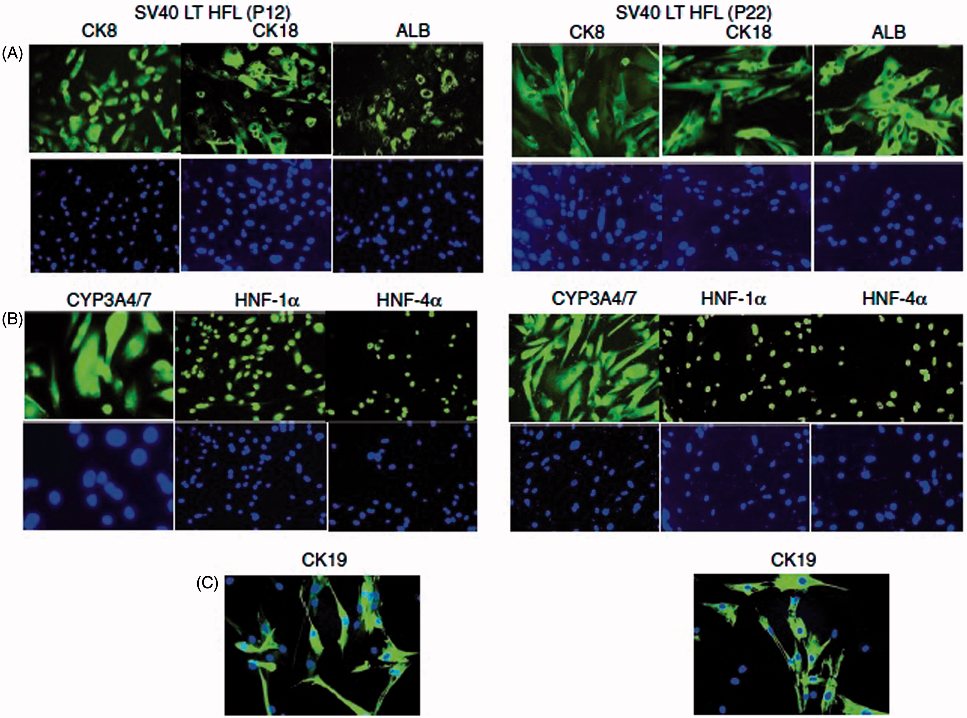
Revised: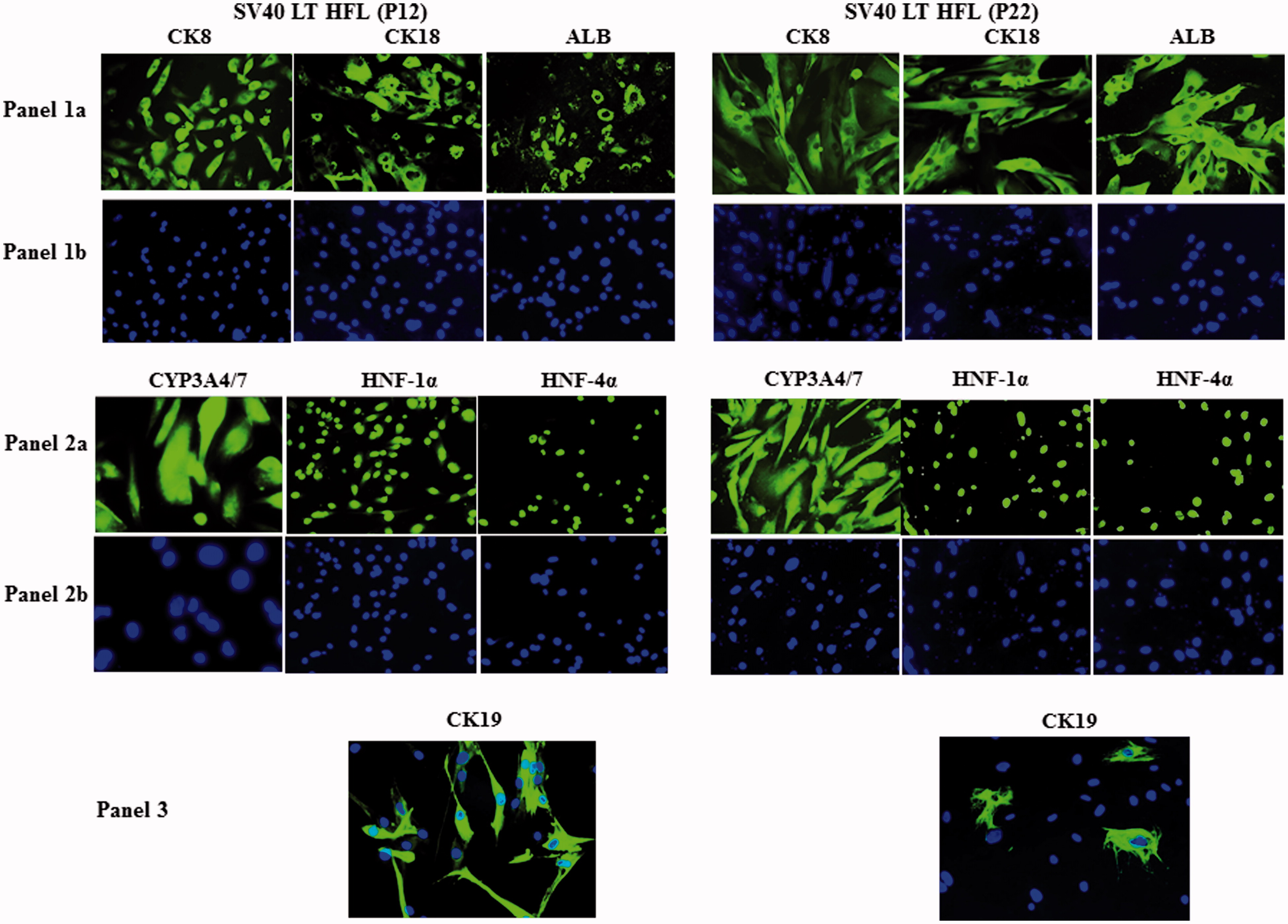
Figure caption:
Characterization of SV40LT-HFL cells. Immunofluorescent staining of SV40LT-HFL cells in passages 12 (left) and 22 (right) showing in A, from left to right, the detection of cytoplasmic staining of hepatic markers CK8, 18 and albumin. The level of expression as judged from the intensity of staining was similar in both passages. In B, the same cells are stained with the nuclear stain, DAPI. In A, from left to right, positive cytoplasmic staining of CYP3A4/7 and nuclear staining of transcription factors HNF-1a and HNF-4a are seen in all cells. In B, the same cells are stained with the nuclear stain, DAPI. In C, a small number of cells staining positive for the hepatobiliary marker CK19 was detected in both early and late passages (magnification 40×).
Figure 5:
Original: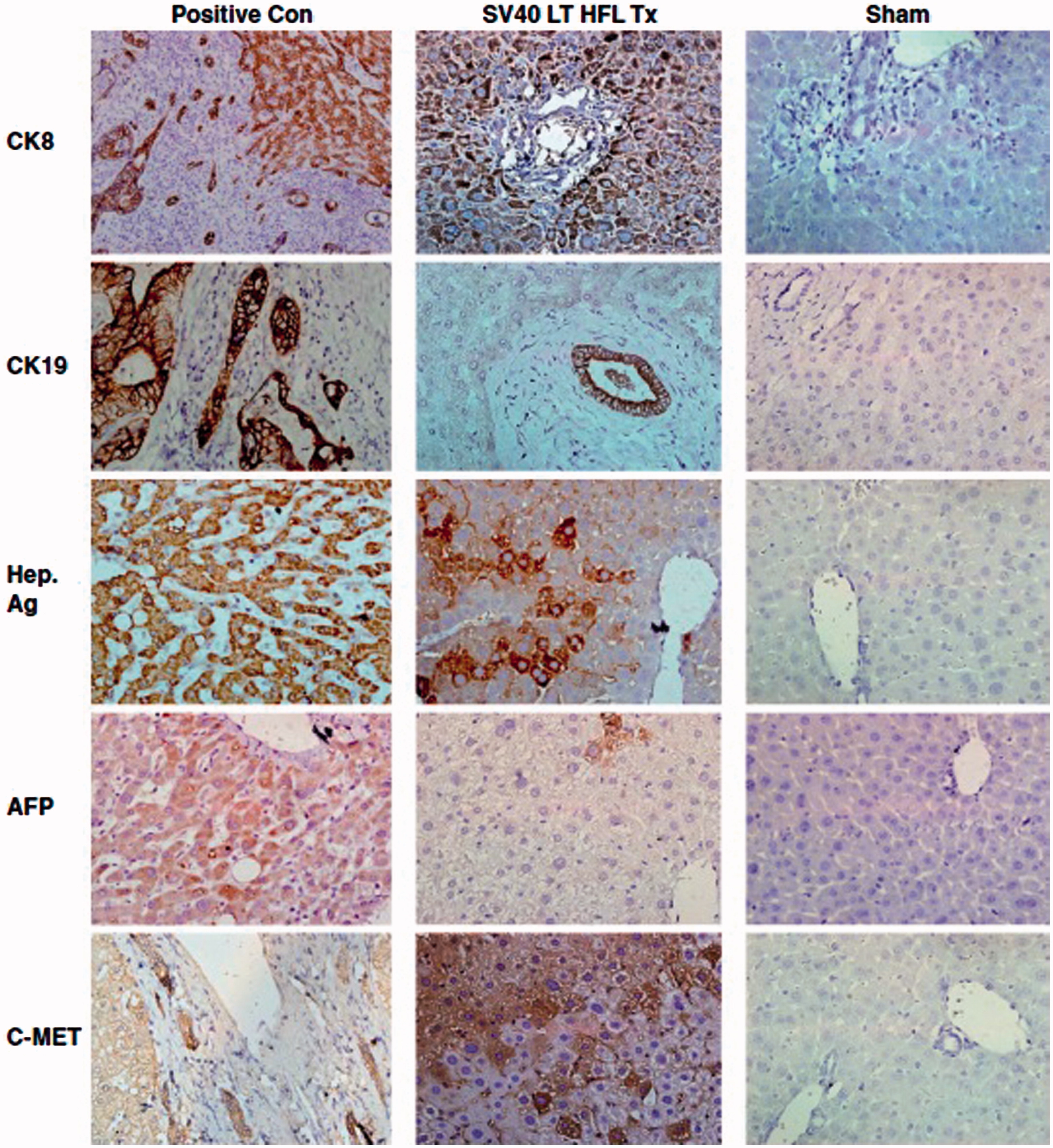
Revised: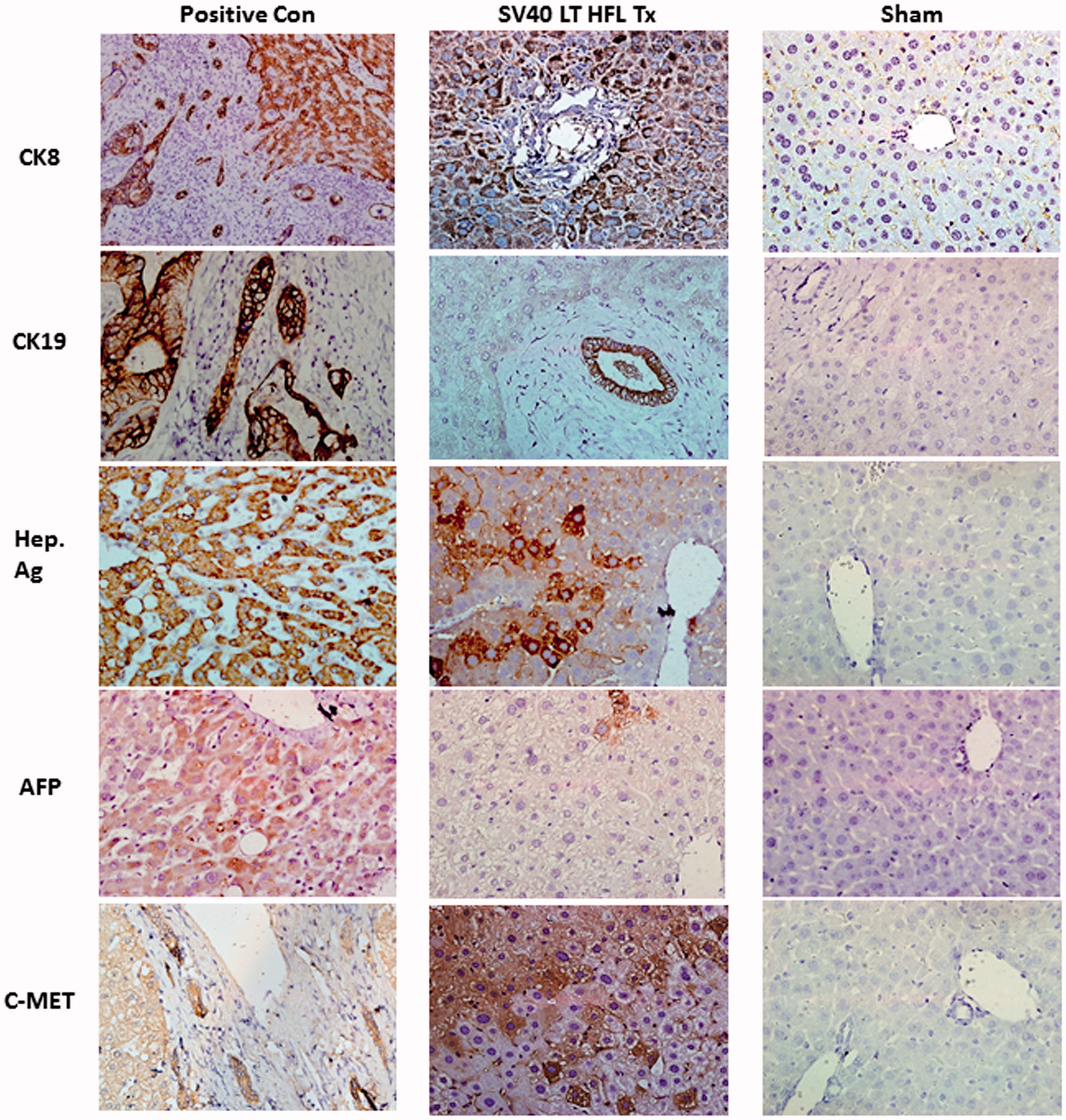
Figure caption:
Expression of human liver-specific markers in the livers of nude mice transplanted with SV40 LT HFLCs. Two-million SV40 LT HFLs were transplanted into the spleen of D-galactosamine-treated nude mice that underwent 30% partial hepatectomy at the time of transplantation. Immunohistochemistry was performed on fresh frozen liver sections of transplanted animals, and small clusters of human CK8-, CK19-, hepatocyte-specific antigen-, and c-Met-expressing, engrafted cells (dark red-brown) with hepatocyte morphology were detected throughout the liver. Some cells expressing alpha-feto protein were also detected. Biopsy sections from patients with liver cancer served as positive control and staining of liver sections from sham-transplanted animals served as negative control. HE was used as counter-stain (magnification 40×). CK, cytokeratin; Hep Ag, hepatocyte-specific antigen; AFP, alpha-feto protein.