Abstract
Xeroderma pigmentosum (XP) is a DNA repair defect syndrome associated with an increased risk to developing skin neoplasms on sun-exposed cutaneous surfaces. This report describes the case of a 15-year-old boy with XP who developed cutaneous angiosarcoma. The patient was cured with surgery alone, despite incomplete resection, and he is alive without evidence of disease 40 months after diagnosis. It is the fourth reported case—and the third in pediatric age—of the association of XP with this soft part sarcoma.
Xeroderma pigmentosum (XP) is a DNA repair defect syndrome with an autosomal recessive transmission affecting approximately 1 in 250,000 people Citation[1]. XP appears as a skin disease associated with photosensitivity and several thousand-fold greater risk of developing multiple skin neoplasms on sun-exposed cutaneous surfaces, especially the face, head, and neck skin. UV light-induced skin damage begins early in life Citation[1].
The pattern of skin cancers includes squamous cell carcinoma, basal cell carcinoma, and melanoma, and ocular malignancies may also develop; a significant, but smaller increase in the risk of other solid tumors has also been reported in XP patients Citation[1–4].
We describe the case of a 15-year-old boy affected by XP who developed a cutaneous angiosarcoma (AS). To our knowledge, this is the fourth reported case of the association of AS with XP, and the third in pediatric age.
CASE REPORT
XP was diagnosed when the boy was 4 years old. The parents were Caucasian and were not blood relatives. No XP cases were recorded in his family. The boy had a history of cutaneous alterations, such as telangiectasias, atrophy, pigment changes, actinic keratosis and many junctional nevus excision from the sun-exposed skin. A deeper investigation was performed and the analysis of DNA repair in the patient's fibroblasts following UV irradiation revealed defects in post-replication repair.
In subsequent years, in spite of measures to ensure protection against the sun, the boy repeatedly underwent the surgical resections of numerous basal and squamous cell carcinomas and cutaneuos melanomas, especially from the neck and face.
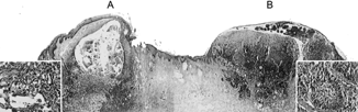
When he was 15 years old, the boy had 6 further cutaneous lesions resected from the face. One of these lesions was located on the nose and appeared as a dark grey and reddish nodule, 1.5 cm in diameter. The specimen from the skin of the nose revealed the presence of two adjacent lesions (). The one was an ulcerated classic basocellular carcinoma, 0.7 cm in diameter. Next and underneath this lesion, the dermis was occupied by a neoplastic nodule, 0.8 cm in diameter, formed partly by vascular channels lined with highly atypical cells and partly by a solid proliferation comprising highly atypical cells with abundant eosinophylic cytoplasms, diagnosed as cutaneous angiosarcoma. This second nodule involved the inked resection margin.
1 Presence of two adjacent cutaneous lesions: a cutaneous angiosarcoma involving the superficial and deep dermis (A) and a classic basocellular carcinoma (B) (HE, 20×; insets 400×).
The other five resected lesions were represented by basocellular carcinomas.
According to the staging systems in use for pediatric soft tissue sarcomas, the cutaneous angiosarcoma was classified as T1AN0M0 (due to the absence of local invasiveness and the size smaller than 5 cm, in the absence of nodal involvement or metastatic spread), and as IRS group II (due to the marginal resection with suspected microscopic residual disease).
Given the location of the AS, it was impossible to implement deep surgery to obtain free margins without mutilating effects, so re-excision was considered unfeasible and was not performed. Post-surgical radiotherapy and chemotherapy were not considered indicated because of the XP and the consequently high risk of developing further malignancies.
At the time of this report, 40 months after the diagnosis of AS, the boy is alive and without evidence of local or distant recurrence of AS.
DISCUSSION
AS is a highly malignant vascular tumor that can occur in any region of the body, but unlike other soft tissue sarcomas that prefer deep sites, most AS arise in the skin and superficial soft tissue, particularly in the head and neck region Citation[5]. AS is very uncommon in pediatric age. The largest pediatric series was reported by the Italian and German Cooperative Group and confirmed the tumor's aggressiveness, high propensity to metastasize, and poor prognosis, as observed in adults, where AS is more frequent Citation[6]. Said report confirmed the pivotal role of surgery in the treatment of AS, though complete resection seemed insufficient in most patients. The role of adjuvant therapies remains uncertain.
The present case was included in the reported Italian and German series: in this subset, most cases were soft tissue AS, and only 2 patients had a cutaneous AS Citation[6].
Cutaneous AS is the most common type of AS in adult, but exceptional in childhood. It arises almost exclusively in sun-exposed skin (especially the scalp) or irradiated areas of individuals over 50 years old, and men more than women; the association with previous radiotherapy, chronic lymphedema, or enviromental toxins is well-known Citation[5].
The clinical presentation is variable and could mimic a benign lesion, with bruise-like areas with indurated border or well-circumscribed purple or brown papules; advanced lesions may be large elevated swelling or masses, with ulcerated overlying skin. The prognosis of cutaneous AS is no different from that of other AS; the only effective treatment approach is wide excision followed by wide-field radiotherapy. In our reported case, the site involved and the boy's syndrome induced us to avoid chemotherapy and especially radiotherapy, despite surgical resection being incomplete. The patient has suffered no local or distant relapse, however, more than 3 years after diagnosis: this is unlikely to be due to the presence of XP, but it is noteworthy that in the few reported patients with AS and XP the outcome seemed better than in other cases of AS () Citation[2–4]. The series is too small to suggest that AS arising in XP may present biological differences and more a favorable behavior, but the findings are of interest.
Reported Cases with Cutaneous AS in XP
Considering the various reported cases, the association between cutaneous AS and XP does not seem accidental. XP is a disorder of post-replication DNA repair following UV exposure Citation[7]. On the other side, cutaneous AS—usually associated with prior irradiation—may arise in sun-exposed skin areas. It may be—but remains to be demonstrated—that the development of cutaneuos AS in these patients correlates with UV-induced DNA damage. Other pathogenetic mechanisms could be involved, however; as a matter of fact, the incidence of non-UV related tumors is higher than normal in XP patients.
In conclusion, our report suggests that cutaneous AS should be considered in the differential diagnosis of superficial lesions in XP patients. The association between AS and XP is evidently rare, but not fortuitous.
The authors are very grateful to Mr Giovanni Roncato for his contribution in the realization of the picture.
REFERENCES
- Lambert W C, Kuo H R, Lambert M W. Xeroderma pigmentosum. Dermatol Clin.. 1995; 13: 169–209. [PUBMED], [INFOTRIEVE]
- Leake J, Sheenan M P, Rampling D, et al, Angiosarcoma complicating xeroderma pigmentosum. Histopatology.. 1992; 21: 179–181
- De Silva B D, Nawroz I, Doherty V R. Angiosarcoma of the head and neck associated with xeroderma pigmentosum variant. Br J Dermatol.. 1999; 141: 166–167. [CROSSREF], [PUBMED], [INFOTRIEVE]
- Ludolph-Hauser D, Thoma-Greber E, Sander C, et al, Mast cells in angiosarcoma complicating xeroderma pigmentosum in a 13 years old girl. J Am Acad Dermatol.. 2000; 43: 300–302
- Weiss S W, Goldblum J R. Malignant vascular tumor. Weiss S W, Goldblum J R. Enzinger and Weiss's Soft Tissue Tumors, ed 4., St Louis, Mosby. 2001; 917–954
- Ferrari A, Casanova M, Bisogno G, et al, Malignant vascular tumors in children and adolescents: a report from the Italian and German Soft Tissue Sarcoma Cooperative Group. Med Pediatr Oncol.. 2002; 39: 109–114. [CROSSREF], [PUBMED], [INFOTRIEVE]
- Kleijer W J, Lohman P HM, Mulder M P, et al, Repair of X-ray damage in DNA of cultivated cells from patients having xeroderma pigmentosum. Mutation Res.. 1970; 9: 517–523. [CROSSREF], [PUBMED], [INFOTRIEVE]