
ABSTRACT
The aim of the present work is to provide information about Enterococcus strains isolated from traditional Turkish cheese samples in Ankara (Turkey), focusing on their prevalence, phenotypic and genotypic characteristics, and antibiotic resistance. A total of 213 probable enterococcal isolates isolated from 215 samples were identified by phenotypic and genotypic methods. As a result of 16S rDNA sequence analysis, 88 of the 213 enterococci strains were identified as Enterococcus faecium and 125 as Enterococcus faecalis. The E. faecalis strains (58.7%) were identified as the dominant species isolated from cheese samples in Turkey. The 213 Enterococcus strains were tested for susceptibility to 12 different antimicrobial agents. The resistance phenotype were as follow: nalidixic acid (100%), kanamycin (98.6%), rifampicin (78.4%), ampicillin (48.8%), ciprofloxacin (45.5%), erythromycin (18.8%), tetracycline (11.7%), penicillin G (5.6%), chloramphenicol (4.2%), gentamycin (3.8%) and streptomycin (1.4%). None of the strains was resistant to vancomycin. E. faecium strains showed more resistant phenotypes than E. faecalis strains as shown by the antibiotic resistance levels. It was also observed that the resistance of E. faecium and E. faecalis strains against the antibiotics was statistically significant (p ˂ 0.05). In total, 100% of E. faecium and 88.8% of E. faecalis strains were resistant to multiple drugs.
Introduction
Enterococcus was first determined as intestinal bacteria by Thiercelinin 1899. In 1906, Andrewes and Horder identified a potentially pathogenic bacterium isolated from a patient with endocarditis and renamed Thiercelin’s enterococcias Streptococcus faecalis.[Citation1] Scheleifer and Kilpper-Balz[Citation2] supported that enterococci were reclassified into their own genus according to the DNA-DNA hybridization and 16S rRNA sequencing, and since then the genus has been valid.[Citation3] So far, the genus Enterococcus were divided into 55 species and 2 subtypes based on 16S rRNA sequences.[Citation4]
Enterococci are an important part of lactic acid bacteria (LAB). Unlike most of the other LAB, European Food Safety Authority recommended that they are not recognized as safe in the qualified presumption of safety.[Citation5] These organisms occur in soil, surface waters, insects, plants, gastrointestinal tract of human and warm-blooded animals.[Citation1] Additionally, they are found in different food sources such as cheeses, meat, vegetables and olives.[Citation6] Enterococcus faecium and Enterococcus faecalis are the most frequently isolated Enterococcus species in food industry.[Citation7,Citation8] These bacteria can play a positive function in cheeses and meats as a starter or probiotic cultures. Enterococcus strains in dairy products have desirable metabolic traits because of their role of ripening and the development of flavour, probably by way of proteolysis, lipolysis, exopolysaccharide production and their diacetyl production by citrate metabolisms.[Citation9] In addition to their technological properties, some enterococci, mainly E. faecalis and E. faecium, produced inhibitory substances called as bacteriocins that are capable of inhibiting the growth of food pathogens and spoilage microorganisms such as Listeria monocytogenes and Staphylococcus aureus including vegetative cells as well as spores.[Citation10] On the other hand, the enterococci species caused nosocomial infections including endocarditis, bacteremia, biliary and urinary tract, central nervous system infections have also led to debates about the use of enterococcal strains as starter cultures. E. faecalis strains (80–90%) are the most prevalent Enterococcus species isolated from human infections, followed by E. faecium (5–20%). The remaining Enterococcus species infrequently cause infections.[Citation11]
Antibiotic resistance are wide-spread in enterococci isolated from foods including meat and dairy products. Increasingly resistance to multiple antibiotics in recent years has also been defined. A bacterial transfer of conjugative plasmids and transposons play an important role in the development of antibiotic resistance.[Citation5] Dissemination of antimicrobial resistance genes is a major concern for public health worldwide. Food is frequently granted as a possible intermediate vector for transmission of antibiotic-resistant genes via enterococcal isolates from animal origin.[Citation12] For the safety reason, an important criteria for the selection of starter cultures in foods is the absence of transferable antibiotic resistance in enterococci.[Citation13] Enterococcus ssp. have intrinsic and acquired resistance to most of the antibiotics used in humans.[Citation14–Citation16] Therefore, treatment of enterococcal infections may be difficult.[Citation10] Enterococci are described to be intrinsically resistant to β-lactam antibiotics, with penicillins (ampicillin, amoxicillin/clavulanic acid, penicillin G, methicillin, piperacillin), followed by carbapenems (imipenem) and cephalosporins (cefoperazone, ceftriaxone). Furthermore, enterococci can also acquire resistance to tetracyclines, quinolones, macrolides, erythromycin, streptogramins, polymixins, clindamycin and glycopeptides (vancomycin).[Citation17,Citation18]
The aim of the present work is to provide information about Enterococcus strains isolated from traditional cheeses produced in Turkey, focusing on prevalence, phenotypic and genotypic characteristics, and their antibiotic resistance.
Materials and methods
Materials
Sampling
A total of 215 traditional cheese samples were randomly purchased from various local bazaars and supermarkets in Ankara, Turkey. The analysed-cheeses consisted of hard, soft and semi-soft ripened cheeses (White, Kasar, Tulum, Ezine, Lor, Orgu and Civil). Samples were transported to the laboratory under cold conditions on the sampling day and processed immediately.
Bacterial strains and culturing
Enterococcal strains isolated in this study and references strains were propagated on Tryptic Soy Broth (TSB) (Merck, Germany) and Brain Hearth Infusion (BHI) Broth (Merck, Germany), respectively. They were grown at 37°C for 24 h. The initial isolates were kept at – 20°C in 30% (v/v) aqueous glycerol (Merck, Germany). Three reference strains (E. faecalis ATCC 29212, Escherichia coli LMG3083 (ETEC) and S. aureus ATCC 6538) were obtained from the culture collection of Prokaryote Genetics Laboratory, Department of Biology, Faculty of Science, Ankara University.
Isolation of enterococci
For isolation of enterococci, 10 g of each sample was homogenized and added to 90 mL of ¼ ringer solution (Merck, Germany) andincubated for 10 min at room temperature to ensure complete homogenization. Then serial dilution of homogenates was prepared up to 10−5 in 0.85% (w/v) NaCL (Merck, Germany) and 100 µL of each dilution was plated on the surface of plates containing Kanamycin Aesculin Azide (KAA) agar (Merck, Germany). The plates were incubated at 37°C for 18–48 h, and two typical colonies on KAA were picked randomly for further identification analysis (as detail below).
Phenotypic characterization
All Enterococcus isolates were subjected to identification according to standard biochemical tests and phenotypic description for identifying as enterococci. These tests were gram staining, catalase production, growth in TSB with 6.5% NaCL, growth at pH 9.6, esculin hydrolysis on Bile Esculin Azide Agar (Merck, Germany) and growth at 10°C–45°C.[Citation9]
Determination of hemolytic activity
The hemolytic activity was determined on TSA containing 5% (w/v) sheep blood plates and kept at 37°C for 24–48 h, under anaerobic condition.[Citation19] The zones around developing colonies formed on the bloody agar were examined. Clear zones around the colonies and green-hued zones were defined as β-hemolytic and α-hemolytic, respectively. No zones around the colonies were characterized as γ-hemolytic. Test was performed in duplicate. E. coli LMG3083 (ETEC) and S. aureus ATCC6538 were used as control bacteria in this study.
Genotypic characterization
The strains were identified by amplification and sequencing of the 16S rRNA gene. Primarily, genomic DNA from enterococcal and control strains were extracted from overnight TSB cultures as previously described.[Citation20] DNA concentration and purity were evaluated spectrophotometrically by NanoDrop ND-2000 spectrofotometer (Thermo Scientific, USA). The DNA was stored at –20°C. Then, the PCR procedures used in this study have been described previously.[Citation21] The pair of universal primers 907r (CCGTCAATTCMTTTRAGTTT) and 27f (AGAGTTTGATCMTGG CTCAG) proposed by Beasley and Saris[Citation22] were used to amplify 16S rRNA gene. Briefly, each PCR assay was performed in a total volume of 50 μL reaction mixtures containing 3 μL bacterial DNA template, 34.75 μL RNase/DNase free water, 0.25 μL Taq DNA polymerase in reaction buffer, 1 μL 2 mM of each dNTP, 4 μL 25 mM MgCl2, 1 μL of each primers (forward and reverse) and 5 μL PCR buffer. PCR amplifications were applied in a ThermoCycler (Techne TC-512, Staffordshire, UK). The PCR were used (1) initial hold of 2 min at 95°C, (2) denaturation step at 95°C/45 s, annealing at 55°C/45 s, extension at 72°C/2 min and (3) final extension step at 72°C/7 min. The PCR products purified using a GeneJET PCR purification kit (Thermo Scientific, USA) were analyzed on 1% agarose gel electrophoresis, stained in ethidium bromide solution and visualized under UV. The size of amplified fragments was determined by comparison with an O’Gene RulerTM 1000-bp DNA ladder (Thermo Scientific, USA). The sequence analysis results were compared with the 16S rRNA sequences in the National Center for Biotechnology Information (NCBI) database using the Basic Local Alignment Search Tool (BLAST) program.
Antimicrobial susceptibility testing
Antimicrobial susceptibility tests were applied to the 213 isolates by disk diffusion method on Mueller Hinton agar (Merck, Germany) with antibiotic disc according to the Clinical Laboratory Standards Instituteguidelines.[Citation23] Twelve different antibiotics were used: penicillin G (10 µg/disc), kanamycin (30 µg/disc), ampicillin (10 µg/disc), rifampicin (5 µg/disc), chloramphenicol (30µg/disc), erythromycin (15 µg/disc), gentamycin (120 µg/disc), tetracycline (30 µg/disc), vancomycin (30 µg/disc), nalidixic acid (30 µg/disc), streptomycin (300 µg/disc) and ciprofloxacin (5 µg/disc). All antibiotic discs were obtained from Bioanalyse (Turkey). E. faecalis ATCC 29212 and S. aureus ATCC 6538 strains were used as positive controls. According to inhibition zone measured, the strains were categorized as suspectible, intermadiate or resistant by taking into account the criteria of the CLSI.
Statistical analysis
SPSS 16 package program was used for statistical analysis. F (ANOVA, Analysis of Variance) was applied to determine the difference between the groups. The level of significance of differences between treatments was determined at p < 0.05.
Results and discussion
Isolation and identification of enterococci
From 215 traditional cheese samples, 213 (99.1%) probable enterococcal isolates were identified (data not shown). The isolation rate of Enterococcus strains in this study was quite high. This result was consistent with the results of previous studies, which reported that the percentage of positive samples of enterococci in dairy products was 72% in France,[Citation5] 83.3% in Brazilian,[Citation8] 93.3% in Egyptian,[Citation10] 100% in Southern Brazil,[Citation11] 94.6% in Spain[Citation24] and 90% in Egyptian.[Citation25] In previous studies in Turkey,[Citation26–Citation29] the prevalence of enterococci in cheese samples was reported to range between 62% and 99%. Enterococci have been recognized as an essential part of the natural microbial floraina variety of cheeses made from raw and pasteurized milk, because of their resistant to unsuitable conditions such as high and low temperature, low pH, high salt concentrations.[Citation5,Citation30] Enterococci likely result from the initial contamination of milk used for the cheese making, and also from the variety of the technological process. Moreover, enterococci are not all elimination by thermal treatments. Their levels depend on the extent of milk contamination, type of cheese, using the starter culture and the technology applied.
Morphological and cultural tests were applied to 213 enterococcal isolates. All of the isolates showed developmental characteristics at pH 9.6, 6.5% NaCL and 10–45°C. Fifty-two isolates were also identified as Gr (+), catalase (-) and esculin hydrolysis (+). Results of the phenotyping tests agreed with the previous studies of Gomes et al.,[Citation8] Rafaat et al.,[Citation10] Oladipo et al.,[Citation13] Lavova et al.[Citation31] When tested for hemolytic activity, 11.3% (24 of 213) enterococcal strains showed β-hemolytic character. While 9.9% strains (21 of 52) exhibited γ-hemolytic character, other 78.8% strains (168 of 213) were α-hemolytic character (data not shown). In this study, higher frequency of α-hemolysin than β-hemolysin was observed, in agreement with Tuncer,[Citation26] Barbosa et al.[Citation32] and Ispirli et al.[Citation33] We also found that β-hemolysin was most prevalent among E. faecium (66.7%, 16 of 24) when compared to E. faecalis (33.3%, 8 of 24). These findings were not similar to the results by Gomes et al.,[Citation8] who found that E. faecalis strains (38.7%) were dominant species to produce β-hemolysis. β-hemolytic isolates are unacceptable in foods and not desirable to use as a starter in the food fermentations. Nevertheless, it should not be forgotten that non-hemolytic Enterococcus spp. isolated from food may not be safe for starter cultures.[Citation34]
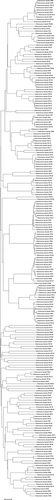
In this study, a total of 213 isolates were identified at species level by 16S rRNA sequence analysis (). Enterococcus strains were identified as, 88 E. faecium (41.3%) and 125 E. faecalis (58.7%). E. faecium and E. faecalis were most commonly isolated from cheeses.[Citation3,Citation29] In our study, E. faecalis was identified as the dominant species. Our findings were in agreement with the results reported by Jamet et al.,[Citation5] Morandi et al.,[Citation7] Abouelnaga et al.,[Citation9] Nieto-Arribas et al.,[Citation24] Templer and Baumgartner.[Citation35] There were same data that E. faecalis isolated from traditional cheeses and was the most frequently detected species in Turkey, as previously reported by Kürekçi et al.,[Citation27] Muş et al.,[Citation29] Toğay et al.,[Citation30] Çıtak et al.[Citation36] In contrast to our findings, previous works[Citation10,Citation11,Citation18,Citation25,Citation28,Citation34,Citation37] have reported that higher incidence of E. faecium strains was isolated from various cheese samples.
Figure 1. Dendrogram of the relationships between strains of enterococci based on the homologies of 16S rRNA sequence analysis. The dendrogram was constructed using the maximum-likelihood method with the MEGA 5.10 software.
Antimicrobial susceptibility
Genetic determinants of resistance in enterococcal strains are generally located in conjugative plasmids or transposons, for the reason that antibiotic resistance in enterococci isolated from foods is a special concern.[Citation8] The results of antibiotic susceptibility testing according to CLSI[Citation23] in the present study is summarized in . Analysis of the antimicrobial susceptibility of the 213 Enterococcus isolates revealed that resistance to nalidixic acid (100%), kanamycin (98.6%, 210 of 213) and rifampicin (78.4%, 167 of 213) were most frequent. Furthermore, resistance to ampicillin (48.8%, 104 of 213), ciprofloxacin (45.5%, 97 of 213), erythromycin (18.8%, 40 of 213) and tetracycline (11.7%, 25 of 213) was also observed, although at slightly lower levels. Additionally, low frequencies of resistance to penicillin G (5.6%, 12 of 213), chloramphenicol (4.2%, 9 of 213), gentamycin (3.8%, 8 of 213) and streptomycin (1.4%, 3 of 213) were detected. None of the analysed strains was resistant to vancomycin.
Table 1. Antimicrobial resistance profiles of enterococcus strains to 12 antimicrobial agentsa.
In 1988, it was first reported to vancomycin resistance enterococci (VRE) in European countries. After this date, VRE have been frequently isolated worldwide. The prevalence of VRE decreased markedly because of banning the use of avoparcin, an analogue of the glycopeptide vancomycin as a growth promoter in animal production, in 1997 in European countries and Turkey.[Citation27] There was no enterococcal isolates found to be resistant to vancomycin in this study. This was in agreement with previous studies.[Citation6,Citation28,Citation38] In contrast to our study, studies reported vancomycin resistance rates among enterococci from dairy products in Turkey ranging between 86.1%[Citation36] and 84.8%.[Citation39] These rates of vancomycin were considerably higher than our results. Enterococci have an intrinsically low resistance to β-lactam antibiotics (e.g. ampicillin and penicillin) because of their use in the treatment of enterococcal infections.[Citation40] In our study, resistance to ampicillin (48.8%) was higher than it was to penicillin G (5.6%). In agreement with the Gaglio et al.,[Citation6] Pesavento et al.,[Citation17] Demirgül and Tuncer,[Citation37] and Bulajic et al.[Citation41] we detected low resistance to penicillin G. The incidence of ampicillin resistance was higher than that reported by Gaglio et al.,[Citation6] Rafaat et al.,[Citation10] and Yuksel et al.[Citation39] Higher levels of ampicillin resistance in enterococci are achieved by increasing levels of penicillin-binding protein 5(PBP5) expressions.[Citation38] In our study, lower incidence of resistance to tetracycline (11.7%) was observed. This type of resistance is frequent in clinical and animal isolates of enterococci. Furthermore, it has largely been identified in clinical isolates.[Citation6,Citation25] As opposed to our study, Jamet et al.,[Citation5] Raafat et al.,[Citation10] and Pesavento et al.[Citation17] found a much higher incidence of resistance to tetracycline 92.4%, 35.3% and 53%, respectively. Of the 213 strains tested, only 9 strains of E. faecalis were resistant to chloramphenicol (4.2%). The use of chloramphenicol in animal husbandry was banned in 2002 in Turkey.[Citation42] This forbidding probably caused the low resistance to this antibiotic in Turkey. The percentage of resistant to chloramphenicol we detected was much lower than those in Turkey reported by Çıtak et al.[Citation36] and Yuksel et al.,[Citation39] who found 79.2% and 46.6%, respectively. Resistance to streptomycin (1.4%) and gentamycin (3.8%) were also observed in this work. Gentamycin and streptomycin have a synergistic effect when used together with a cell wall active agents.[Citation43] Around 96.2% (205 of 213) of enterococcal isolates were susceptible to gentamycin, in accordance with 94.9% and 83.6% of susceptible isolates reported by Jamet et al.[Citation5] and Bulajic et al.[Citation41] In our study, 98.6% of the enterococcal isolates were resistance to kanamycin. This result was in agreement with Jamet et al.,[Citation5] Kürekçi et al.[Citation27] and Yuksel et al.,[Citation39] who found 81%, 84.2% and 100%, respectively. The highest resistance was observed for nalidixic acid (100%) which is not surprising as most enterococci were reported to have intrinsic resistance to nalidixic acid.[Citation43] The incidence of resistance to nalidixic acid in all enterococcal isolates was similar to that reported by Yuksel et al.,[Citation39] but higher than that reported by Furlaneto-Maia et al.[Citation11] Rifampicin resistance is widespread in enterococci. It inhibits transcription of mRNA.[Citation43] A high level resistance to rifampicin (78.4%) was noticed in this study. Our findings also indicated a higher incidence of resistant to ciprofloxacin (45.5%) whereas other reports obtained the opposite results.[Citation6,Citation10,Citation25,Citation27] Resistant to ciprofloxacin may be a result of the overwhelming use of antibiotics in human and veterinary medicine.[Citation32]
Multi-drug resistance defined as resistance to three or more antimicrobial agents was found in 70.90% (151of 213) of Enterococcus strains in this study. A summary of multiple drugs among the enterococcal strains was reported in . As enterococci are naturally resistant to nalidixic acid, it was not used in the definition of multiple-drug resistance. Abouelnaga et al.,[Citation9] Rafaat et al.,[Citation10] Yüceer and Tuncer[Citation44] detected the same results with 83%, 78% and 68% respectively. In contrast, lower incidence (24.59%) were found by Bulajic et al.[Citation41] A total of 199 Enterococcus strains displayed resistance to at least two antibiotics (from 2 to 5 out of the 12 antibiotics tested). Of the 88 E. faecium strains (100%) showed resistant profile against three or more tested antibiotics. Out of 125 strains, 111 E. faecalis strains (88.8%) were resistant to them. Previous studies [8, 17, 27, 29, 39] indicated that E. faecalis strains showed more resistance phenotype than E. faecium. Our findings in this study are interesting that when the antibiotic resistance levels of the enterococcal species identified were compared; E. faecium strains showed more resistance phenotype than E. faecalis strains. Similar result was also reported by Furlaneto-Maia et al.[Citation11] and Delpech et al.[Citation18] The results of this study confirmed that the resistance of E. faecium and E. faecalis strains against antibiotics was found statistically significant (p ˂ 0.05).
Table 2. Multiple antibiotic resistance in Enterococcus strains excluding nalidixic acid.
Conclusion
This study focused on the isolation and identification of strains which belong to genus Enterococcus isolated from traditional cheeses retailed in Turkey and then to determine their antibiotic resistance. The results of this study indicated that E. faecalis was found to be the most frequently isolated species. Antibiotic resistance has been a growing concern for a number of years. Our results clearly showed that E. faecalis and E. faecium strains isolated from Turkish traditional cheese samples in Ankara could be considered a potential source for the dissemination of antibiotic resistance. In addition, the high prevalence of multi-drug resistance among Enterococcus species is a serious threat to public health.
Acknowledgments
We thank Prof. Dr. Mustafa AKCELIK (Ankara University) for supplying references strains and providing support to carry out this research.
Additional information
Funding
Related Research Data
References
- Moreno, M. R. F.; Sarantinopoulos, P.; Tsakalidou, E.; De Vuyst, L., The Role and Application of Enterococci in Food and Health. International Journal of Food Microbiology 2006, 106, 1–24. DOI: 10.1016/j.ijfoodmicro.2005.06.026.
- Schleifer, K. H.; Kilpper-Bälz, R. Transfer of Streptococcus Faecalisand Streptococcus Faeciumto the Genus Enterococcus Nom. Rev. As Enterococcus Faecaliscomb. Nov. And Enterococcus Faeciumcomb. Nov. International Journal of Systematic Bacteriology 1984, 34, 31–34.
- Ogier, J. C.; Serror, P., Safety Assessment of Dairy Microorganisms: The Enterococcus Genus. International Journal of Food Microbiology 2008, 126, 291–301. DOI: 10.1016/j.ijfoodmicro.2007.08.017.
- LPSN bacterio.net, List of Prokaryotic Names with Standing in Nomenclature. http://www.bacterio.net/enterococcus.html, Access date: Feb 15, 2017).
- Jamet, E.; Akary, E.; Poisson, A. A.; Chamba, J. F.; Bertrand, X.; Serror, P., Prevalence and Characterization of Antibiotic resistantEnterococcus Faecalis in French Cheeses. Food Microbiology 2012, 31, 191–198. DOI: 10.1016/j.fm.2012.03.009.
- Gaglio, R.; Couto, N.; Marques, C.; Lopes, M. F. S.; Moschetti, G.; Pompa, C.; Settani, L., Evaluation of Antimicrobial Resistance and Virulence of Enterococci from Equipment Surfaces, Raw Marterials, and Traditional Cheeses. International Journal of Food Microbiology 2016, 236, 107–114. DOI: 10.1016/j.ijfoodmicrobiol.2016.07.020.
- Morandi, S.; Brasca, M.; Andrighetto, C.; Lombardi, A.; Lodi, R., Technological and Molecular Characterization of Enterococci Isolated from North-West Italian Dairy Products. International Dairy Journal 2006, 16, 867–875. DOI: 10.1016/j.idairyj.2005.09.005.
- Gomes, B. C.; Esteves, C. T.; Palazzo, I. C. V.; Darini, A. L. C.; Felis, G. E.; Sechi, L. A.; Franco, B. D. G. M.; De Martinis, E. C. P., Prevalence and Characterization of Enterococcus Spp. Isolated from Brazilian Foods. Food Microbiology 2008, 25, 668–675. DOI: 10.1016/j.fm.2008.03.008.
- Abouelnaga, M.; Lamas, A.; Quintela-Baluja, M.; Osman, M.; Miranda, J. M.; Cepeda, A.; Franco, C. M., Evaluation of the Extent of Spreading of Virulence Factors and Antibiotic Resistance in Enterococci Isolated from Fermented and Unfermented Foods. Annalen Microbiology 2016, 66, 577–585. DOI: 10.1007/s13231-015-1138-6.
- Raafat, S. A.; Abo-Elmagd, E. K.; Awad, R. A.; Hassan, E. M. Prevalence of Vancomycin Resistant Enterococci in Different Food Samples. Egyptian Journal of Medical Microbiology 2016, 25(4), 47–55.
- Furlaneto-Maia, L.; Rocha, K. R.; Henrique, F. C.; Giazzi, A.; Furlaneto, M. C., Antimicrobial Resistance in Enterococcus Sp. Isolated from Soft Cheese in Southern Brazil. Advanced Microbiology 2014, 4, 18–175. DOI: 10.4236/aim.2014.43023.
- Lopes, M. F. S.; Ribeiro, T.; Abrantes, M.; Marques, J. J. F.; Tenreiro, R.; Crespo, M. T. B., Antimicrobial Resistanceprofiles of Dairy and Clinical Isolates and Type Strains of Enterococci. International Journal of Food Microbiology 2005, 103, 191–198. DOI: 10.1016/j.ijfoodmicro.2004.12.025.
- Oladipo, I. C.; Sanni, A. I.; Swarnakar, S., Pheotypic and Genomic Characterization of Enterococcus Species from Some Nigerian Fermented Foods. Food Biotechnology 2013, 27, 39–53. DOI: 10.1080/08905436.2012.755627.
- Sallem, R. B.; Klibi, N.; Klibi, A.; Said, B. L.; Dziri, R.; Boudabous, A.; Torres, C.; Slama, K. B., Antibiotic Resistance and Virulence of Enterococci Isolates from Healty Humans in Tunisis. Annalen Microbiology 2016, 66, 717–725. DOI: 10.1007/13213-015-1157-3.
- Franz, C. M.; Huch, M.; Abriouel, H.; Holzapfel, W.; Galvez, A. Enterococci as Probiotics and Their Implications in Food Safety. Int. Journal of Food Microbiology 2011, 151(2), 125–140. DOI:10.1016/j.ijfoodmicro.2011.08.014.
- Camargo,C, H.; Bruder-Nascimento, A.; Lee, S. H. I.; Junior, A. F.; Kaneno, R.; Rall, V. L. M. Prevalence and Phenotypic Characterization of Enterococcus Ssp. Isolated from Food in Brazil. Brazilian Journal of Microbiology 2014, 45(1), 111–115.
- Pesavento, G.; Calonico, C.; Ducci, B.; Magnannini, A.; Lo Nostro, A., Prevalence and Antibiotic Resistance of Enterococcus Spp. Isolated from Retail Cheese, Ready-To-Eat Salads, Ham, and Raw Meat. Food Microbiology 2014, 41, 1–7. DOI: 10.1016/j.fm.2014.01.008.
- Delpech, G.; Pourcel, G.; Schell, C.; Luca, M. D.; Basualdo, J.; Bernstein, J.; Grenovero, S.; Sparo, M. Antimicrobial Resistance Profiles of Enterococcus Faecalis and Enterococcus Faecium Isolated from Artisanal Food of Animal Origin in Argentina. Foodborne Pathogens Disease 2012, 9(10), 939–944. DOI:10.1089/fpd.2012.1192.
- Valenzuela, A. S.; Omar, N. B.; Abriouel, H.; Lopez, R. L.; Veljovic, K.; Canamero, M. M.; Topisirovic, M. K. L.; Galvez, A., Virulence Factors, Antibiotic Resistance, and Bacteriocins in Enterococci from Artisan Foods of Animal Origin. Food Control 2009, 20, 381–385. DOI: 10.1016/j.foodcont.2008.06.004.
- Cancilla, M. R.; Powell, I. B.; Hillier, A. J.; Davidson, B. E. Rapid Genomic Fingerprinting of Lactococcus Lactis Strains by Arbitrarily Primed Polymerase Chain Reaction with 32P and Fluorescent Labels. Applied and Environmental Microbiology 1992, 58(5), 1772–1775.
- Blaiotta, G.; Pepe, O.; Mauiello, G.; Villani, F.; Andolfi, R.; Moschetti, G. 16S-23S rDNA Intergenic Spacer Region Polymorphism of Lactococcus Gavriae, Lactococcus Raffinolactis and Lactococcus Lactis as Revealed by PCR and Nucleotide Sequence Analysis. Systematic and Applied Microbiology 2002, 25(4), 520–527. DOI:10.1078/07232020260517652.
- Beasley, S. S.; Saris, P. E. J. Nisin-Producing Lactococcus Lactis Strains Isolated from Human Milk. Applied and Environmental Microbiology 2004, 70(8), 5051–5053. DOI:10.1128/AEM.70.8.5051–5053.2004.
- CLSI. Performance Standards for Antimicrobial Susceptibility Testing: Twenty First Informational Supplement; Clinical and Laboratory Standards Institute, M100-S21: Wayne, PA, USA, 2011.
- Neito-Arribas, P.; Sesana, S.; Poveda, J. M.; Chicon, R.; Cabezas, L.; Palop, L. Enterococcus Populations in Artisanal Manchego Cheese: Biodiversity, Technological and Safety Aspects. Food Microbiology 2011, 28(5), 891–899. DOI:10.1016/j.fm.2010.12.005.
- Hammad, A. M.; Hassan, H. A.; Shimamoto, T., Prevalence, Antibiotic Resistance and Virulence of Enterococcus Spp. In Egyptian Fresh Raw Milk Cheese. Food Control 2015, 50, 815–820. DOI: 10.1016/j.foodcont.2014.10.020.
- Tuncer, Y.;. Some Technological Properties of Phenotypically Identified Enterococcal Strains Isolated from Turkish Tulum Cheese. African Journal of Biotechnology 2009, 8(24), 7008–7016.
- Kürekçi, C.; Önen, P.; Yipel, M.; Aslantaş, Ö.; Gündoğdu, A. Characterization of Phenotypic and Genotypic Antibiotic Resistance Profile of Enterococci from Cheeses in Turkey. Korean Journal of Food Science Animal Resources 2016, 36(3), 352–358. DOI:10.5851/kosfa.2016.36.3.352.
- Yoğurtçu, N. N.; Tuncer, Y. Antibiotic Suspectibility Patterns of Enterococcus Strains Isolated from Turkish Tulum Cheese. International Journal of Dairy Technology 2013, 66, 2, 236–242. DOI:10.1111/1471-0307.12014.
- Muş, T. E.; Cetinkaya, F.; Cıbık, R.; Soyutemiz, G. E.; Simsek, H.; Coplu, N. Antibiotic Resistance Profiles of Enterococci from Foods of Animal Origin in Turkey. Acta Vet Hungarian 2017, 65(4), 461–467. DOI:10.1556/004.2017.044.
- Toğay, S. Ö.; Keskin, A. Ç.; Açık, L.; Temiz, A., Virulence Genes, Antibiotic Resistance and Plasmid Profiles of Enterococcus Faecalis and Enterococcus Faecium from Naturally Fermented Turkish Foods. Journal of Applied Microbiology 2010, 109, 1084–1092. DOI: 10.1111/j.1365-2672.2010.04763.x.
- Lavova, M.; Bezekova, J.; Canigova, M.; Krocko, M.; Domig, K. J. Species Identification of Enterococci by Biochemical Test and Molecular-Genetic Methods. Potravinarstvo 2014, 8, 1, 124–129. DOI:10.5219/364.
- Barbosa, J.; Gibbs, P. A.; Teixeira, P., Virulence Factors among Enterococci Isolated from Traditional Fermented Meat Products Produced in the North of Portugal. Food Control 2010, 21, 651–656. DOI: 10.1016/j.foodcont.2009.10.002.
- Ispirli, H.; Demirbaş, F.; Dertli, E., Characterization of Functional Properties of Enterococcus Spp. Isolated from Turkish White Cheese. LWT-Food Science and Technology 2017, 75, 358–365. DOI: 10.1016/j.lwt.2016.09.010.
- De Vuyst, L.; Moreno, M. R.; Revets, H., Screening for Enterocin and Detection of Hemolysin and Vancomycin Resistance in Enterococci of Different Origins. International Journal of Food Microbiology 2003, 84, 299–318. DOI: 10.1016/S0168-1605(02)00425-7.
- Templer, S. P.; Baumgartner, A. Enterococci from Appenzeller and Schabziger Raw Milk Cheese: Antibiotic Resistance, Virulence Factors, and Persistence of Particular Strains in the Products. Journal of Food Protection 2007, 70, 2, 450–455.
- Çıtak, S.; Yucel, N.; Orhan, S. Antibiotic Resistance and Incidence of Enterococcus Species in Turkish White Cheese. International Journal of Dairy Technology 2004, 57, 1, 27–31. DOI:10.1111/j.1471-0307.2004.00122.x.
- Demirgül, F.; Tuncer, Y. Detection of Antibiotic Resistance and Resistance Genes in Enterococci Isolated from Sucuk, a Traditional Turkish Dry-Fermented Sausage. Korean Journal of Food Science Animal Resources 2017, 37, 5, 670–681. DOI:10.5851/kofsa.2017.37.5.670.
- Vrabec, M.; Lovayova, V.; Dudrikova, K.; Gallo, J.; Dudrikova, E., Antibiotic Resistance and Prevalence of Enterococcus Spp. And Escherichia coli Isolated from Bryndza Cheese. Italian Journal of Animal Science 2015, 14, 609–614. DOI: 10.4081/ijas.2015.3968.
- Yuksel, F. N.; Akcelik, N. Akcelik, M.Incidence of Antibiotic Resistance Virulence Determinants in Enterococcus Faecium and Enterococcus Faecalis Strains, Isolated from Traditional Cheeses in Turkey. Molecular Genetics, Microbiology Virology 2015, 30, 4, 206–215. DOI:10.3103/S089141681504014X.
- Garrido, A. M.; Galvez, A.; Pulido, R. P., Antimicrobial Resistance in Enterococci. Journal of Infectious Diseases Therapies 2014, 2, 150. DOI: 10.4172/2332-0877.1000150.
- Bulajić, S.; Tambur, Z.; Opačić, D.; Miljkovic-Semlimovic, B.; Doder, R.; Cenic-Milosevic, D. Characterization of Antibiotic Resistance Phenotypes and Resistance Genes in Enterococcus Spp. Isolated from Cheeses. Archives Biology Sciences 2015, 67, 1, 139–146. DOI:10.2298/ABS140426016B.
- Kasimoglu-Doğru, A.; Gencay, Y. E.; Ayaz, N. D., Prevalence and Antibiotic Resistance Profiles of Enterococcus Species in Chicken at Slaughter Level; Absence of vanA and vanB Genes in E. Faecalis and E. Faecium. Research Vetnary Sciences 2010, 89, 153–158. DOI: 10.1016/j.rvsc.2010.02.005.
- Miller, W. R.; Munita, J. M.; Arias, C. A. Mechanisms of Antibiotic Resistance in Enterococci. Expert Reviews Anti Infectious Therapy 2014, 12, 10, 1221–1236. DOI:10.1586/14787210.2014.956092.
- Yüceer, Ö.; Tuncer, B. Ö. Determination of Antibiotic Resistance and Biogenic Amine Production of Lactic Acid Bacteria Isolated from Fermented Turkish Sausage (Sucuk). Journal of Food Safety 2015, 35, 276–285. C-1: Include complete journal titles in all cases. DOI:10.1111/jfs.12177.