
ABSTRACT
Auricularia auricula is one of the most popular edible mushrooms in China. Limited information is available regarding genes and proteins related to A. auricula development from mycelium to mature fruiting body and response to environmental stimuli. With the development of proteomics, this technology platform for addressing this problem is available. A critical step of a good proteome is protein extraction which can gives the best quality and a wide coverage of total proteins. To establish an effective protein extraction protocol suitable for proteomics analysis in mycelium and fruiting body of A. auricula, four previously reported protein extraction protocols including direct extraction, trichloroacetic acid (TCA) precipitation, TCA/acetone precipitation and phenol-based extraction, were compared based on the protein yield and 2-DE patterns. The efficiency of the four methods was evaluated by comparing protein yield and 2-DE patterns. Of the four methods, both TCA precipitation and phenol-based extraction method for A. auricula mycelia protein extraction yielded the good protein separation pattern and higher number of protein spots in 2-DE analysis. Furthermore, phenol-based extraction is superior to the other tested methods for A. auricula fruiting body proteome analysis .
Introduction
Auricularia auricula forms mutualistic symbioses with many tree species, commonly known as tree-ear, which is an edible mushroom found worldwide. Several studies have reported diverse biological activities of this mushroom, including anti-tumor and anti-coagulant activity owing to its enrichment of polysaccharide and polyphenolic compounds.[Citation1–Citation3] The consumption of A. auricula may potentially contribute to the decreased risk of hypercholesterolemia and improve heart function.[Citation4,Citation5] Much attention has been paid to the bioactive compounds isolation, identification, and characterization in A. auricula, due to their numerous health beneficial effects.[Citation4,Citation6–Citation9] With the development of modern chromatographic and spectrometric techniques, bioactive compounds in A. auricula has been analyzed easier than before[Citation10–Citation13], but the regulation of this metabolic process is poorly understood and will require targeted, well-designed and appropriately research to gain more information.
The wild A. auricula could not meet production requirements due to the high demand and big market of A. auricula, nowadays A. auricula has been implemented artificial cultivated.[Citation14,Citation15] The quality and quantity of artificial cultivated A. auricula is susceptible to temperature and humidity, however, the molecular mechanisms underlying this phenomenon remain to be elucidated.[Citation16] Notably, A. auricula is a non-model fungi species, only a few of genes were identified, including eight laccase genes, 18S ribosomal RNA gene and two glyceraldehyde-3-phosphate dehydrogenase (GAPDH) genes.[Citation17,Citation18] It is clearly insufficient to establish the genetic and molecular knowledge of A. auricula based on the gene function, especially the molecular mechanism of bioactive compound metabolism and physiological responses to the environment.
Many different research experiments that were not dependent on genomic sequence were carried with the development of proteomics in the past decade.[Citation19,Citation20] Utilizing appropriate proteomics approaches and analysis tools, the variation in protein expression induced by changing environmental conditions, different developmental stages, or tissue types can be examined.[Citation21–Citation24] Some software programs has been used to analyze the proteome, which included but are not limited to proteome profiling, protein quantification, protein modification, and protein-protein interaction.[Citation25] To find the meaningful biological changes and advancing biological discoveries in A. auricula, the further development and application of proteomic methodologies are necessary. Two-dimensional gel electrophoresis (2-DE) is a powerful tool to separate the different expression protein that was applied for many fields such as animals, plants, and microorganisms.[Citation26,Citation27] The quantities and qualities of protein extraction are one of the most critical processes of 2-DE which impact on the isoelectric focusing on first dimension and sodium dodecyl sulphate (SDS) electrophoresis on second dimension.[Citation28,Citation29] Unfortunately, there is no general method of protein extraction method that can be suitable for all kinds of organisms.[Citation30] Because it contain lots of interfering elements, i.e. polysaccharide and melanin that can interfere with 2-DE analysis, A. auricula is studied as a good representative of recalcitrant species.[Citation31] The basic principles of protein methods should be followed in those protocols, (1) Improve sample protein solubility as far as possible, extract from the maximum amount of total protein, and reduce protein loss; (2) Decrease the artificial modification of protein; (3) Separate the interaction of proteins and other biological macromolecules, and make sure the proteins in a completely denatured state. To meet those principles, many protein extraction protocols have been published for many fungi, including direct extraction, precipitation extraction, and phenol-based extraction.[Citation3,Citation19,Citation27,Citation32,Citation33]
The growth of mycelium and fruiting body formation is the important development stage of A. auricula. To find a proper protein extraction protocol for mycelium and fruiting body, we compared four previously reported protein extraction protocols, including direct extraction, trichloroacetic acid (TCA) precipitation extraction, TCA in acetone precipitation extraction, and phenol-based extraction. The efficiency and compatibility of the four methods was evaluated by comparing protein yield and separation patterns analysis in order to produce gels that contain the good resolution and high protein spot coverage for further proteomic research.
Materials and method
Material
A. auricula DPM strain using in this research was obtained from the experimental forestry farm of Heilongjiang Academy of Forestry. It is cultured on the liquid medium. (17.27 g/L yeast extracts, 1.92 g/L tyrosine, 3.84 g/L lactose, 1 g/L NaCl, 2 g/L MgSO4, 0.5 mg/L biotin, 1 g/L KH2PO4) at 25°C, 150 rpm for seven days, then the mycelia of DPM ware collected through a nylon mesh to remove medium and washed three times by deionized water. The fruiting bodies of A. auricula were also collected from experimental forestry farm of Heilongjiang Academy of Forestry. The fresh fruiting bodies and mycelia were flash frozen in liquid nitrogen, then ground to a fine powder using a mortar and pestle, respectively. The fine powder of fruiting bodies and the mycelia were stored in −80°C before protein extraction.
Protein extraction
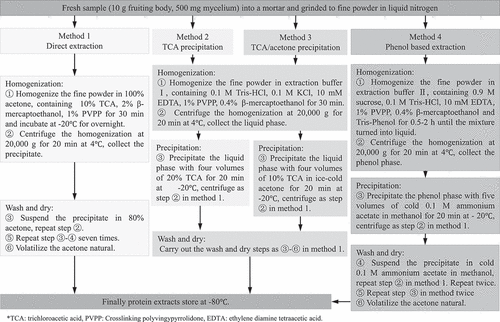
The amount of powdered fruiting body and mycelia was respectively 10 g and 500 mg for each protein extraction protocol. We chose four different protein extraction methods used in microorganism and higher plants, including direct extraction (Method 1), TCA precipitation extraction (Method 2), TCA/acetone precipitation extraction (Method 3), and phenol-based extraction (Method 4). An overview of the four protein extraction methods is presented in . Each method was repeated three times and protein extracts were stored at −80°C.
Figure 1. Graphical representative of four protein extraction methods.
Protein quantification and purification
The protein powder was dissolved in lysis solution (7 M urea, 2 M thiourea, 4% 3-[(3- Cholamidopropyl) dimethylammonio] propanesulfonate (CHAPS), 40 mM dithiothreitol (DTT), 2% pharmalyte, 4% protease inhibitor). Protein concentrations were determined by use of a 2-D Quant Kit (GE Healthcare, Little Chalfont, UK). The dialyzed protein solution cleaning was performed with a 2-D Clean-Up kit (GE Healthcare, Little Chalfont, UK) which can remove interfering substances (salt or charged detergents) from protein solution.
Sodium dodecyl sulphate-polyacrylamide gel electrophoresis (SDS-PAGE)
In a total 80 μg protein extracts were mixed with 4.5 μL SDS Sample Buffer (0.5 M Tris-HCl (pH 6.8), Glycerol 85%, 10% (w/v) SDS, and 0.1% bromophenol blue) and 3 μL β-mercaptoethanol. The protein samples were boiled in sample buffer and loaded on a 10% SDS-PAGE (20 cm wide × 20 cm tall) using 1× SDS electrophoresis buffer (2.5 mM Tris, 19.2 mM Glycine, 0.01% SDS) as a running buffer. The gel was pre-run for 20 min at 100 V before samples were loaded and run at 180 V for 3 h. Protein bands following Coomassie Brilliant Blue R250 staining to visualize.
Two-dimensional polyacrylamide gel electrophoresis (2-DE)
Two-dimensional electrophoresis of protein extracted was performed using a GE Healthcare 2-DE system on the basis of manufacturer’s manuals. 1300 μg protein sample was loaded by rehydration to immobilize Dry Strips (pH 3–10 linear, 24 cm) (GE Healthcare, Waukesha, WI, USA) individually. The separation parameters on an IPGphor II unit (GE Healthcare, Waukesha, WI, USA) were performed as follows: 30 V for 8 h, 50 V for 4 h, 100 V for 1 h, 300 V for 1 h, 500 V for 1 h, 1,000 V for 1 h, and 8,000 V for 12 h. IPG buffer was rehydrated using hydration buffer (8 M urea, 2% CHAPS, 20 mM DTT) containing 0.6% (v/v). When isoelectric focusing completed, the strips were equilibrated with 10 mL equilibration buffer I containing 6 M urea, 2% SDS, 2.5 mM Tris-HCl (pH 8.8), 30% glycerol, and 1% DTT for 15 min, then with 10 mL equilibration buffer II containing 6 M urea, 2% SDS, 2.5 mM Tris-HCl (pH 8.8), 30% glycerol, and 4% 2-iodoacetamide (IAA) for 15 min. The second dimension separation of proteins was performed on SDS-PAGE gel (12.5% polyacrylamide) with EttanTM Daltsix apparatus (GE Healthcare, Waukesha, WI, USA). The electrophoresis was carried out at 16 °C and 3.5 W/gel for pre-electrophoresis and then 17.5 W/gel until the bromophenol blue dye arrived at the bottom of the gels. With the use of Coomassie Brilliant Blue R250 staining, the protein samples in gel were visualized and acquired using an ImageScanner (GE Healthcare, Waukesha, WI, USA). ImageMaster 2D Platinum Software Version 7.0 (GE Healthcare, Waukesha, WI, USA) was conducted to analysis the 2-DE gel images.
Statistical and image analysis
All statistical calculations were made by SPSS 17.0 software package for Windows (SPSS Inc., Chicago, IL, USA).
Results
Evaluation of protein yields based on different extraction methods
Four different protein extraction methods were evaluated with their consistency and reproducibility in extracting total protein samples from mycelium and fruiting body of A. auricula. The protein yield was based as the quantity of protein extracted from 500 mg of fresh mycelia or 10 g of fruiting body. There was no significant difference in the protein yield extracted via direct extraction method (Method 1), TCA precipitation method (Method 2) and TCA/acetone precipitation method (Method 3) from mycelium and fruiting body (). Highest protein yields of 5.21 mg/g and 0.27 mg/g from mycelium and fruiting body, respectively, were obtained using direct extraction method (Method 1). In contrast, the phenol-based extraction (Method 4) gave the lowest protein yield with 3.89 mg/g from mycelium and 2.0 mg/g from fruiting body (). The protein extracted from mycelium was found to be consistently higher in concentration compared to the protein extracted from fruiting body of A. auricula () in four extraction methods.
Table 1. Protein yields obtained from mycelium and fruiting body of A. auricula using four different extraction methods.
Evaluation of SDS-PAGE patterns based on different extraction methods
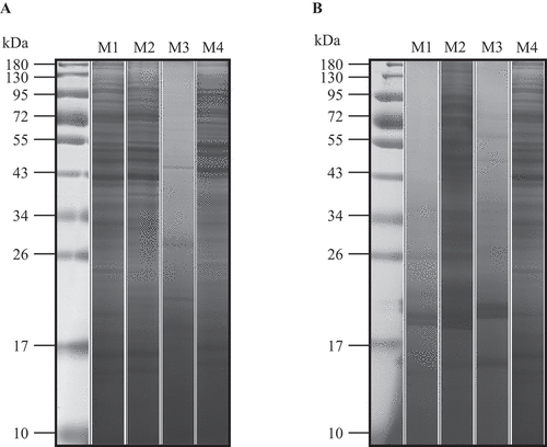
The four protocols compared in this study were evaluated using one dimensional SDS gel polyacrylamide electrophoresis (1D-SDS-PAGE) (). According to their molecular weight, the proteins separated were resolved between 10 and 180 kDa that revealing an overview of the total protein profile. Except TCA/acetone precipitation method (Method 3), the SDS-PAGE profile of proteins extracted from mycelium clearly showed the majority of the protein bands were common in the other three protocols (). Although the lowest yield of protein from fruiting body using phenol-based extraction (Method 4), the proteins extracted represented the highest number of bands resolved in the SDS-PAGE (). Sharp bands were observed from fruiting body with phenol-based extraction (). TCA precipitation method (Method 2) resulted in unclear protein separation patterns, and the gel image produced using direct extraction method (Method 1) and TCA/acetone precipitation method (Method 3) only managed to resolve low molecular weight proteins (). The extraction method which could cater to include total proteins would be valuable for subsequent analyses.
Figure 2. SDS-PAGE seperation of proteins from mycelium (A) and fruiting body (B) using different protein extraction methods, including direct extraction (Method 1), TCA precipitation extraction (Method 2), TCA/acetone precipitation extraction (Method 3), and phenol-based extraction (Method 4). Totally, 80 μg proteins are resolved using 12.5% polyacrylamide gel and visualized with CBB.
Evaluation of 2-DE patterns based on different extraction methods
After scrutinizing the 1D-PAGE profiles, the mycelium protein obtained from direct extraction method (Method 1), TCA precipitation method (Method 2) and phenol-based extraction (Method 4), and the fruiting body proteins obtained from TCA precipitation method (Method 2) and phenol-based extraction method (Method 4) were then run on 2-DE. Proteins extracted using different extraction protocols from mycelium and fruiting body of A. auricula were first separated according to their isoelectric points, followed by an electrophoretic separation based on their respective molecular weights. Extracted proteins were focused on 24 cm pH 3–10 IPG strips and run on 12.5% polyacrylamide gels. The gels were stained using Coomassie Brilliant Blue R250 to visualize the protein spots (, ). The number of protein spots on each gel was quantified with ImageMaster 2D gel software (). The mycelium proteins extracted using direct extraction method (Method 1) did not produce good 2-DE images, compared to TCA precipitation (Method 2) and phenol-based extraction method (Method 4) (). Although protein yield was the highest using direct extraction method, 2-DE images showed the lowest number of protein spots, with 144 spots detected in mycelium (). Efficient protein separation and spot detection were observed in the 2-DE images obtained from mycelium using TCA precipitation (Method 2) and phenol-based extraction method (Method 4), 1365 spots and 1380 spots were successfully detected (, ). Streaking or smearing of protein spots were not detected from any of these two extraction methods for mycelium ().
Table 2. Total numbers of detectable spots from mycelium and fruiting body based on different protein extraction methods.
Figure 3. 2-DE analysis of extracted proteins from mycelium tissues. Equal amounts (1300 μg) of proteins were loaded on 24 cm pH 3–10 IPG strips. (A) direct extraction method, (B) TCA precipitation extraction method, (C) phenol-based extraction method.
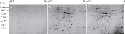
Figure 4. 2-DE analysis of extracted proteins from fruiting body. Equal amounts (1300 μg) of proteins were loaded on 24 cm pH 3–10 IPG strips. (A) TCA precipitation extraction method, (B) phenol/ammonium acetate extraction method, (C) TCA precipitation extraction in combination with protein purification (D) phenol-based extraction in combination with protein purification.
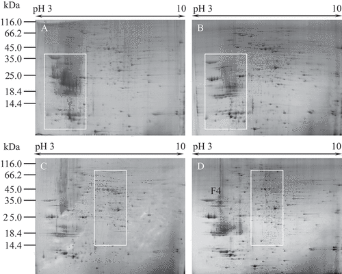
The 2-DE pattern obtained with TCA precipitation (Method 2) was very similar to that obtained with the phenol-based extraction (Method 4) from A. auricula fruiting body, 1035 spots and 1038 spots from A. auricula fruiting body were successfully detected (, B, ). However, compared with phenol-based extraction, extra vertical streaks were detected in the acidic region (pI 4 to 5) of the gel from TCA precipitation method. In addition, TCA precipitation resulted in protein spots that were diffuse and poorly resolved at the acidic ends and low molecular mass regions indicated by red box (, B). To address those issues, a method for protein purification was presented to fruiting body protein. After the purification, phenol-based extraction resulted in higher numbers of spots in 2-DE images compared with TCA precipitation extraction that produced more even spot distribution across the near-neutral region indicated by white box (, , ). A total of 759 protein spots were detectable from TCA precipitation extraction and 833 spots were detectable from phenol-based extraction. The vertical streaks and spot diffuse were almost removed after protein purification. These results indicate that protein purification steps result in the loss of small amounts of proteins but produced better 2-DE images for proteomic analysis.
Discussion
Over the past decade, major advancements in omic technologies (e.g., genomics, proteomics, and metabolomics) have made high-throughput monitoring of a variety of molecular and organismal processes possible. Among the approaches for studying regulatory mechanisms in biological systems, proteomic analysis is directly and efficiently. Large numbers of developmental studies on plant cell division, elongation, differentiation, and formation of various organs were carried out with the growing interest in plant proteomics continually produces.[Citation34] At the present time, very few proteomic studies have been carried out in edible mushroom. This is probably on account of the fact that no appropriate techniques for obtaining high-quality protein extracts was available and a limited number of the genome sequence has been generated for these organisms. A. auricula is a species of non-toxic edible ear-shaped macrofungus, containing carbohydrates and pigments which can interference protein extraction efficiency[Citation18,Citation35] that possibly explain the absence of data in the literature for obtaining quality proteins and studies of successful proteomic analysis of A. auricula. The pigments in A. auricula had been confirmed to a polyphenolic compounds[Citation13], thereby PVPP was added to extraction buffer which be shown to be effective in chelating and precipitating existing polyphenolic compounds to resolved the pigments interference.[Citation36]
Because of the direct effect on the purity of protein which influences the reproducibility and reliability, the results of proteomic procedure mainly depend of the extraction methods. In this study, we evaluated the effects of four protein extraction methods including direct extraction (Method 1), TCA precipitation extraction (Method 2), TCA/acetone precipitation extraction (Method 3), and phenol-based extraction (Method 4). In the four methods, the maximum amount of protein was obtained by direct extraction, which had the simplest way and the least purification steps, whereas it contained more impurities that led to fewer bands in SDS-PAGE and poor 2-DE pattern ( and ).
TCA (10% w/v) in acetone was used to precipitating protein in the initially protein extraction protocol to extract wheat seeding proteins.[Citation37–Citation39] Acidic and/or hydrophobic conditions could help to concentrate proteins and remove contaminants in denatured protein is the principle of this protocol.[Citation40] A precipitation step was utilized to concentrate the proteins and to remove them salts, lipids, and sugars from sample in the meantime. Compare with direct TCA precipitation of protein extracts, the concentration of TCA in acetone is 10%, which is usually more effective than either TCA or acetone alone.[Citation28] A published 2-DE protocol for fruiting tissue (low protein sources, high lipid, and high acidity) proteins extraction used TCA/acetone as the protein precipitant for fruiting tissue samples and has been reproduced successfully for 2-DE.[Citation41] However, in our study we found fruiting body proteins did not solubilize after TCA/acetone precipitation, the TCA/acetone precipitation method result in the poor SDS-PAGE profile of proteins extracted from mycelium and fruiting body. Direct TCA precipitation method has been successfully generate high-quality protein from mycelia and fruiting body. Although TCA precipitation worked well, compared with phenol-based extraction, extra vertical streaks were detected in the acidic region (pI 4 to 5) of the gel from TCA precipitation method with fruiting sample (). To address those issues, a method for protein purification was presented to fruiting body protein. The vertical streaks and spot diffuse almost removed after protein purification, but possible protein loss due to multiple cleanup steps.
The phenol-based extraction has been widely used for total protein extraction from plant tissues.[Citation42–Citation46] It is time-consuming but effective for plant material that contained large amounts of polysaccharides in tissues.[Citation47,Citation48] Proteins dissolve in phenol and lipids leaving water-soluble substances (carbohydrates, nucleic acids, etc.) in the aqueous phase, thus proteins in phenol phase are purified and concentrated together with subsequent methanol precipitation.[Citation49] Another advantage of phenol-based extraction is that it minimizes protein degradation often encountered during sample preparation, due to endogenous proteolytic activity.[Citation49] In our study, proteins from mycelia in the phenol-based extraction had a similar result in good SDS-PAGE and 2-DE, whereas the procedures was more complicated than TCA precipitate. With the fruiting body sample, well-resolved 2-DE patterns were obtained by phenol-based extraction with/without extra cleanup step. In general o, phenol protocol is more efficient adjective than TCA precipitation, but when a sample preparation procedure is being designed, its toxicity and time-consuming nature should be considered.
Conclusion
In this study, we have observed that the protein yields via TCA precipitation method was higher than that via phenol-based extraction in A. auricula mycelia and fruit-body. The protein of A. auricula mycelia via TCA precipitation and phenol-based extraction methods had no obvious difference in SDE-PAGE and 2-DE proteomics analysis in the attached file with editor mode. Furthermore, protein extraction methods involving phenol-based extraction were found to be superior to the other tested methods for A. auricula fruiting body proteome analysis in term of 2-DE proteomics analysis. The present study shows the insight into understanding of protein extraction efficiency using different methods with A. auricula. The techniques described here appeared reproducible and robust. Our results should aid future proteomic studies of A. auricula.
Additional information
Funding
References
- Misaki, A.; Kakuta, M. Kikurage (Tree-Ear) and Shirokikurage (White Jelly-Leaf): Auricularia Auricula and Tremella Fuciformis. Food Reviews International 1995, 11, 211–218.
- Ma, Z.; Wang, J.; Zhang, L.; Zhang, Y.; Ding, K. Evaluation of Water Soluble β-d-glucan from Auricularia Auricula-Judae, as Potential Anti-Tumor Agent. Carbohydrate Polymers 2010, 80, 977–983. DOI: 10.1016/j.carbpol.2010.01.015.
- Li, H.; Wang, J.; Wang, J.; Geng, G.; Ju, H.; Creamer, R. Protein Extraction Methods for the Two-Dimensional Gel Electrophoresis Analysis of the Slow Growing Fungus Undifilum Oxytropis. Afrcan Journal of Microbiology Research 2012, 6, 757–763.
- Chen, G.; Luo, Y.-C.; Ji, B.-P.; Li, B.; Guo, Y.; Li, Y.; Su, W.; Xiao, Z.-L. Effect of Polysaccharide from Auricularia Auricula on Blood Lipid Metabolism and Lipoprotein Lipase Activity of ICR Mice Fed a Cholesterol‐Enriched Diet. Journal of Food Science 2008, 73, H103–H108.
- Wu, Q.; Tan, Z.; Liu, H.; Gao, L.; Wu, S.; Luo, J.; Zhang, W.; Zhao, T.; Yu, J.; Xu, H. Chemical Characterization of Auricularia Auricula Polysaccharides and Its Pharmacological Effect on Heart Antioxidant Enzyme Activities and Left Ventricular Function in Aged Mice. International Journal of Biological Macromolecules 2010, 46, 284–288. DOI: 10.1016/j.ijbiomac.2010.01.016.
- Zeng, W.-C.; Zhang, Z.; Gao, H.; Jia, L.-R.; Chen, W.-Y. Characterization of Antioxidant Polysaccharides from Auricularia Auricular, Using Microwave-Assisted Extraction. Carbohydrate Polymers 2012, 89, 694–700. DOI: 10.1016/j.carbpol.2012.03.078.
- Khaskheli, S. G.; Zheng, W.; Sheikh, S. A.; Khaskheli, A. A.; Liu, Y.; Soomro, A. H.; Feng, X.; Sauer, M. B.; Wang, Y.-F.; Huang, W. Characterization of Auricularia Auricula Polysaccharides and Its Antioxidant Properties in Fresh and Pickled Product. International Journal of Biological Macromolecules 2015, 81, 387–395. DOI: 10.1016/j.ijbiomac.2015.08.020.
- Prados-Rosales, R.; Toriola, S.; Nakouzi, A.; Chatterjee, S.; Stark, R.; Gerfen, G.; Tumpowsky, P.; Dadachova, E.; Casadevall, A. Structural Characterization of Melanin Pigments from Commercial Preparations of the Edible Mushroom Auricularia Auricula. Journal of Agriculture Food Chemistry 2015, 63, 7326. DOI: 10.1021/acs.jafc.5b02713.
- Zhang, M.; Xiao, G.; Thring, R. W.; Chen, W.; Zhou, H.; Yang, H. Production and Characterization of Melanin by Submerged Culture of Culinary and Medicinal Fungi Auricularia Auricular. Applied Biochemistry and Biotechnology 2015, 176, 253–266. DOI: 10.1007/s12010-015-1571-9.
- Oke, F.; Aslim, B. Protective Effect of Two Edible Mushrooms against Oxidative Cell Damage and Their Phenolic Composition. Food Chemistry 2011, 128, 613–619. DOI: 10.1016/j.foodchem.2011.03.036.
- Xu, S.; Xu, X.; Zhang, L. Branching Structure and Chain Conformation of Water-Soluble Glucan Extracted from Auricularia Auricula-Judae. Journal of Agriculture Food Chemistry 2012, 60, 3498–3506. DOI: 10.1021/jf300423z.
- Li, C.; Mao, X.; Xu, B. Pulsed Electric Field Extraction Enhanced Anti-Coagulant Effect of Fungal Polysaccharide from Jew’s Ear (Auricularia Auricula). Phytochemical Analysis 2013, 24, 36. DOI: 10.1002/pca.2376.
- Sun, S.; Zhang, X.; Sun, S.; Zhang, L.; Shan, S.; Zhu, H. Production of Natural Melanin by Auricularia Auricula and Study on Its Molecular Structure. Food Chemitry 2016, 190, 801–807. DOI: 10.1016/j.foodchem.2015.06.042.
- Hanson, A. M.; Hodge, K. T.; Porter, L. M. Mycophagy among Primates. Mycologist 2003, 17, 6–10. DOI: 10.1017/S0269915X0300106X.
- Comte, A. D.; Laessoe, T. The Edible Mushroom Book; Penguin: Denmark 2008.
- Zhang, Y.; Yao, A.; Song, K. Torrefaction of Cultivation Residue of Auricularia Auricula-Judae to Obtain Biochar with Enhanced Fuel Properties. Bioresource Technology 2016, 206, 211. DOI: 10.1016/j.biortech.2016.01.099.
- Fan, X.; Zhou, Y.; Xiao, Y.; Xu, Z.; Bian, Y. Cloning and Characterization of Two Allelic Glyceraldehyde-3-Phosphate Dehydrogenase Genes in Auricularia Auricula-Judae. World Journal of Microbiology & Biotechnology 2014, 30, 181–189. DOI: 10.1007/s11274-013-1436-8.
- Fan, L.; Zhang, S.; Yu, L.; Ma, L. Evaluation of Antioxidant Property and Quality of Breads Containing Auricularia Auricula Polysaccharide Flour. Food Chemistry 2007, 101, 1158–1163. DOI: 10.1016/j.foodchem.2006.03.017.
- Kim, Y.; Nandakumar, M. P.; Marten, M. R. Proteomics of Filamentous Fungi. Trends in Biotechnology 2007, 25, 395–400. DOI: 10.1016/j.tibtech.2007.07.008.
- Champagne, A.; Boutry, M. Proteomics of Nonmodel Plant Species. Proteomics 2013, 13, 663–673. DOI: 10.1002/pmic.201200312.
- Al‐Obaidi, J. R.;. Proteomics of Edible Mushrooms: A Mini‐Review. Electrophoresis 2013, 37, 1257–1263. DOI: 10.1002/elps.201600031.
- Rigobellomasini, M.; Penteado, J. C.; Masini, J. C. Monolithic Columns in Plant Proteomics and Metabolomics. Analytical and Bioanalytical Chemistry 2013, 405, 2107–2122. DOI: 10.1007/s00216-012-6574-6.
- Sanchez-Lucas, R.; Mehta, A.; Valledor, L.; Cabello-Hurtado, F.; Romero-Rodrıguez, M. C.; Simova-Stoilova, L.; Demir, S.; Rodriguez-de-Francisco, L. E.; Maldonado-Alconada, A. M.; Jorrin-Prieto, A. L.; et al. A Year (2014-2015) of Plants in Proteomics Journal Progress in Wet and Dry Methodologies, Moving from Protein Catalogs, and the View of Classic Plant Biochemists. Proteomics 2016, 16, 866–876. DOI: 10.1002/pmic.201500351.
- Voelckel, C.; Gruenheit, N.; Lockhart, P. Evolutionary Transcriptomics and Proteomics: Insight into Plant Adaptation. Trends in Plant Science 2017, 22, 462–471. DOI: 10.1016/j.tplants.2017.03.001.
- Zhang, Y.; Fonslow, B. R.; Shan, B.; Baek, M. C.; Iii, J. R. Y. Protein Analysis by Shotgun/Bottom-Up Proteomics. Chemistry Reviews 2013, 113, 2343–2394. DOI: 10.1021/cr3003533.
- Andersen, J. S.; Wilkinson, C. J.; Mayor, T.; Mortensen, P.; Nigg, E. A.; Mann, M. Proteomic Characterization of the Human Centrosome by Protein Correlation Profiling. Nature 2003, 426, 570–574. DOI: 10.1038/nature02166.
- Rampitsch, C.; Bykova, N. V.; McCallum, B.; Beimcik, E. V. A.; Ens, W. Analysis of the Wheat and Puccinia Triticina (Leaf Rust) Proteomes during a Susceptible Host‐Pathogen Interaction. Proteomics 2006, 6, 1897–1907. DOI: 10.1002/pmic.200500351.
- Görg, A.; Obermaier, C.; Boguth, G.; Csordas, A.; Diaz, J. J.; Madjar, J. J. Very Alkaline Immobilized pH Gradients for Two-Dimensional Electrophoresis of Ribosomal and Nuclear Proteins. Electrophoresis 1997, 18, 328–337. DOI: 10.1002/elps.1150180306.
- Gomez-Vidal, S.; Tena, M.; Lopez-Llorca, L. V.; Salinas, J. Protein Extraction from Phoenix Dactylifera L Leaves, a Recalcitrant Material, for Two-Dimensional Electrophoresis. Electrophoresis 2008, 29, 448–456. DOI: 10.1002/elps.200700380.
- Shaw, M. M.; Riederer, B. M. Sample Preparation for Two‐Dimensional Gel Electrophoresis. Proteomics 2003, 3, 1408–1417. DOI: 10.1002/pmic.200300471.
- Granier, F.;. Extraction of Plant Proteins for Two‐Dimensional Electrophoresis. Electrophoresis 1988, 9, 712–718. DOI: 10.1002/elps.1150091106.
- Fernánde-Acero, F. J.; Jorge, I.; Calvo, E.; Vallejo, I.; Carbú, M.; Camafeita, E.; López, J. A.; Cantoral, J. M.; Jorrín, J. Two‐Dimensional Electrophoresis Protein Profile of the Phytopathogenic Fungus Botrytis Cinerea. Proteomics 2006, 6, S1.
- Isola, D.; Marzban, G.; Selbmann, L.; Onofri, S.; Laimer, M.; Sterflinger, K. Sample Preparation and 2-DE Procedure for Protein Expression Profiling of Black Microcolonial Fungi. Fungal Biology 2011, 115, 971–977. DOI: 10.1016/j.funbio.2011.03.001.
- Takáč, T.; Pechan, T.; Šamaj, J. Differential Proteomics of Plant Development. Journal of Proteomics 2011, 74, 577–588. DOI: 10.1016/j.jprot.2011.02.002.
- Zou, Y.; Xie, C.; Fan, G.; Gu, Z.; Han, Y. Optimization of Ultrasound-Assisted Extraction of Melanin from Auricularia Auricula Fruit Bodies. Innovative Food Science & Emerging Technology 2010, 11, 611–615. DOI: 10.1016/j.ifset.2010.07.002.
- Usuda, H.; Shimogawara, K. Phosphate Deficiency in Maize VI Changes in the Two-Dimensional Electrophoretic Patterns of Soluble Proteins from Second Leaf Blades Associated with Induced Senescence. Plant Cell and Physiology 1995, 36, 1149–1155. DOI: 10.1093/oxfordjournals.pcp.a078861.
- Damerval, C.; De Vienne, D.; Zivy, M.; Thiellement, H. Technical Improvements in Two‐Dimensional Electrophoresis Increase the Level of Genetic Variation Detected in Wheat‐Seedling Proteins. Electrophoresis 1986, 7, 52–54. DOI: 10.1002/(ISSN)1522-2683.
- Nandakumar, M. P.; Shen, J.; Raman, B.; Marten, M. R. Solubilization of Trichloroacetic Acid (TCA) Precipitated Microbial Proteins via NaOH for Two-Dimensional Electrophoresis. Journal of Proteome Research 2003, 2, 89–93.
- Lau, B. Y. C.; Deb-Choudhury, S.; Morton, J. D.; Clerens, S.; Dyer, J. M.; Ramli, U. S. Method Developments to Extract Proteins from Oil Palm Chromoplast for Proteomic Analysis. SpringerPlus 2015, 4, 1. DOI: 10.1186/s40064-015-1576-4.
- Contreras, L.; Ritter, A.; Dennett, G.; Boehmwald, F.; Guitton, N.; Pineau, C.; Moenne, A.; Potin, P.; Correa, J. A. Two-Dimensional Gel Electrophoresis Analysis of Brown Algal Protein Extracts. Journal of Phycology 2008, 44, 1315–1321. DOI: 10.1111/j.1529-8817.2008.00575.x.
- Barraclough, D.; Obenland, D.; Laing, W.; Carroll, T. A. Method for Two-Dimensional Protein Electrophoresis of Fruit Samples. Postharvest Biology Technology 2004, 32, 175–181. DOI: 10.1016/j.postharvbio.2003.11.002.
- Hurkman, W. J.; Tanaka, C. K. Solubilization of Plant Membrane Proteins for Analysis by Two-Dimensional Gel Electrophoresis. Plant Physiology 1986, 81, 802–806.
- Delaplace, P.; Wal, F. V. D.; Dierick, J. F.; Cordewener, J. H. G. Potato Tuber Proteomics: Comparison of Two Complementary Extraction Methods Designed for 2-DE of Acidic Proteins. Proteomics 2006, 6, 6494–6497. DOI: 10.1002/pmic.200600493.
- Vincent, D.; Wheatley, M. D.; Cramer, G. R. Optimization of Protein Extraction and Solubilization for Mature Grape Berry Clusters. Electrophoresis 2006, 27, 853–1865. DOI: 10.1002/elps.200500698.
- Ahsan, N.; Lee, D. G.; Lee, S. H.; Lee, K. W.; Bahk, J. D.; Lee, B. H. A. Poteomic Screen and Identification of Waterlogging-Regulated Proteins in Tomato Roots. Plant Soil 2007, 295, 31–57. DOI: 10.1007/s11104-007-9258-9.
- Sheoran, I. S.; Ross, A. R. S.; Olson, D. J. H. Compatibility of Plant Protein Extraction Methods with Mass Spectrometry for Proteome Analysis. Plant Science 2009, 176, 9–104. DOI: 10.1016/j.plantsci.2008.09.015.
- Saravanan, R. S.; Rose, J. K. A Critical Evaluation of Sample Extraction Techniques for Enhanced Proteomic Analysis of Recalcitrant Plant Tissues. Proteomics 2004, 4, 2522–2532. DOI: 10.1002/pmic.200300789.
- Zheng, Q.; Song, J.; Doncaster, K.; Rowland, E.; Byers, D. M. Qualitative and Quantitative Evaluation of Protein Extraction Protocols for Apple and Strawberry Fruit Suitable for Two-Dimensional Electrophoresis and Mass Spectrometry Analysis. Journal of Agriculture Food Chemistry 2007, 55, 1663–1673. DOI: 10.1021/jf062850p.
- Schuster, A. M.; Davies, E. Ribonucleic Acid and Protein Metabolism in Pea Epicotyls I the Aging Process. Plant Physiology 1983, 73, 809–816.