
ABSTRACT
The common view of art and science as polar opposites along the educational spectrum can sometimes mask the degree to which they inform one another. In fact, art can also serve as a way to foster interest in querying the natural world, ultimately allowing us to recruit highly creative individuals to join the scientific community. We have experienced firsthand how cellular processes, such as autophagy, which are not usually highlighted or described in detail in foundational cell biology textbooks, have served as an on-ramp for artists at the undergraduate and high school levels in the context of scientific research and science outreach, respectively. We discuss our experiences in this article and highlight the ways in which art’s many dimensions are well-suited, not only for forging connections between scientists and their communities but also for encouraging creativity in the way scientists engage with visually and conceptually complex phenomena, such as autophagy.
Abbreviations: AP-3: adaptor protein complex 3; Atg27: autophagy related protein 27; STEAM: science, technology, engineering, arts, and mathematics; STEM: science, technology, engineering and math.
Introduction
In the field of macroautophagy/autophagy, illustrations have allowed us not only to model the microscopic components that carry out autophagy as we currently understand them but also to find additional ways to put these models to the test at the bench. In fact, cellular processes such as autophagy, which are not usually highlighted and described in detail in foundational cell biology textbooks, can serve to capture the attention of students and scholars at different stages in their careers [Citation1,Citation2]. This is synergistic with the use of science, technology, engineering, arts, and mathematics (STEAM)-based pedagogies as a means to recruit highly creative and innovative individuals into science, technology, engineering and math (STEM) fields [Citation3,Citation4].
Inspiring young artists to engage with science
We designed a program entitled the Cell Art Collaborative around the notion of STEAM-focused learning [Citation5]. The program begins with high school students spending a day in February on High Point University’s campus and conducting lab experiments with current undergraduate biology students. The high school students then go on to produce works of art inspired by the science-based activities in which they participated. In April of the same spring semester, students showcase their works of art during an on-campus exhibit, where they also meet and interact with an invited scientist-artist guest speaker. Past guest speakers have included scientist-artist and Professor at the University of Wisconsin-Madison, Dr. Ahna Skop, who spoke about the ways in which her art and science connect and inspire one another. This outreach program guides students to explore an expanded field of interests, especially in regards to those who do not identify themselves as scientists. Our hope is to engage students while illustrating different ways to enter the scientific community.
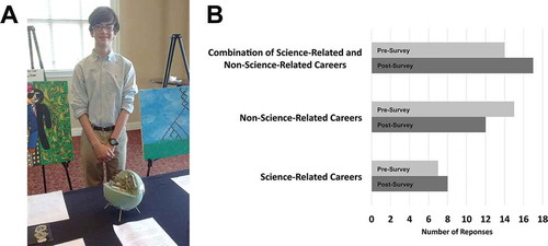
In one of our most recent art exhibit day events, high school student artist Noah Franks produced a cellular model out of recyclable materials. The idea for his larger statement on the importance of waste management in human society was sparked by learning about the existence of autophagy as a recycling pathway at the cellular, microscopic scale (). We observe that high school students consistently find interesting and personalized ways to interpret and communicate the science they were exposed to into art. Data that we have collected shows that high school participants, after engaging with our program, show an increased interest in careers that incorporate both science and art (). Pre-survey data show high school student artists had a baseline interest in STEM careers that increased with participation in the program. This interest might be an untapped resource in similar populations of students as we look to recruit creative students with different mindsets towards science and careers in STEM.
Figure 1. The Cell Art Collaborative Program. (A) recycell by Noah Franks (miscellaneous recyclable materials). This human cell model made from recyclable materials is symbolic of a prominent world issue and how what we learn about cells can help us solve important societal problems. The recyclable composition of the piece relates to the problem of extra waste, which is emphasized in the continents of the earth (made out of aluminum foil) on the exterior of the human cell model. The U.S. is the #1 trash-producing country in the world, highlighting the need to find ways to efficiently manage and recycle/reuse waste. Cells have efficient ways to dispose of their waste or damaged components. One of these processes is autophagy, during which old or damaged cellular parts are broken down into their building blocks, which are employed to build new ones. Cells and their processes should be an example to us. Just as all the parts of a cell work together to keep the cell useful and healthy, we should work together to keep our planet clean. (B) The Cell Art Collaborative Program helped high school participants refine their career interests. During the first year of the program, students were asked to describe their future career preferences before (pre-survey, light grey bars) and after (post-survey, dark grey bars) participating in the program. Answers were coded and quantified (High Point University Institutional Review Board approval #201511-418)
The Cell Art Collaborative program has consistently been implemented by undergraduate students who have dual interest in art and science. The successes of the first years of the event were achieved in part through the efforts of then-undergraduate student Casey Garr, now a graduate of the master’s program in biomedical visualization at The University of Illinois, Chicago, who works as a medical illustrator. This is an example of how a STEAM outreach program has supported individuals who have something unique to contribute to science. She has gone on to focus on biomedical animations, 3D renderings, and illustrations designed to inform, teach, and explain complex scientific topics (). Casey’s depiction of autophagy in highlights the formation of autophagosomes, the autophagic hallmark [Citation6]. In doing this, she captures in three dimensions the unique feature of a double-membraned or bilayered autophagosome, distinguishing both the inner and outer membranes and the intersection between them during autophagosome formation. This figure is an outstanding example of art as a tool for communication in that it perfectly conveys an aspect of autophagy that is usually not explicitly illustrated in autophagy diagrams.
Figure 2. Lipid biosynthesis and trafficking during autophagosome biogenesis by Casey Garr. The diagram was adapted from Goyal et al. [Citation3] and created using Autodesk 3ds Max. To create the protein models in the diagram, 3D data was collected from the Protein Data Bank. This data was utilized in the Visual Molecular Dynamics program to create 3D models, which were then transferred to and rendered in Autodesk 3ds Max. Due to the unavailability of complete 3D data, artistic license was used in the rendering of some proteins (Atg9 and Atg27)
![Figure 2. Lipid biosynthesis and trafficking during autophagosome biogenesis by Casey Garr. The diagram was adapted from Goyal et al. [Citation3] and created using Autodesk 3ds Max. To create the protein models in the diagram, 3D data was collected from the Protein Data Bank. This data was utilized in the Visual Molecular Dynamics program to create 3D models, which were then transferred to and rendered in Autodesk 3ds Max. Due to the unavailability of complete 3D data, artistic license was used in the rendering of some proteins (Atg9 and Atg27)](/cms/asset/593bb69d-a45a-4c6a-867b-4562a2143d74/kaup_a_1769972_f0002_c.jpg)
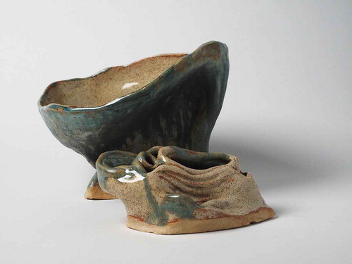
Casey’s multifaceted role in the program was subsequently taken on by another student interested in a science-driven art career, Candyce Sturgeon. Like Casey, Candyce aspires to enter into the field of medical illustration, and was able not only to help in the planning of experiments for the event, but also to design artwork to advertise the events and plan the art exhibit. Candyce’s work on the Cell Art Collaborative sparked her interest in bench research. Alongside her labmates, she recently published an autophagy-related scientific paper [Citation7]. Participating in the publishing process was a successful culmination not only of her bench work, but also of the creative process through which they pursued their research project and packaged it into a manuscript. Candyce’s work at the bench has also fueled her artwork with ideas (). Candyce’s sculpture pictured in captures the vacuole and the late Golgi, the donor and target organelles of the AP-3 pathway in budding yeast [Citation8]. Her interest in the AP-3 pathway stems from her Atg27-related work at the bench, a protein cargo of the AP-3 pathway [Citation9–11].
Figure 3. AP-3 Pathway: A sculpture by Candyce Sturgeon (clay). Inspired by Atg27 being an AP-3 cargo, Candyce set out to use clay to represent the donor and target organelles of the AP-3 pathway, the late Golgi and the vacuolar membrane
Conclusions
Our experiences provide examples consistent with the evidence in the educational literature that STEAM-based learning environments produce graduates who excel in their professional lives. These examples illustrate how the recruitment of highly creative students into STEM fields through connections to art can be a first step in defining a specialized career path that leads to a valuable and unique contribution to science. STEAM represents a tool to tailor methods of teaching to take advantage of a wider range of student talents and interests, preparing them to go forth into society as the creative thinkers and problem-solvers the world needs.
Acknowledgments
We thank the American Society for Cell Biology for funding (COMPASS Outreach grant) to implement the pilot Cell Art Collaborative (CAC) event (originally known as Emerging Undergraduate Research-Inspired Cell Art! Or EURICA!). We thank High Point University’s Department of Biology, Wanek School of Natural Sciences, and Cultural Grants Program for resources and funding. Data presented in was collected with the approval of High Point University Institutional Review Board #201511-418. We thank Professor Benita Vanwinkle for assistance in capturing the sculpture image.
Disclosure statement
No potential conflict of interest was reported by the authors.
References
- Klionsky DJ. The” found-art vacuole”-people learn in different ways. Autophagy. 2019;15:1493–1494.
- Klionsky DJ, Kumar A. A systems biology approach to learning autophagy. Autophagy. 2006;2:12–23.
- Klionsky DJ. An interactive exercise to learn eukaryotic cell structure & organelle function. Am Biol Teach. 1999;61:539–542.
- Falkenberg C, Holmes R, Natalizio B, et al. STEAM: using the Arts to Train Well-Rounded and Creative Scientists. J Microbiol Biol Educ. 2018;19:1–7.
- Budzinski C, Garr C, Hegedus T, et al. The art-science connection: students create art inspired by extracurricular lab investigations. Sci Teach. 2016;83:25–31.
- Goyal S, Robinson MR, Segarra VA, et al. Intracellular lipid homeostasis and trafficking in autophagy. In: Cellular Metabolism. London: IntechOpen. 2019. p. 1–16.
- Clemmer D, Khawaja A, Penton M, et al. Kinetic assay of starvation sensitivity in yeast autophagy mutants allows for the identification of intermediary phenotypes. BMC Res Notes. 2019;12:1–5.
- Stepp JD, Huang K, Lemmon SK. The yeast adaptor protein complex, AP-3, is essential for the efficient delivery of alkaline phosphatase by the alternate pathway to the vacuole. J Cell Biol. 1997;139:1761–1774.
- Segarra VA, Boettner DR, Lemmon SK. Atg27 tyrosine sorting motif is important for its trafficking and Atg9 localization. Traffic. 2015;16:365–378.
- Ma M, Burd CG, Chi RJ. Distinct complexes of yeast Snx4 family SNX‐BARs mediate retrograde trafficking of Snc1 and Atg27. Traffic. 2017;18:134–144.
- Suzuki SW, Emr SD. Membrane protein recycling from the vacuole/lysosome membrane. J Cell Biol. 2018;217:1623–1632.