
1. Prader-Willi syndrome
Prader-Willi syndrome (PWS) is a complex, multisystem neurodevelopmental disorder that occurs with a frequency of approximately 1/10,000 to 1/30,000 [Citation1]. PWS results from the loss of expression of paternally derived genes by a variety of mechanisms, which include paternally inherited deletions (70–75%), maternal uniparental disomy (20–30%), and imprinting defects (2–5%) [Citation2,Citation3]. DNA methylation analysis will correctly diagnose more than 99% of cases, and is the most sensitive genetic test available [Citation1–Citation4]. The genetic anomalies lead to a distinctive phenotype that includes central hypotonia, cognitive delay, developmental disabilities, behavioral problems, obesity, growth hormone deficiency, hypogonadism, and life-threatening hyperphagia [Citation1].
Infants with PWS have feeding difficulties, initially necessitating assisted feeding, often via gastrostomy tube or nasogastric tube. The feeding issues gradually improve and are followed by weight gain without a change in calories (18 months to 3 years of age) and subsequently, by an increased interest in food in early childhood (4–5 years of age) [Citation5]. Hyperphagia and food-related behavior problems often evolve by 8–9 years of age, and are characterized by the lack of a normal satiety response, food preoccupations, and problematic food-seeking behaviors [Citation5]. Although symptom severity varies, hyperphagia poses persistent, life-long risks to the health and safety of affected individuals. While strict environmental control and diet modifications may help reduce caloric intake and effectively manage weight for some individuals with PWS, the persistent drive for food prevents individuals with PWS from achieving a high quality of life or being well-integrated into their communities.
Individuals with PWS have a characteristic eating pattern [Citation6]. They exhibit a slower initiation of eating with a longer meal duration and no deceleration in the amount eaten over the entire mealtime [Citation6]. They have an earlier return of hunger after completion of a meal, and will often start asking about the next meal while still eating. Left to their own devices, individuals with PWS will consume three times more food than obese control subjects [Citation6,Citation7]. They have a strong preference for higher-carbohydrate foods, and are more likely to consume inedible items (e.g. toothpaste, lotion) than obese controls [Citation6,Citation7]. This hyperphagia results in food-seeking behavior, often with hoarding or foraging for food, and stealing of food or money to buy food. In most individuals, gastric emptying is delayed, and vomiting is rare [Citation8]. Thus, the drive to eat in individuals with PWS remains significantly elevated even in the face of delayed gastric emptying, which is unusual when compared to individuals with diet-induced obesity.
Individuals with PWS have a very high risk for obesity, both due to the hyperphagia and eating behaviors, as well as from decreased total caloric requirement due to decreased muscle mass and lower resting energy expenditure. The obesity in PWS is primarily central (abdomen, buttocks, and thighs) in both sexes, and interestingly, there is less visceral fat in obese individuals with PWS than would be expected for the degree of obesity [Citation9,Citation10]. Although there are many common clinical features exhibited by people with PWS, the hyperphagia is the unifying symptom in this diagnosis [Citation11], in addition to being the most life-limiting feature.
The underlying molecular mechanisms that contribute to excessive weight gain and hyperphagia in PWS remain incompletely understood. Appetite and satiety issues are regulated by a complex interplay between gut hormones and hypothalamic neuropeptides. Most gut-related appetite-regulating hormones are normally expressed in PWS, with the notable exception being ghrelin, a growth hormone secretagogue that is high in fasting states and decreases with eating [Citation12,Citation13]. Highly elevated levels of ghrelin have been identified in individuals with PWS [Citation12,Citation13]. Ghrelin levels decrease after intake of a meal, as is normal, but still remain comparatively high [Citation14]. However, it has been shown that circulating ghrelin levels are elevated in young children with PWS long before the onset of hyperphagia, especially during the early phase of poor appetite and feeding [Citation15], so it seems unlikely that high ghrelin levels are directly responsible for the switch to the hyperphagic nutritional phases in PWS. Thus, the underlying metabolic/hormonal cause of the hyperphagia remains unclear.
Functional MRI studies have shown abnormally high activation of the reward pathways in the brain for food-related stimuli, as well as hyperfunction of the areas that drive eating behavior (e.g. amygdala) in individuals with PWS [Citation16]. Functional MRI studies have also shown abnormal functional resting state connectivity between the hypothalamus and areas of the brain involved in processing satiety responses [Citation17]. It is hypothesized that the combination of the increased reward value for food and the abnormalities in the hypothalamus-neural connections in PWS cause the characteristic hyperphagia.
Growing evidence suggests that the oxytocin-producing neurons in the hypothalamus are a critical piece of the puzzle of hyperphagia in PWS. Oxytocin is a nine-amino acid neuropeptide hormone that is predominantly produced in the hypothalamic paraventricular nucleus (PVN) and supraoptic nucleus. Oxytocin plays a critical role in regulation of food intake via the nucleus tractus solitarius, the dorsal motor nucleus of the vagus nerve and the area postrema, as well as in reward processing of food and eating behaviors via the ventral tegmental area, nucleus accumbens, and the nucleus stria terminalis (reward pathways) [Citation17]. Oxytocin administration causes a restraint of food intake by enhancing the activity of brain regions that exert cognitive control, while concomitantly increasing the activity of structures that process food reward value [Citation18]. Peripheral administration of oxytocin also results in activation of neurons in hindbrain areas (e.g. dorsal vagal complex) linked to the control of meal size and forebrain areas (e.g. hypothalamus, amygdala) linked to control of body weight [Citation18]. Administration of exogenous oxytocin circumvents the decrease in metabolic rate that typically accompanies weight loss, likely by inducing lipolysis and fat oxidation [Citation19]. Typically, oxytocin is also produced and released in local tissues, such as the gastrointestinal tract, where it has autocrine and paracrine effects [Citation20]. Whether this peripheral oxytocin production is present in individuals with PWS is unknown.
It is known that the volume of the PVN and total number of cells present in the PVN were lower in the brains of the individuals with PWS from autopsy studies [Citation21]. The number of paravocellular oxytocin neurons was significantly decreased in the PWS brains, which suggests that these cells might be connected to decreased satiety in PWS [Citation21]. Additionally, there are fairly low levels of oxytocin found in the cerebrospinal fluid of individuals with PWS [Citation22]. Taken together, these findings suggest that lack of appropriate oxytocin levels could be a reason for the lack of satiety in PWS. As intranasal oxytocin can cross the blood-brain-barrier and have direct central effects by binding to oxytocin receptors throughout the brain, exogenous administration of oxytocin is being extensively studied in individuals with PWS.
2. Oxytocin and appetite
Interestingly, oxytocin has two opposite effects on feeding behavior depending on the timing of administration. In infancy, oxytocin induces suckling and helps improve feeding in PWS; whereas later in life, oxytocin is a potent anorexigenic hormone which is integral to the complex neural and gut networks associated with the homeostatic control of food intake, satiety, and energy balance [Citation23,Citation24]. Studies have demonstrated that administration of intranasal oxytocin in adults suppresses food intake and acts most potently in obese individuals, as obesity seems to be a particularly oxytocin-sensitive state [Citation25]. In the periphery, oxytocin is involved in electrolyte homeostasis, gastric motility, glucose homeostasis, adipogenesis, and osteogenesis, while within the brain it is involved in food reward and food choice, as well as satiety [Citation26]. Oxytocin preferentially suppresses intake of sweet-tasting carbohydrates while improving glucose tolerance and supporting bone remodeling [Citation27]. In neurotypical individuals, oxytocin release is stimulated by changes in osmolality, distension of the stomach, a diet high in sweet-tasting carbohydrates, and ingestion of a satiating amount of food [Citation28]. Exogenous administration of oxytocin decreases the appetite for carbohydrates (particularly sweet ones) and for non-nutritive sweeteners like saccharin [Citation27]. Typically, once the plasma osmolality, toxicity, or gastrointestinal stretch parameters reach levels that jeopardize the internal mileu, oxytocin ensures that feeding termination occurs, regardless of whether the energy needs of the individual have been met [Citation28].
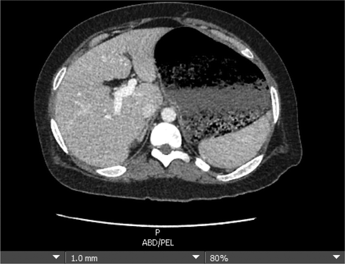
The mechanisms of oxytocin release are clearly abnormal in individuals with PWS. Reports indicate that 3–6% of mortality in PWS is due to gastric necrosis as a result of severe gastric distension following binges of food intake () [Citation29]. Additionally, many studies have shown that individuals with PWS have preferences for sweet tastes and for calorie-dense foods over lower calorie foods, but do not feel satiated at termination of a meal and can consume tens of thousands of calories at a time during a food binge [Citation30]. All of these findings indicate that the normal mechanisms of oxytocin release are not present in individuals with PWS. These findings, coupled with the decreased number of oxytocin neurons and low levels of oxytocin in the cerebrospinal fluid of individuals with PWS, suggest that administration of exogenous oxytocin could be beneficial in helping to control food intake in this syndrome.
Figure 1. Severe gastric dilation in 21 year old male with PWS following food binge. Residual food is seen within the lumen of the extremely dilated stomach.
3. Oxytocin and weight control
Animal studies have consistently shown that oxytocin administration causes early termination of food intake, thereby causing weight loss [Citation31]. One study of rhesus monkeys with diet-induced obesity found that oxytocin administration caused 27% reduction in food intake with 3.3% weight loss and 14% increased energy expenditure [Citation32]. While studies in humans are still scarce, early findings suggest that intranasal oxytocin treatment reduces the drive to eat for pleasure, decreases reward-induced eating, improves cognitive control of food choices, and improves energy expenditure [Citation27]. One study found that 8 weeks of intranasal oxytocin treatment in obese adults resulted in 9 kg of body weight loss and a decrease in waist and hip circumference [Citation33].
Preclinical studies show that oxytocin increases lipolysis and fat oxidation, thereby reducing visceral fat, liver fat, and triglycerides [Citation34] A single dose of intranasal oxytocin can result in a decrease in respiratory quotient, indicating that oxytocin administration is causing an increase in fat oxidation [Citation35]. Oxytocin also directly increases glucose uptake in skeletal muscles by activating AMP-activated protein kinase (AMPK), which may explain how oxytocin administration helps to preserve lean muscle mass and to maintain energy expenditure despite weight loss [Citation36]. In addition to these direct metabolic benefits of exogenous oxytocin administration, there may also be complimentary effects on weight control due to oxytocin downregulating the hypothalamic–pituitary–adrenal axis [Citation37]. There are oxytocin receptors located in the anterior pituitary and adrenal glands, and administration of oxytocin reduces levels of adrenocorticotropic hormone and cortisol [Citation38]. Although oxytocin administration reduces cortisol levels, hypoadrenalism has not been reported as a consequence of treatment. Further studies are needed to determine whether the effects of oxytocin on the hypothalamic–pituitary–adrenal axis lead to an improved metabolic profile with longer treatment trials in humans.
4. Intranasal oxytocin in PWS
Thus far, studies of the effects of intranasal oxytocin on hyperphagia in individuals with PWS have had conflicting results. Three studies have shown potential benefits of oxytocin on appetite in children and adults with PWS, whereas one study showed no benefits of intranasal oxytocin on hyperphagia [Citation24,Citation39–Citation41]. It has been postulated that the study finding no positive results of intranasal oxytocin in adults and adolescents with PWS may have been due to an overdose of the hormone [Citation41]. In animal models, there is considerable evidence that oxytocin can bind to and affect receptors for the closely related peptide, arginine vasopressin (AVP); particularly the V1A receptor subtype [Citation42]. At higher levels, oxytocin and AVP are partial agonists for their homologous receptors [Citation43]. Thus, the high doses of IN-OT used in the study by Einfeld et al. could have saturated the OT receptors and bound to the AVP receptors, thus resulting in lack of benefit of the oxytocin administration. As individuals with PWS have reduced oxytocin receptors [Citation21], even a minimal overdose of exogenously administered oxytocin could presumably result in increased binding of the oxytocin to the AVP receptors, which could create negative/unwanted behaviors. Therefore, dose-finding studies are essential before clinicians prescribe this medication.
Ideally, dose-finding studies would utilize measurement of oxytocin levels after treatment to correlate oxytocin levels with the outcome measure (hyperphagia decrease in this case). After intranasal administration of oxytocin, levels significantly increase in both plasma and cerebral spinal fluid [Citation44]. Plasma concentrations of oxytocin peak at 15 min after intranasal administration and decrease after 75 min, whereas cerebral spinal fluid concentrations can take up to 75 min to reach a significant level [Citation44]. However, recent studies have demonstrated that there is no correlation between plasma oxytocin levels and oxytocin concentration in the cerebral spinal fluid [Citation45]. The currently available oxytocin assays all have deficits and all are likely measuring both oxytocin as well as other molecules erroneously tagged as oxytocin [Citation46]. Therefore, the measurements of oxytocin levels after intranasal administration in all of the published studies are likely invalid, making it difficult to know whether dosing plays a role in the conflicting results seen in studies of intranasal oxytocin in individuals with PWS.
Regardless, the possibilities of appetite-reduction, weight loss, improved metabolic profile, and decreased affinity for sweet-tasting carbohydrates with oxytocin administration in individuals with PWS are impressive to ponder. It is possible that administration of exogenous oxytocin to individuals with PWS could decrease the most devastating and universal feature of this syndrome – hyperphagia. Clearly additional research needs to be done in this vulnerable population, but oxytocin treatment offers potential hope for patients and families affected by this devastating disease.
5. Expert opinion
The severe, excessive appetite and the resultant obesity combined with a lower metabolic rate, poor gastrointestinal motility, anxiety, and compulsive behaviors make PWS an exceedingly difficult syndrome to manage. As oxytocin helps regulate both food intake and metabolism, treatment with intranasal oxytocin in the correct dose may improve a variety of symptoms in PWS, and thus improve quality of life for affected individuals and their families. As the correct dosing regimen remains to be elucidated, additional research is necessary before widespread treatment is undertaken.
Declaration of Interest
J Miller has received research funding from NIH, Prader-Willi syndrome Association U.S.A., Foundation for Prader-Willi Research, Zafgen Pharmaceuticals, Ferring Pharmaceuticals, Rhythm Pharmaceuticals, and GLWL. The authors have no other relevant affiliations or financial involvement with any organization or entity with a financial interest in or financial conflict with the subject matter or materials discussed in the manuscript apart from those disclosed. Peer reviewers on this manuscript have no relevant financial or other relationships to disclose
References
- Driscoll DJ, Miller JL, Schwartz S, et al. In: Adam MP, Ardinger HH, Pagon RA, et al., editors. GeneReviews® [Internet]. Seattle (WA): University of Washington, Seattle;1998 Oct 6. [cited 2017 Dec 14]. p. 1993–2018.
- Sanjeeva GN, Maganthi M, Kodishala H, et al. Clinical and molecular characterization of Prader-Willi syndrome. Indian J Pediatr. 2017 Nov;84(11):815–821.
- Bittel DC, Kibiryeva N, Sell SM, et al. Whole genome microarray analysis of gene expression in Prader-Willi syndrome. Am J Med Genet A. 2007;1(5):415–421.
- Buiting K, Cassidy SB, Driscoll DJ, et al. Clinical utility gene card for: Prader-Willi syndrome. Eur J Hum Genet. 2014;22:9.
- Miller JL, Lynn CH, Driscoll DC, et al. Nutritional phases in Prader-Willi syndrome. Am J Med Genet A. 2011;155A(5):1040–1049.
- Martínez Michel L, Haqq AM, Wismer WV. A review of chemosensory perceptions, food preferences and food-related behaviours in subjects with Prader-Willi syndrome. Appetite. 2016 Apr;1(99):17–24.
- Joseph B, Egli M, Koppekin A, et al. Food choice in people with Prader-Willi syndrome: quantity and relative preference. Am J Ment Retard. 2002 Mar;107(2):128–135.
- Hoybye C, Barkeling B, Naslund E, et al. Eating behavior and gastric emptying in adults with Prader-Willi syndrome. Ann Nutr Metab. 2007;51(3):264–269.
- Tanaka Y, Abe Y, Oto Y, et al. Characterization of fat distribution in Prader-Willi syndrome: relationships with adipocytokines and influence of growth hormone treatment. Am J Med Genet A. 2013 Jan;161A(1):27–33.
- Sode-Carlsen R, Farholt S, Rabben KF, et al. Höybye C body composition, endocrine and metabolic profiles in adults with Prader-Willi syndrome. Growth Horm IGF Res. 2010 Jun;20(3):179–184.
- Heymsfield SB, Avena NM, Baier L, et al. Hyperphagia: current concepts and future directions proceedings of the 2nd international conference on hyperphagia. Obesity (Silver Spring). 2014 Feb;22(Suppl 1):S1–S17.
- Kuppens RJ, Diène G, Bakker NE, et al. Elevated ratio of acylated to unacylated ghrelin in children and young adults with Prader-Willi syndrome. Endocrine. 2015 Dec;50(3):633–642.
- Haqq AM, Grambow SC, Muehlbauer M, et al. Ghrelin concentrations in Prader-Willi syndrome (PWS) infants and children: changes during development. Clin Endocrinol (Oxf). 2008 Dec;69(6):911–920.
- Bizzarri C, Rigamonti AE, Luce A, et al. Children with Prader-Willi syndrome exhibit more evident meal-induced responses in plasma ghrelin and peptide YY levels than obese and lean children. Eur J Endocrinol. 2010 Mar;162(3):499–505.
- Kweh FA, Miller JL, Sulsona CR, et al. Hyperghrelinemia in Prader-Willi syndrome begins in early infancy long before the onset of hyperphagia. Am J Med Genet A. 2015;167A(1(Jan):69–79.
- Holsen LM, Zarcone JR, Brooks WM, et al. Neural mechanisms underlying hyperphagia in Prader-Willi syndrome. Obesity (Silver Spring). 2006 Jun;14(6):1028–1037.
- Lukoshe A, van Dijk SE, van den Bosch GE, et al. Altered functional resting-state hypothalamic connectivity and abnormal pituitary morphology in children with Prader-Willi syndrome. J Neurodev Disord. 2017 Feb;9(1):12.
- Spetter MS, Feld GB, Thienel M, et al. Oxytocin curbs calorie intake via food-specific increases in the activity of brain areas that process reward and establish cognitive control. Sci Rep. 2018 Feb 9;8(1):2736.
- Olszewski PK, Klockars A, Levine AS. Oxytocin and potential benefits for obesity treatment. Curr Opin Endocrinol Diabetes Obes. 2017 Oct;24(5):320–325.
- Ohlsson B, Truedsson M, Djerf P, et al. Oxytocin is expressed throughout the human gastrointestinal tract. Regul Pept. 2006 Jul 15;135(1–2):7–11.
- Swaab DF, Purba JS, Hofman MA. Alterations in the hypothalamic paraventricular nucleus and its oxytocin neurons (putative satiety cells) in Prader-Willi syndrome: a study of five cases. J Clin Endocrinol Metab. 1995 Feb;80(2):573–579.
- Martin A, State M, Anderson GM, et al. Cerebrospinal fluid levels of oxytocin in Prader-Willi syndrome: a preliminary report. Biol Psychiatry. 1998 Dec 15;44(12):1349–1352.
- Tauber M, Boulanouar K, Diene G, et al. The use of oxytocin to improve feeding and social skills in infants with Prader-Willi syndrome. Pediatrics. 2017;139(2):Feb.
- Kuppens RJ, Donze SH, Hokken-Koelega AC. Promising effects of oxytocin on social and food-related behaviour in young children with Prader-Willi syndrome: a randomized, double-blind, controlled crossover trial. Clin Endocrinol (Oxf). 2016 Dec;85(6):979–987.
- Schorr M, Marengi DA, Pulumo RL, et al. Oxytocin and its relationship to body composition, bone mineral density, and hip geometry across the weight spectrum. J Clin Endocrinol Metab. 2017 Aug 1;102(8):2814–2824.
- Iwasaki Y, Maejima Y, Suyama S, et al. Peripheral oxytocin activates vagal afferent neurons to suppress feeding in normal and leptin-resistant mice: a route for ameliorating hyperphagia and obesity. Am J Physiol Regul Integr Comp Physiol. 2015 Mar 1;308(5):R360–9.
- Striepens N, Schröter F, Stoffel-Wagner B, et al. Scheele D oxytocin enhances cognitive control of food craving in women. Hum Brain Mapp. 2016 Dec;37(12):4276–4285.
- Ho JM, Blevins JE. Coming full circle: contributions of central and peripheral oxytocin actions to energy balance. Endocrinology. 2013 Feb;154(2):589–596.
- Stevenson DA, Heinemann J, Angulo M, et al. Gastric rupture and necrosis in Prader-Willi syndrome. J Pediatr Gastroenterol Nutr. 2007 Aug;45(2):272–274.
- Glover D, Maltzman I, Williams C. Food preferences among individuals with and without Prader-Willi syndrome. Am J Ment Retard. 1996 Sep;101(2):195–205.
- Roberts ZS, Wolden-Hanson T, Matsen ME, et al. Chronic hindbrain administration of oxytocin is sufficient to elicit weight loss in diet-induced obese rats. Am J Physiol Regul Integr Comp Physiol. 2017 Oct 1;313(4):R357–R371.
- Blevins JE, Graham JL, Morton GJ, et al. Chronic oxytocin administration inhibits food intake, increases energy expenditure, and produces weight loss in fructose-fed obese rhesus monkeys. Am J Physiol Regul Integr Comp Physiol. 2015 Mar 1;308(5):R431–-8.
- Ott V, Finlayson G, Lehnert H, et al. Oxytocin reduces reward-driven food intake in humans. Diabetes. 2013 Oct;62(10):3418–3425.
- Cai D, Purkayastha S. A new horizon: oxytocin as a novel therapeutic option for obesity and diabetes. Drug Discov Today Dis Mech. 2013 Jun 1;10(1–2):e63–e68.
- Maejima Y, Iwasaki Y, Yamahara Y, et al. Peripheral oxytocin treatment ameliorates obesity by reducing food intake and visceral fat mass. Aging (Albany NY). 2011 Dec;3(12):1169–1177.
- Lee ES, Uhm KO, Lee YM, et al. Oxytocin stimulates glucose uptake in skeletal muscle cells through the calcium-CaMKK-AMPK pathway. Regul Pept. 2008 Nov 29;151(1–3):71–74.
- Stanić D, Plećaš-Solarović B, Mirković D, et al. Oxytocin in corticosterone-induced chronic stress model: focus on adrenal gland function. Psychoneuroendocrinology. 2017 Jun;80:137–146.
- Wirth MM, Gaffey AE, Martinez BS. Effects of intranasal oxytocin on steroid hormones in men and women. Neuropsychobiology. 2015;71(4):202–211.
- Tauber M, Mantoulan C, Copet P, et al. Oxytocin may be useful to increase trust in others and decrease disruptive behaviours in patients with Prader-Willi syndrome: a randomised placebo-controlled trial in 24 patients. Orphanet J Rare Dis. 2011 Jun 24;6:47.
- Miller JL, Tamura R, Butler MG, et al. Oxytocin treatment in children with Prader-Willi syndrome: a double-blind, placebo-controlled, crossover study. Am J Med Genet A. 2017 May;173(5):1243–1250.
- Einfeld SL, Smith E, McGregor IS, et al. A double-blind randomized controlled trial of oxytocin nasal spray in Prader Willi syndrome. Am J Med Genet A. 2014 Sep;164A(9):2232–2239.
- Francis SM, Sagar A, Levin-Decanini T, et al. Oxytocin and vasopressin systems in genetic syndromes and neurodevelopmental disorders. Brain Res. 2014 Sep;11(1580):199–218.
- Song Z, Albers HE. Cross-talk among oxytocin and arginine-vasopressin receptors: relevance for basic and clinical studies of the brain and periphery. Front Neuroendocrinol. 2017 Oct 18.
- Striepens N, Kendrick KM, Hanking V, et al. Elevated cerebrospinal fluid and blood concentrations of oxytocin following its intranasal administration in humans. Sci Rep. 2013 Dec;6(3):3440.
- Kagerbauer SM, Martin J, Schuster T, et al. Plasma oxytocin and vasopressin do not predict neuropeptide concentrations in human cerebrospinal fluid. J Neuroendocrinol. 2013 Jul;25(7):668–673.
- Szeto A, McCabe PM, Nation DA, et al. Evaluation of enzyme immunoassay and radioimmunoassay methods for the measurement of plasma oxytocin. Psychosom Med. 2011 Jun;73(5):393–400.