3,489
Views
7
CrossRef citations to date
0
Altmetric
Research Article

Physicochemical properties and oxygen affinity of glutaraldehyde polymerized crocodile hemoglobin: the new alternative hemoglobin source for hemoglobin-based oxygen carriers

, , , , , & show all
Pages 852-861 | Received 14 Nov 2018, Accepted 29 Jan 2019, Published online: 15 Mar 2019

Abstract

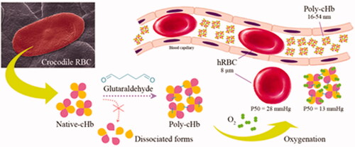
Hemoglobin-based oxygen carriers (HBOCs) are modified stroma-free hemoglobin molecules used in developing a blood substitute for therapeutic usage. In order to prevent hemoglobin dissociation, glutaraldehyde (GTA) was used to generate high-molecular weight heterogeneous crocodile hemoglobin (Poly-cHb). This work, Poly-cHb was created using various GTA concentrations, ranging from 0.025-0.150% (v/v). Physicochemical properties were investigated that were comparable GTA polymerized human hemoglobin (Poly-hHb). This study has revealed that GTA polymerization increases the molecular size of Native-cHbs from 14.10 nm over a range from 16.31 to 54.27 nm. Moreover, this polymerization alters the secondary structure and heme environment by decreasing the helicity ratio from 1.00 to 0.95 at the highest condition and exhibits hypochromic shift of the Soret band to be 0.88 times lower than the native. However, all Poly-cHbs still possessed higher oxygen affinity than that of Poly-hHbs with average P50 values of 13 and 21 mmHg, respectively. Although, polymerization affected the overall Poly-cHb structure slightly, but compensated by decreasing the denaturation level to lower than 10%. Thus, it is interesting to note that Poly-cHb may advantageously provide effective oxygen carriage and ability for pasteurization, which may benefit the search for new alternative hemoglobin sources for HBOC development.

GRAPHICAL ABSTRACT

Introduction

Hemoglobin (Hb) has been investigated mainly for its function as an oxygen transport protein. One molecule of natural tetrameric hemoglobin consists of two identical α- and β-polypeptide chains, each incorporating an iron-containing heme as a prosthetic group. The oxygen-binding ability of hemoglobin is mediated by the redox mechanism of the iron atom, in which its reduced state (Fe2+) is acceptable for oxygen binding. However, a reversible oxidized state (Fe3+), a nonfunctional methemoglobin could occur normally via autoxidation. In the sense of Hb’s biochemical activity, NAD-cytochrome b5 reductase plays an important role in maintaining the reduced state of Hb in red blood cells (RBCs) [Citation1,Citation2].

So far, hemoglobin has been utilized to prepare oxygen carrying compounds called hemoglobin-based oxygen carriers (HBOCs), which aim to overcome limitations of blood transfusions [Citation3–5]. Stored RBC products are restricted to a short shelf-life through loss in cell membrane integrity, unleashing hemoglobin and its negatively charged regulator (2,3-bisphosphoglycerate; 2,3-BPG) leading to ineffective RBCs [Citation6,Citation7]. Moreover, blood compatibility, pathological contamination, immunosuppression, and acute lung injury are critical issues which have received much attention for safe blood substitute usage [Citation8,Citation9].

Stroma-free hemoglobin (SFH) has many benefits, such as not requiring blood group compatibility, availability in sterilization and being highly effective at oxygen delivery in plasma. On the other hand, living outside the RBC may affect the oxygen unloading capacity of SFH because of lacking in appropriate regulation of 2,3-BPG. Consequently, the stroma-free hemoglobin exhibits higher oxygen affinity than living in its natural environment and changes its oxygen unloading capacity. These advantages were found to be solved by the use of animal hemoglobin, such as bovine hemoglobin (bHb), which its oxygen affinity is not mainly regulated by 2,3-BPG but highly sensitive to chloride ion [Citation10]. Likewise, the crocodilian hemoglobin (cHb), its oxygen affinity is markedly reduced by bicarbonate ion instead of 2,3-BPG which resulted in a controllable oxygen releasing ability outside red blood cells’ environment [Citation11]. These unique characteristics lead them to possess different oxygen affinity with P50 values of 13.8, 11.3 and 3.9 mmHg for hHb [Citation12], bHb [Citation13] and cHb [Citation14], respectively. While basic physical properties are not significantly different with molecular weight ranged 53.0–64.5 kDa and the number of amino acid residues of α and β chains are 141 and 146 residues, respectively [Citation14–16]. However, unmodified SFH favorably dissociates into monomers and dimers, resulting in a short circulatory life, renal toxicity via precipitation, alteration in blood viscosity and colloidal osmotic pressure (COP), inducing vasoconstriction and hypertension and many adverse effects [Citation17–20].

To prevent these issues, Hb must be firstly modified to reduce its dissociation. Inter/intra molecular crosslinking of Hb using glutaraldehyde (GTA) have been created to generate high-molecular weight Hb polymer products. PolyBvHb (Hemopure®, Biopure Corporation, Cambridge, Massachusetts, USA) is known as GTA αα intra/intermolecular crosslinked bovine hemoglobin and was developed to treat acute anemia patients during surgery [Citation21]. Likewise, PolyHb (PolyHeme®, Northfield Laboratories Inc., Evanston, IL), GTA polymerized human hemoglobin has also been developed to treat trauma and acute blood loss [Citation22]. These products reflect the effectiveness of GTA as a crosslinking agent. On the other hand, most HBOCs are made of human Hb (hHb) derived from outdated human blood, which is restricted for human blood supply. Thus, finding new approaches to create HBOCs from new Hb sources may overcome these limitations.

Crocodile Hb (cHb) has been reported to possess higher oxygen affinity than that of hHb [Citation23]. This characteristic may be beneficial in the sense of having high-residual oxygen affinity after passing through a strong chemical modification. Since, it is sensitively regulated by bicarbonate ions instead of 2,3-BPG [Citation11]. Thus, the oxygen binding and releasing ability of modified stroma-free cHb in blood circulation could be properly controllable. In Thailand, Siamese crocodile (Crocodylus siamensis) blood is an abundant waste product in the crocodile meat and leather industries. Therefore, it may meet requirements for HBOC development. Hence, this study aimed to produce modified stroma-free cHb through one-step polymerization using GTA to yield a high-molecular weight of Hb polymer (Poly-Hb). Then, its physicochemical properties were investigated, comparing them to hHb. The results obtained reveal the suitable properties of this new Hb source and may give us a new interesting alternative for HBOC development.

Experimental procedures

Materials

Crocodile blood was supplied by a slaughterhouse of Sriracha Moda Co., Ltd. (Chon Buri, Thailand) and the outdated human blood was obtained from the Central Blood Bank (Faculty of Medicine, Khon Kaen University, Khon Kaen, Thailand). The 50% (v/v) GTA was purchased from Fluka analytical (Munich, Germany) and all chemicals were purchased as analytical grade.

Preparation of glutaraldehyde polymerized Hb (Poly-Hb)

Hemoglobin extraction

Crocodile blood was collected and managed following the method of Pata et al. [Citation24]. Then, Hb was extracted from RBCs by the method of Jandaruang et al. [Citation25] with minor modifications. Briefly, whole blood was centrifuged at 8000×g for 2 min (4 °C) (High speed refrigerated centrifuge, Himac CR 22GII, Hitachi, Japan) to collect the RBCs. The collected cells were re-suspended in 1× phosphate-buffered saline (PBS, pH 7.4). Then, other contaminants were removed using centrifugation at 3000×g for 5 min (4 °C). The RBCs were further lysed in cold double distilled water with vigorous shaking. Material was stored at 4 °C for 10 min. Then, hemoglobin solution was obtained after cell debris was removed by centrifugation at 10,000×g for 20 min (4 °C).

Glutaraldehyde (GTA) polymerization

Five milliliters of 10 mg/ml Hb solution were prepared in double distilled water. Crosslinking conditions were divided into two groups. First, Hb solution was mixed with 5% (v/v) GTA to obtain 0.025, 0.050, 0.075, 0.100, 0.125, and 0.150% (v/v) GTA final concentrations. The reaction mixtures were gently shaken on ice for 2 h. Second, 0.175, 0.200, 0.225, 0.250, 0.275 and 0.300% (v/v) GTA polymerized Hb (Poly-Hb) solution was prepared and shaken on ice for 4 h. Then, 1 ml of 1 M Tris-HCl (pH 8.0) was added to quench the reaction. The Poly-Hb solutions were collected after centrifugation at 8000×g for 5 min. Unreacted GTA was removed from Poly-Hb samples using dialysis against double distilled water for 24 h at 4 °C.

Protein pattern and molecular weight determination

The sodium dodecyl sulfate acrylamide gel electrophoresis (SDS-PAGE) technique was carried out to examine the protein pattern and molecular weight [Citation26]. SDS gels were prepared with 14% separating gel and 4% stacking gel (30% T acrylamide, 1.5 M Tris-HCl (pH 8.8), 1.5 M Tris-HCl (pH 6.8), 10% SDS, 10% APS, TEMED, and DDW). Protein samples were prepared by mixing with 2 × SDS loading buffer (0.5 M Tris-HCl (pH 6.8), 0.5% (w/v) bromophenol blue, 2% (w/v) SDS, 10% (v/v) glycerol, 10% (v/v) β-mercaptoethanol) at a ratio of 1:1. After the protein mixture was boiled for 5 min, it was allowed to be separated under an electric field of 120 volts for 2 h (Mini Protein® 3 Cell and PowerPac™ Basic, Bio-Rad, California, USA). The low-molecular weight calibration kit (Amersham™, GE healthcare, Chicago, IL) was used as a protein standard marker. The SDS gels were stained by Coomassie brilliant blue R-250 and the protein patterns were observed after several changes of destaining solution. The average particle size of Poly-Hb samples was measured using Zetasizer Nano-S90 Ver. 7.11 (Malvern Instruments Ltd, Malvern, UK).

Ultraviolet-visible (UV-Vis) spectrophotometry and circular dichroism (CD) spectroscopy

The Hbs (Native-Hbs and Poly-Hbs) at a concentration of 0.125 mg/ml were prepared in 1× PBS (pH 7.4). Absorbance was measured in the wavelength range of 200–250 nm (far UV region) and over 250–700 nm (near UV to visible light region) for CD analysis (J-815 CD spectrometer, Jasco, Japan) and UV-Vis spectrophotometry analysis (SpectraMax M5, Molecular device Inc., California, USA), respectively. All samples were analyzed using a 1 cm path length cuvette. To investigate the secondary structure changes of protein, the raw CD data in units of mdeg was converted to units of θ (deg•cm2/dmol).

Oxygen affinity measurement

The oxygen affinity was performed by the conventional method of Asakura et al. [Citation27] with minor modifications. Briefly, four milliliters of 35 µM (heme) Hb sample was prepared in 1× PBS, containing 71.4 mM of Cl (pH 7.4) in the Thunberg tube. Deoxyhemoglobin was obtained by flushing Argon gas repeatedly. Then, oxygenation was performed by adding a known volume of air to the Thunberg tube. The incubation at 25 °C for 5 min was done before the absorption spectra of 400–700 nm were measured (Spectronic 200, Thermo Fisher Scientific, Waltham, MA). The changes of deoxyhemoglobin spectra to oxyhemoglobin spectra were observed. The percentage of oxygen saturation (%Y) and partial pressure of the oxygen (pO2) was calculated by the following equations, %Y = (D/DHbO2) × 100, Absorbance of Hb = (A577 of oxygenated peak – A577 of deoxygenated peak) + (A555 of deoxygenated peak – A555 of oxygenated peak)], and pO2 = (1/Vc) × [PaV0 – 760(1.36XY/100)]. In these equations, D is the absorbance of Hb under each round of examination, DHbO2 is the absorbance of Hb at the complete oxygenation step, pO2 is the partial pressure of oxygen at 25 °C, Pa is the oxygen pressure in the air at atmospheric conditions (150 mmHg), V0 is the volume of introduced air (ml), Vc is the volume of the gas phase inside the Thunberg tube (ml) and X is the weight of Hb in solution (g). The oxygen affinity and cooperative activity were reported in terms of P50 value and Hill’s cooperativity coefficient (n value) which was obtained from the oxygen equilibrium curve (OEC) and Hill’s plot, respectively.

The Bohr effect of selected polymerized cHb was observed in 50 mM Tris-HCl buffer (pH 6.9–8.4) and the Bohr coefficient (δH+) was calculated by the following equation, δH+ = ΔlogP50/ΔpH.

Thermal denaturation

The precipitation test, a semi-quantitative analysis, was performed to determine thermal denaturation for Hb [Citation28]. Eleven milliliters of 0.1 M sodium phosphate buffer (pH 7.4), containing 0.03 g Hb, was prepared. One milliliter of Hb solution was divided into 12 aliquots in tight-capped microcentrifuged tubes. All of them were heated at 63 °C using a constant-temperature water bath (SW22 Shaking Water Bath, Seelbach, Germany) for 0, 2, 4, 6, 8, 10, 15, 20, 25, 30, 35, and 40 min, then immediately cooled on ice for 5 min. Each aliquot was centrifuged at 10,000 rpm for 10 min (MiniSpin® Microcentrifuge, Eppendorf®, Hamburg, Germany) to remove the precipitate. The absorbance of supernatant was measured at 418 nm (Spectronic 200, Thermo Fisher Scientific, India) and the percentage of denaturation was calculated by % Denaturation = [(A0 − At)/A0] × 100, where A0 is the absorbance of the unheated aliquot and At is the absorbance of the heated aliquot at each heating time.

Statistical analysis

All samples were analyzed in triplicate and statistical analysis was performed using SPSS version 19.0 (IBM SPSS statistic software, IBM Inc., New York, USA). The data variation and multiple comparison tests were analyzed using one-way ANOVA and Duncan’s new multiple range test, respectively. p Values of < 0.05 was considered to be significant. The data are expressed as mean ± standard deviation (SD). The graphic analyses for OEC were performed by GraphPad Prism7 (GraphPad Software Inc., California, USA).

Results

Size determination

SDS-PAGE analysis

The Poly-Hbs possessed various heterogeneous molecular weight distributions on SDS gels (). The heterogeneous molecular weight distributions also increased depending on the increase of the GTA concentrations in a dose-dependent manner. Native-cHb revealed double bands of Hb monomer at 15 and 16 kDa, which corresponds to α and β subunits, respectively [Citation29]. The protein band of 29 kDa was also observed as an Hb dimer and may be carbonic anhydrase [Citation30]. However, Poly-cHbs revealed the heterogeneous molecular weight products of protein dimer (29–30 kDa), trimer and tetramer (45–75 kDa), and diffused banding patterns (>75 kDa) ( and ). In contrast, a slight decrease in the intensity of the α subunit band was observed along with an increase of the GTA concentration. In contrast, the Native-hHb revealed the single band of an Hb monomer at 13–15 kDa (α and β subunit). The Hb dimer was observed at 31 kDa and the other unidentified bands were observed at 24 and 60 kDa. It is important to note that Poly-hHb patterns were similar to Poly-cHb patterns, which exhibited heterogeneous molecular weight products ( and ). However, a large number of precipitates were observed in Poly-Hb samples derived from the GTA concentration ranging from 0.175–0.300% (v/v). Thus, only Poly-Hb samples in the GTA concentration range of 0.025–0.150% (v/v) were properly selected for further experiments.

Figure 1. Protein patterns of poly-cHbs with a GTA concentration range from 0.025 to 0.300% (A and B). Poly-hHbs with a GTA concentration range of 0.025–0.300% (C and D).

Figure 1. Protein patterns of poly-cHbs with a GTA concentration range from 0.025 to 0.300% (A and B). Poly-hHbs with a GTA concentration range of 0.025–0.300% (C and D).

Particle size analysis

The variation of Poly-Hb product size was determined using particle size analysis (Supplementary Figure S1). The results demonstrate that the average particle size of Poly-cHbs and Poly-hHbs had obviously increased and was greater than that of Native-cHb (14.10 nm) and Native-hHb (9.41 nm) at 16.31, 20.39, 24.82, 28.83, 37.95 and 54.27 nm and 9.73, 12.42, 13.66, 17.32, 15.95 and 18.96 nm, for 0.025–0.150% (v/v) GTA, respectively (). Moreover, the large particle size of Poly-Hbs tended to reduce the diffusion property in aqueous solution, as shown in . However, this study indicates that the average particle size of Poly-cHbs is bigger than Poly-hHbs and its native.

Table 1. The average particle sizes and diffusion properties of hemoglobin samples.

Structure analysis

CD Analysis

CD analysis was performed to investigate the secondary structural changes of Poly-Hbs compared to that of their native structures. Throughout GTA polymerization, the helical structure of all Poly-cHbs exhibited double minimum peaks at 212 and 221 nm, which are similar to the native spectrum (). In contrast, all Poly-hHbs revealed small shifts of double minimum peaks from 216 to 212 nm and 227 to 224 nm, respectively, compared with the native spectrum (). Secondary structure contents revealed that native-cHb contains 36.73% of an α-helix structure, 25.17% of β-strand and β-turn structures and 38.10% of other structures (). The increase in GTA concentration resulted in a decrease in the α-helix structure. The enhancing effect of β-strand and β-turn structures has been detected (). A similar trend of structural changes was also observed in Poly-hHbs. Furthermore, the secondary structure has also been expressed in the ratio. It is noted that the ratio of α-helix structure was slightly decreased to 0.95 at the highest GTA concentration. On the other hand, the ratio of β-strand and β-turn structures continually increased to 1.11 at the highest GTA concentration (). A similar trend of this ratio was also observed in Poly-hHbs (). In addition, the Soret bands of Poly-cHbs and Poly-hHbs were seen to be slightly shifted from their native positions, at 416 ± 1 nm and 411 ± 2 nm, respectively, but no significant differences were observed. Moreover, an alteration of CD ellipticity in the range of 250–300 nm of both Poly-cHbs and Poly-hHbs was observed.

Figure 2. Circular dichroism spectra of cHbs (A and B) and hHbs (C and D) were recorded at 25 °C in the far-UV region of 200–250 nm and near-UV region of 250–480 nm.

Figure 2. Circular dichroism spectra of cHbs (A and B) and hHbs (C and D) were recorded at 25 °C in the far-UV region of 200–250 nm and near-UV region of 250–480 nm.

Figure 3. Ratio of secondary structure changes of cHbs (A) and hHbs (B). The decrease of α-helix structure and increase of β-sheet and β-turn structure have been observed along with the increase of GTA concentration.

Figure 3. Ratio of secondary structure changes of cHbs (A) and hHbs (B). The decrease of α-helix structure and increase of β-sheet and β-turn structure have been observed along with the increase of GTA concentration.

Table 2. Secondary structure contents of hemoglobin samples.

UV-Vis analysis

A hypochromic shift of the Soret band of both Native-cHb (416 nm) and Native-hHb (415 nm) was observed (). Poly-cHbs exhibited an extreme decrease in the absorbance intensity at 416 nm, which correlated to the increase of GTA concentration (). However, Poly-hHbs showed a slight decrease in absorbance intensity at 415 nm (). Moreover, the highest GTA concentration exhibited a decrease of the Soret band intensity ratio to 0.88 and 0.94 for Poly-cHbs and Poly-hHbs, respectively (). In addition, UV-Vis analysis revealed a hyperchromic shift at 274 nm in both Poly-cHbs and Poly-hHbs, which indicated a destruction of the hydrophobic interaction inside Hb molecules. However, all Hbs showed characteristic of Oxy-Hb with no change in α and β bands of both cHbs (α = 578 nm and β = 544 nm) and hHbs (α = 578 nm and β = 542 nm) ().

Figure 4. UV-Vis spectra of cHb (A) and hHb samples (B) were recorded at 250–480 nm at 25 °C.

Figure 4. UV-Vis spectra of cHb (A) and hHb samples (B) were recorded at 250–480 nm at 25 °C.

Figure 5. Ratio of the Soret band intensity of cHb and hHb samples.

Figure 5. Ratio of the Soret band intensity of cHb and hHb samples.

Figure 6. UV-Vis spectra of cHb (A) and hHb samples (B) were recorded at 475–700 nm at 25 °C.

Figure 6. UV-Vis spectra of cHb (A) and hHb samples (B) were recorded at 475–700 nm at 25 °C.

Oxygen affinity and Bohr effect

To investigate the key biochemical function of Hb samples, oxygen affinity has been measured and presented in oxygen equilibrium curves (OEC) (). Native-hHb possessed lower oxygen affinity than that of Native-cHb, resulting in a right-shifted characteristic of OEC and showed P50 values of 12.40 and 6.86 mmHg, respectively ( and ). In contrast, Poly-Hbs were found to be not significantly different among their groups ( and ). Interestingly, Poly-cHbs and Poly-hHbs possessed lower oxygen affinity than that of their natives at approximately 2.0 and 1.7 times, respectively (). However, Poly-cHbs still retained higher oxygen affinity than Poly-hHbs. Moreover, GTA polymerization also resulted in decrease of Hill’s cooperativity coefficient (n) for all Poly-Hbs ().

Figure 7. Oxygen equilibrium curves of Native-Hb samples (A), Poly-cHb samples (B), and Poly-hHb samples (C).

Figure 7. Oxygen equilibrium curves of Native-Hb samples (A), Poly-cHb samples (B), and Poly-hHb samples (C).

Figure 8. The ratio of P50 values of cHb and hHb samples.

Figure 8. The ratio of P50 values of cHb and hHb samples.

Table 3. The P50 value and Hill’s cooperativity coefficient (n) of Hb samples.

The P50 values at different pH were measured to investigate the Bohr effect of cHb affected by GTA polymerization. As pH values increased from 6.9, 7.4, 7.9 and 8.4, the P50 values of Native-cHb decreased from 5.25, 3.87, 3.58 and 1.89 mmHg, respectively [Citation14]. While, the P50 values of 0.025%GTA-cHb decreased from 17.77, 16.49, 15.98 and 15.41 mmHg, respectively. This result demonstrated that lower oxygen affinity of Native-cHb and Poly-cHb was obtained in an acidic condition. The Bohr coefficients (δH+) were −0.27 and −0.04 for Native cHb and 0.025%GTA-cHb (), respectively.

Table 4. The Bohr coefficient (δH+) of native-cHb and 0.025%GTA-cHb.

Thermal denaturation

Thermal denaturation of Hb samples was performed to elucidate the stability of Poly-Hbs comparing to their natives. The results demonstrate that Native-cHb and Native-hHb presented % denaturations of 18 and 63, respectively. Interestingly, higher stability has been obtained after GTA polymerization. In detail, the maximum % denaturation of most Poly-cHbs and Poly-hHbs were not more than 10 and 30%, respectively, during incubation time ().

Figure 9. Thermal denaturation analysis of cHb samples (A) and hHb samples (B).

Figure 9. Thermal denaturation analysis of cHb samples (A) and hHb samples (B).

Discussion

GTA is an effective bifunctional crosslinking reagent mainly used in chemical modifications of various proteins. Reactive aldehyde groups of this compound covalently link to amine groups of lysine or hydroxylysine in protein molecules and create more stability than natural proteins [Citation31,Citation32]. The nonspecific intramolecular and intermolecular crosslinking of GTA generates heterogeneous products of Hb. In the present study, Poly-Hbs possessed a high-molecular weight greater than 75 kDa, even in the denaturation condition of the SDS gel. This result reflects not only the intramolecular, but also the intermolecular cross-linking behaviors of GTA. Moreover, the result indicates that GTA polymerizes on an α subunit more often than on a β subunit, which may correlate to the number of lysine residues on the surface of each subunit. In addition, crosslinking compatibility also depends on the distance between two lysine residues. Both of the crosslinking characteristics of GTA yield an increase in the particle size. A previous study has reported that the HBOCs prepared using GTA exhibit a size that correlates with our results [Citation33].

The alterations of protein structure and heme characteristics were investigated to observe the Hb structure changes via chemical modification effects. According to the CD spectra, Poly-Hbs slightly lose their native helical structure, which indicates the destruction of hydrogen bonding stability between amide hydrogen and carbonyl oxygen atoms [Citation34]. Moreover, the alteration of CD ellipticity at 250–300 nm has been presented, which may imply that GTA polymerization disrupts the conformation transition stability of all Poly-Hbs [Citation12]. In addition, UV-Vis spectrophotometry analysis informs us with more details, in the sense of the hydrophobic interaction and the heme environment. According to the UV-Vis spectra, the hyperchromic shifts at 274 nm, which indicate aromatic residues (phenyl groups of tyrosine and tryptophan amino acids) of both intramolecular and intermolecular subunits, were forced to be exposed on the protein surface and encountered the surrounding environment [Citation35]. This increasing in an absorbance intensity was found to correlate with the increasing of GTA concentration which exhibited a disruption of hydrophobic interaction inside Hb molecules. In addition, this hyperchromic shift could be influenced by the light absorption and light scattering characteristics of larger particle size [Citation36]. Moreover, an apparent hypochromic shift of the Soret band was influenced by the interaction changes between heme groups and surrounding aromatic residues. This result perfectly supports a suggestion of helicity destruction because the helicity of Hb is maintained partially by the heme pocket environments [Citation37–39]. Altogether, these results point to the fact that the overall structure of cHb is more influenced by GTA polymerization than that of hHb. This alteration directly drops the oxygen binding affinity of Poly-Hbs, which is approximately two times lower than their natives. However, it is important to note that Poly-cHbs still possesses higher oxygen binding affinity than that of Poly-hHbs. Interestingly, the P50 of Poly-cHbs is almost the same as the P50 of Native-hHb. This evidence leads to the possibility of using Poly-cHb as a blood substitute for humans.

Furthermore, the Hill’s cooperativity coefficient was decreased in a dose-dependent manner by GTA. This result and CD analysis imply that GTA polymerization causes a strong perturbation of the quaternary transition (R-state and T-state) stability of Hb. We suggest that less flexibility on the subunit interfaces via polymerization may disrupt the conformational transition changes and decrease its subunit cooperativity. In addition, our polymerization process was performed in air-equilibrated condition causing large amount of oxygenated Hb (R-state). Thus, this strategy may provide numerous products of stabilized oxygenated Poly-Hbs (destabilize the T-state) which resulted in decrease in cooperative activity of the Hb mixture [Citation12].

The Bohr effects are involved in oxygen affinity modulation which is inversely related to the acidity of Hb [Citation40]. The cHb species have been reported to possess unique characteristic of oxygen affinity regulation and allosteric role by pH effects and bicarbonate ions better than that of organic phosphate as mostly vertebrate [Citation23]. The study revealed that oxygen affinity of cHb will be slightly dropped when pH was more acidic. This pH effect may point out the unique cHb’s property which supports crocodile to survive under water for a long period of time. Moreover, GTA polymerization compromised the Bohr effect of cHb by reducing the Bohr coefficient. This result may indicate that polymerization disrupted the conformational changes of cHb which occurred through proton exchanges.

In the case of HBOC development, the thermal stability of chemically crosslinked Hbs is critically evaluated as a potential blood substitute. Commonly, thermal energy enhances the destruction of Hb stability by destroying hydrophobic drive, hydrogen bonding and the heme moiety, resulting in protein aggregation and precipitation [Citation41]. Interestingly, GTA polymerization markedly stabilizes overall Hb structure from the precipitation. This evidence suggests that GTA may act as a strong linker molecule that helps to fasten the subunits via covalent bonds resulting in requiring higher thermal energy to melt it.

In summary, the physicochemical properties of Poly-cHbs are reported, compared with Poly-hHbs. The one-step polymerization generates the high-molecular weight of the heterogeneous Hb complexes. Investigation of crude products reveals that GTA alters the helical structure and conformational transition stability of Hbs. This characteristic directly influences the heme environment and subunit cooperativity. However, Poly-cHbs still retains oxygen favorable affinity, which is higher than that of Poly-hHbs and closely similar to Native-hHb. Thus, Poly-cHbs may benefit in being a new Hb source for HBOC development.

Acknowledgements

I appreciate to especially thank Sriracha Moda Co., Ltd. (Chon Buri, Thailand) for supplying the crocodile blood sample and Department of Biochemistry, Faculty of Science, Khon Kaen University for providing the scientific instruments.

Disclosure statement

No potential conflict of interest was reported by the authors.

Additional information

Funding

This study was financially supported by the Office of the Higher Education Commission; the Army Research and Development Office; Protein and Proteomics Research Center for Commercial and Industrial Purposes; and Research Fund for Supporting Lecturer to Admit High Potential Student to study and Research on His Expert Program Year 2017, Khon Kaen University [Grant number 601T106].

References

  • Bettati S, Viappiani C, Mozzarelli A. Hemoglobin, an “evergreen red protein”. Biochim Biophys Acta. 2009;1794:1317–1324.
  • Mollan TL, Alayash AB. Redox reactions of hemoglobin: mechanisms of toxicity and control. Antioxid Redox Signal. 2013;18:2251–2253.
  • Weiskopf RB. Hemoglobin-based oxygen carriers: disclosed history and the way ahead: the relativity of safety. Anesth Analg. 2014;119:758–760.
  • Buehler PW, D′Agnillo F, Schaer DJ. Hemoglobin-based oxygen carriers: from mechanisms of toxicity and clearance to rational drug design. Trends Mol Med. 2010;16:447–457.
  • Frietsch T, Lenz C, Waschke KF. Artificial oxygen carriers. Eur J Anaesthesiol. 1998;15:571–584.
  • Hess JR. An update on solutions for red cell storage. Vox Sang. 2006;91:13–19.
  • Macdonald S. Red cell 2,3-diphosphoglycerate and oxygen affinity. Anaesthesia. 1977;32:544–553.
  • Harris DR, Palmer AF. Novel strategies for transporting cellular hemoglobin-based oxygen carriers in the systemic circulation. Transfus Alternat Transfus Med. 2007;9:237–245.
  • Booth C, Allard S. Blood transfusion. Medicine. 2017;45:244–250.
  • Fronticelli C, Sato T, Orth C, et al. Bovine hemoglobin as a potential source of hemoglobin-based oxygen carriers: crosslinking with bis(2,3-dibromosalycyi)fumarate. Biochim Biophys Acta. 1986;874:76–81.
  • Komiyama NH, Miyazaki G, Tame J, et al. Transplanting a unique allosteric effect from crocodile into human haemoglobin. Nature. 1995;373:244–246.
  • Wang Q, Sun L, Ji S, et al. Reversible protection of Cys-93(β) by PEG alters the structural and functional properties of the PEGylated hemoglobin. Biochim Biophys Acta. 2014;1844:1201–1207.
  • Webster KD, Dahhan D, Otto AM, et al. Inside-out PEGylation of bovine β-cross-linked hemoglobin. Artif Organs. 2017;41:351–358.
  • Jandaruang J, Siritapetawee J, Songsiriritthigul C, et al. Purification, characterization, and crystallization of Crocodylus siamensis. Protein J. 2014;33:377–385.
  • Eike JH, Palmer AF. Effect of Cl- and H+ on the oxygen binding properties of glutaraldehyde-polymerized bovine hemoglobin-based blood substitutes. Biotechnol Prog. 2004;20:1543–1549.
  • Yu Z, Friso G, Miranda JJ, et al. Structural characterization of human hemoglobin crosslinked by bis(3,5-dibromosalicyl) fumarate using mass spectrometric techniques. Protein Sci. 2008;6:2568–2577.
  • Modery-Pawlowski CL, Tian LL, Pan V, et al. Synthetic approaches to RBC mimicry and oxygen carrier systems. Biomacromolecules. 2013;14:939–948.
  • McCarthy MR, Vandegriff KD, Winslow RM. The role of facilitated diffusion in oxygen transport by cell-free hemoglobins: implications for the design of hemoglobin-based oxygen carriers. Biophys Chem. 2001;92:103–117.
  • Winslow RM. Current status of blood substitute research: towards a new paradigm. J Intern Med. 2003;253:508–517.
  • Bunn HF, Esham WT, Bull RW. The renal handling of hemoglobin. I. Glomerular filtration. J Exp Med. 1969;129:909–923.
  • Levy JH, Goodnough LT, Greilich PE, et al. Polymerized bovine hemoglobin solution as a replacement for allogeneic red blood cell transfusion after cardiac surgery: results of a randomized, double-blind trial. J Thorac Cardiovasc Surg. 2002;124:35–42.
  • Silverman TA, Weiskopf RB. Hemoglobin-based oxygen carriers: current status and future directions. Anesthesiology. 2009;111:946–963.
  • Grigg GC, Wells RMG, Beard LA. Allosteric control of oxygen binding by haemoglobin during embryonic development in the crocodile Crocodylus porosus: the role of red cell organic phosphate and carbon dioxide. J Exp Biol. 1993;175:15–32.
  • Pata S, Yaraksa N, Daduang S, et al. Characterization of the novel antibacterial peptide Leucrocin from crocodile (Crocodylus siamensis) white blood cell extracts. Dev Comp Immunol. 2011;53:545–553.
  • Jandaruang J, Siritapetawee J, Thumanu K, et al. The effects of temperature and pH on secondary structure and antioxidant activity of Crocodylus siamensis hemoglobin. Protein J. 2012;31:43–50.
  • Dimino ML, Palmer AF. Purification of bovine hemoglobin via fast performance liquid chromatography. J Chromatogr B Analyt Technol Biomed Life Sci. 2007;856:535–357.
  • Asakura T, Kawai Y, Yoneyama Y, et al. Use of sodium borohydride in determination of oxygen dissociation curves of hemoglobin. Anal Biochem. 1964;7:393–400.
  • Olsen KW. Thermal denaturation procedures for hemoglobin. Meth Enzymol. 1994;231:514–524.
  • Anwised P, Kabbua T, Temsiripong T, et al. Molecular cloning and expression of α-globin and β-globin genes from crocodile (Crocodylus siamensis). Protein J. 2013;32:172–182.
  • Alayash AI, Summers AG, Wood F, et al. Effects of glutaraldehyde polymerization on oxygen transport and redox properties of bovine hemoglobin. Arch Biochem Biophys. 2001;391:225–234.
  • Silva CJSM, Sousa F, Gübitz G, et al. Chemical modifications on proteins using glutaraldehyde. Food Technol Biotechnol. 2004;42:51–56.
  • Migneault I, Dartiguenave C, Bertrand MJ, et al. Glutaraldehyde: behavior in aqueous solution, reaction with proteins, and application to enzyme cross-linking. BioTechniques. 2004;37:790–820.
  • Wu M, Feng K, Li Q, et al. Glutaraldehyde-polymerized hemoglobin and tempol (PolyHb-tempol) has superoxide dismutase activity that can attenuate oxidative stress on endothelial cells induced by superoxide anion. Artif Cells Nanomed Biotechnol. 2018;46:47–55.
  • Bhomia R, Trivedi V, Coleman NJ, et al. The thermal and storage stability of bovine haemoglobin by ultraviolet-visible and circular dichroism spectroscopies. J Pharm Biomed Anal. 2016;6:242–248.
  • Noble JE, Bailey MJA. Quantitation of protein. Meth Enzymol. 2009;463:73–95.
  • Saian E, Gharibshahi E, Naghavi K. Size-controlled and optical properties of monodispersed silver nanoparticles synthesized by the radiolytic reduction method. IJMS. 2013;14:7880–7896.
  • Hsu MC, Woody RW. The origin of the heme cotton effects in myoglobin and hemoglobin. J Am Chem Soc. 1971;93:3515–3525.
  • Wang Y, Zhang S, Zhang J, et al. Structural, functional and physiochemical properties of dextran-bovine hemoglobin conjugate as a hemoglobin-based oxygen carrier. Process Biochem. 2017;60:67–73.
  • Hargrove MS, Olson JS. The stability of holomyoglobin is determined by heme affinity. Biochemistry. 1996;35:11310–11318.
  • Riggs AF. The Bohr effect. Annu Rev Physiol. 1988;50:181–204.
  • Pace CN, Shirley BA, McNutt M, et al. Forces contributing to the conformational stability of proteins. Faseb J. 1996;10:75–83.