3,406
Views
28
CrossRef citations to date
0
Altmetric
Research Article

Salidroside represses proliferation, migration and invasion of human lung cancer cells through AKT and MEK/ERK signal pathway

, &
Pages 1014-1021 | Received 27 Jan 2019, Accepted 06 Feb 2019, Published online: 18 Mar 2019

Abstract

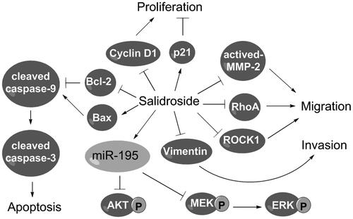
Salidroside, a glycoside of tyrosol, is isolated from Rhodiola rosea and shows anti-cancer functions in several cancers. However, the potentials of salidroside in the migration and invasion of lung cancer cells and its underlying mechanisms remain unknown. We aimed to investigate the functions and mechanisms of salidroside in non-small cell lung cancer (NSCLC). Human NSCLC cell line A549 was treated with different doses of salidroside. Cell viability, colony formation, apoptosis, migration and invasion were detected by CCK-8, crystal violet-staining assay, flow cytometry and transwell assay, respectively. qRT-PCR and western blot analysis were performed to assess the regulatory effects of salidroside on miR-195 expression and the activation of AKT and the MEK/ERK signal pathway. We found that, salidroside remarkably reduced cell viability, colony formation and Cyclin D1 expression, but increased p21 expression and apoptosis in A549 cells. Additionally, salidroside inhibited the migration and invasion of A549 cells by regulating expressions of migration- and invasion-related proteins. Finally, salidroside inhibited phosphorylation of AKT, MEK and ERK by upregulating miR-195 expression in A549 cells. In conclusion, salidroside inhibited the survival, migration and invasion of NSCLC cells. Salidroside blocked AKT and the MEK/ERK signal pathway by upregulating miR-195 expression in A549 cells.

GRAPHICAL ABSTRACT

View addendum:
Expression of Concern

Introduction

Globally, lung cancer is the most common cancer among men, with high incidence and mortality [Citation1]. It is the fourth in incidence, and second in mortality among women [Citation1]. Despite the availability of new diagnostic and advancement in surgical techniques, the 5-year survival rate of lung cancer remains at 15.6% in the United States [Citation1,Citation2], about 9.5% in England [Citation3] and only 8.9% in Europe, China, and other developing countries [Citation1]. Lung cancers are malignancies that arise from epithelial cells. According to histological type, lung cancers are categorized into two classes: small cell lung cancer (SCLC) and non-small cell lung cancer (NSCLC). NSCLC is the most common form of lung cancer and accounts for about 80–85% of all cases. Among the various types of therapy, including surgery, radiotherapy, chemotherapy and targeted therapy, systemic chemotherapy is the main treatment for lung cancer [Citation4,Citation5]. Thus, innovative drugs are urgent required to improve the outcome for patients with NSCLC.

Rhodiola rosea is a medicinal herb that is widely distributed at cold and high altitudes regions in Asia, eastern North America and Eastern Europe [Citation6]. Salidroside (p-hydroxyphenethyl-β-d-glucoside, C14H20O7: 300.30) is extracted from dried roots, stems or whole grass of Rhodiola rosea. Sachalinensis is used as an antidepressant, anti-aging, antiviral, anti-inflammatory agent, resisting anoxia and preventing high-altitude sickness [Citation7–12]. Especially, salidroside has been found to exert anti-cancer functions in breast cancer [Citation13], bladder cancer [Citation14], colon cancer [Citation15], fibrosarcoma [Citation16], neuroblastoma [Citation17] and glioma [Citation18] in vitro. In the regard of lung cancer, despite two recent reports showing that salidroside reduces cell viability and ROS generation, and induces cell cycle arrest and apoptosis in human lung cancer cells [Citation19,Citation20], the potential effects of salidroside on migration and invasion of lung cancer cells have not been fully clarified. Moreover, the underlying anti-cancer mechanisms of salidroside remain largely unknown.

In the present study, we aimed to evaluate the effects of salidroside on inhibiting NSCLC cell proliferation, colony formation, apoptosis, migration and invasion, and revealed the underlying molecular mechanisms involved in its anti-cancer effects. Our study indicates that salidroside has an anti-cancer effect on NSCLC cells maybe through upregulating miR-195 expression, and thereby modulating AKT and the MEK/ERK signal pathways. Salidroside may be an effective drug for the treatment of NSCLC.

Materials and methods

Cell culture and treatment

Human NSCLC cell line A549 was purchased from American Type Culture Collection (Catalogue No.: CCL-185TM; ATCC, Manassas, VA) and was cultured in RPMI 1640 medium (Gibco, Grand Island, NY) supplemented with 10% foetal bovine serum (FBS, Gibco), 100 IU/ml penicillin G (Gibco) and 100 μg/ml streptomycin (Gibco) at 37 °C with 5% CO2.

Salidroside (purity ≥98%) was purchased from Sigma-Aldrich (Catalogue No.: 43866; Sigma-Aldrich, St. Louis, MO). A stock with a concentration of 8 mM was prepared by dissolving Salidroside in dd water and filtered through a 0.22-µm filter before use. Salidroside with different concentrations (0.8, 8, 80, 800 and 8000 μM) was used to treat cells for 48 h [Citation19].

Cell viability assay

Briefly, A549 cells (5000/well) were seeded in 96-well plates. Cell viability was assessed by a Cell Counting Kit-8 (CCK-8, Dojindo Molecular Technologies, Gaithersburg, MD). After different doses of salidroside treatments, the cultures were subjected to 10 μl/well of CCK-8 solution and were incubated for 4 h at 37 °C with humidified 5% CO2. The absorbance was measured at 450 nm using a Microplate Reader (Bio-Rad, Hercules, CA).

Colony formation assay

A549 cells (1000/well) were seeded in 6-well plates and cultured for 24 h. Then, the cells were incubated with or without salidroside (800 μM) for 48 h. The medium was then removed, and cells were washed in PBS and incubated in complete medium for an additional 10 d. The colonies were washed with PBS and fixed in 10% formalin for 10 min at room temperature. After a rinse with PBS, the colonies were stained with crystal violet for 15 min followed by washing with PBS for three times. The visible colonies were counted.

Cell apoptosis assay

Cell apoptosis assay was performed using Annexin V-FITC/PI apoptosis kit (BioVision, Milpitas, CA, USA). After treatment with salidroside (800 μM) for 48 h, cells for apoptosis detection were collected and resuspended in 500 μl of binding buffer. Then, cells were stained with 5 μl of Annexin V-FITC and 5 μl of PI. After reaction for 5 min in the dark at room temperature, samples were analyzed by using a flow cytometer (Becton-Dickinson, San Jose, CA) to quantify early apoptotic cells (Annexin-V positive and PI negative).

Migration and invasion assays

Cell migration and invasion assays were performed using a 24-wells transwell culture chamber with 8 μm PET membranes (Millipore, Bedford, MA). For the migration assay, A549 cells (1 × 105) with or without salidroside (800 μM) treatment for 48 h were suspended in 200 μl of serum-free medium on the upper compartment, and 600 μl of complete medium containing 10% FBS (Gibco) was added to the lower compartment. After incubation at 37 °C for 24 h according to the manufacturer’s protocol, the chamber was carefully removed. Non-traversed cells on the upper surface of the membrane were wiped off carefully with a cotton swab. Traversed cells on the lower side were fixed with methanol for 30 min followed by staining with crystal violet for 20 min. The numbers of traversed cells were counted microscopically in five randomly chosen fields.

For the invasion assay, diluted Matrigel (BD, San Jose, CA) was added vertically at the centre of the membrane in the upper chamber and incubated at 37 °C for 4 h to facilitate gel formation. After salidroside treatment (800 μM, 48 h), cells (5 × 104) were incubated in the upper chambers with 200 μl of serum-free medium. The subsequent steps were same as those of the migration assay described above. The data are presented as relative migration or invasion rates.

Cell transfection

miR-195 inhibitor and inhibitor control were synthesized by GenePharma Co. (Shanghai, China). Cell transfections were conducted using Lipofectamine 3000 reagent (Invitrogen, Carlsbad, CA) following the manufacturer’s protocol. After 72 h of transfection, cells were collected for subsequent analyses.

Real-time quantitative reverse-transcriptase PCR (qRT-PCR)

Total RNA was extracted from cells using Trizol reagent (Invitrogen) according to the manufacturer’s instructions. To test the mRNA levels of Cyclin D1, we used the ExiLERATE universal cDNA synthesis and SYBR® Green Master Mix Kits (Exiqon, Vedbaek, Denmark) in turn according to the manufacturer’s protocols. The GAPDH (Exiqon) was used for normalizing. To test the expression level of miR-195 in cells, the Taqman MicroRNA Reverse Transcription Kit and Taqman Universal Master Mix II (Applied Biosystems, Foster City, CA) were used according to manufacturer’s protocols. U6 was used as an internal standard. The changes in expressions were calculated using the 2−ΔΔCt method.

Western blot

The proteins used for western blot were extracted by using RIPA lysis buffer (Beyotime Biotechnology, Shanghai, China) and quantified using the BCA Protein Assay Kit (Solarbio, Beijing, China). Protein bands were separated by sodium dodecyl sulphate-polyacrylamide gel electrophoresis (SDS-PAGE) followed by transferring to a polyvinylidene difluoride (PVDF) membrane (Millipore). After blocking with 5% skim milk (Nestlé, Shuangcheng, China) in Tris-Buffered Saline with Tween (TBST) for 2 h, the membrane was incubated with primary antibodies at 4 °C overnight. After washing, the membrane was incubated with secondary antibodies marked by horseradish peroxidase for 1 h at room temperature. All antibodies were purchased from Abcam (Cambridge, UK). Primary antibodies included anti-Cyclin D1(ab134175, 1:20000), anti-p21(ab109199, 1:1000), anti-Bcl-2 (ab59348, 1:1000), anti-Bax (ab182733, 1:2000), anti-pro caspase-3 (ab44976, 1:500), anti-cleaved caspase-3 (ab13847, 1:500), anti-caspase-9 (ab202068, 1:2000), anti-active-MMP2 (ab37150, 1:1000), anti-RhoA (ab54835, 1:500), anti-ROCK1 (ab45171, 1:5000), anti-Vimentin (ab8978, 1:1000), anti-AKT (ab32505, 1:2000), anti-p-AKT (ab38449, 1:1000), anti-p-MEK1 (ab96379, 1:1000), anti-MEK1 (ab32088, 1:1000), anti-p-ERK (ab214362, 1:1000), anti-ERK (ab32537, 1:1000) and anti-β-actin (ab8224, 1:1000) antibodies. β-Actin antibody was used as the endogenous protein for reference. Secondary antibodies included goat anti-mouse IgG (ab6789, 1:5000) and goat anti-rabbit IgG (ab6721, 1:5000). After rinsing, the membrane carrying blots and antibodies were treated with enhanced chemiluminescence (ECL) reagents (GE Healthcare, Little Chalfont, UK) according to the manufacturer’s instructions to develop the signals. The intensity of the bands was determined by ImageJ software (version: 1.4.3.67; National Institutes of Health, Bethesda, MD, USA).

Statistical analysis

All experiments were repeated three times. Statistical analyses were performed using Graphpad version 6 statistical software (GraphPad, San Diego, CA). The data of experiments are presented as the mean ± SD. The p values were calculated by using unpaired two-tailed t-test or multiple t-tests. A p value of <.05 indicated a statistically significant result.

Results

Salidroside inhibited cell proliferation of A549 cells

A549 cells were treated with different concentrations of salidroside (0, 0.8, 8, 80, 800 and 8000 μM) for 48 h, and the viability of cells was detected by the CCK-8 assay. We found that salidroside reduced the viability of A549 cells in a dose-dependent manner (; p < .05 or p < .01). The IC50 value (the effective dose that inhibits 50% of viability) of salidroside was calculated as 6.2 μM for A549 cells. Salidroside with a concentration of 800 μM was used to treat cells for the subsequent experiments.

Figure 1. Salidroside inhibited the proliferation of A549 cells. A549 cells were treated with increasing doses of salidroside (0.8, 8, 80, 800 and 8000 μM) for 48 h, and non-treated cells were acted as control. (A) Cell viability was detected by CCK-8 assay. A549 cells were stimulated with salidroside (800 μM) for 48 h, and non-treated cells were acted as control. (B) Cell colony formation was detected by staining with crystal violet. (C) mRNA expression level of Cyclin D1 was measured by qRT-PCR. (D) Protein expression levels of Cyclin D1 and p21 were determined by western blot. Data are presented as the mean ± SD of at least three independent experiments. *p < .05, **p < .01, ***p < .001.

Figure 1. Salidroside inhibited the proliferation of A549 cells. A549 cells were treated with increasing doses of salidroside (0.8, 8, 80, 800 and 8000 μM) for 48 h, and non-treated cells were acted as control. (A) Cell viability was detected by CCK-8 assay. A549 cells were stimulated with salidroside (800 μM) for 48 h, and non-treated cells were acted as control. (B) Cell colony formation was detected by staining with crystal violet. (C) mRNA expression level of Cyclin D1 was measured by qRT-PCR. (D) Protein expression levels of Cyclin D1 and p21 were determined by western blot. Data are presented as the mean ± SD of at least three independent experiments. *p < .05, **p < .01, ***p < .001.

Next, after salidroside (800 μM, 48 h) treatment, cell colony formation was detected by staining with crystal violet. As shown in , salidroside significantly reduced the colony formation of A549 cells (p < .001).

The effects of salidroside on the expressions of Cyclin D1 and p21 in A549 cells were also analyzed. As shown in , salidroside reduced the expression of Cyclin D1 at both mRNA (p < .001) and protein levels in A549 cells. Western blot showed that salidroside increased the protein expression of p21 in A549 cells. These results indicated that salidroside might suppress A549 cell growth.

Salidroside induced cell apoptosis of A549 cells

To reveal the effect of salidroside on the apoptosis of A549 cells, we performed the flow cytometry and detected the expressions of apoptosis-related proteins by western blot. After salidroside (800 μM) treatment for 48 h, salidroside significantly increased the rate of apoptotic cells (; p < .01). Western blot analysis showed that salidroside caused a decrease in the expression of the antiapoptotic protein Bcl-2 (p < .01) and increases in the expressions of proapoptotic protein Bax (p < .01), executioner cleaved caspase-3 (p < .001) and initiator cleaved caspase-9 (p < .001, ). These results indicated that salidroside induced the apoptosis of A549 cells.

Figure 2. Salidroside promoted A549 cell apoptosis. A549 cells were stimulated with salidroside (800 μM) for 48 h, and non-treated cells acted as control. (A) Percentage of apoptotic cells was determined by flow cytometry. (B) Expression levels of apoptosis-related proteins were tested by western blot. Data are presented as the mean ± SD of at least three independent experiments. **p < .01, ***p < .001.

Figure 2. Salidroside promoted A549 cell apoptosis. A549 cells were stimulated with salidroside (800 μM) for 48 h, and non-treated cells acted as control. (A) Percentage of apoptotic cells was determined by flow cytometry. (B) Expression levels of apoptosis-related proteins were tested by western blot. Data are presented as the mean ± SD of at least three independent experiments. **p < .01, ***p < .001.

Salidroside inhibited cell migration and invasion of A549 cells

To reveal the effect of salidroside on the migration of A549 cells, we performed transwell assay and detected migration-related proteins by western blot. As shown in , salidroside caused significant decreases in the migration rate of A549 cells (p < .05). Western blot analysis displayed that salidroside caused decreases in the expressions of active-MMP2, RhoA and ROCK1 (all p < .05, ). These results indicated that salidroside inhibited the migration of A549 cells perhaps by decreasing the expressions of active-MMP2, RhoA and ROCK1.

Figure 3. Salidroside inhibited the migration and invasion of A549 cells. A549 cells were stimulated with salidroside (800 μM) for 48 h, and non-treated cells acted as control. (A) Cell migration was detected by the transwell assay. (B) Expression levels of migration-related proteins were measured by western blot. (C) Cell invasion was detected by the transwell assay (with Matrigel). (D) Expression levels of invasion-related proteins were determined by western blot. Data are presented as the mean ± SD of at least three independent experiments. *p < .05.

Figure 3. Salidroside inhibited the migration and invasion of A549 cells. A549 cells were stimulated with salidroside (800 μM) for 48 h, and non-treated cells acted as control. (A) Cell migration was detected by the transwell assay. (B) Expression levels of migration-related proteins were measured by western blot. (C) Cell invasion was detected by the transwell assay (with Matrigel). (D) Expression levels of invasion-related proteins were determined by western blot. Data are presented as the mean ± SD of at least three independent experiments. *p < .05.

To determine the effects of salidroside on cell invasion and its potential mechanism, transwell assay with Matrigel and western blot analysis were performed. As shown in , salidroside caused a significant decrease in the invasion rate of A549 cells (p < .05). Western blot analysis displayed a decrease in vimentin expressions (p < .05) caused by salidroside. These results indicated that salidroside inhibited the invasion of A549 cells perhaps by decreasing vimentin expression.

Salidroside deactivated AKT and the MEK/ERK signal pathway

In the next step, we sought to investigate the regulatory mechanisms of salidroside in A549 cells. As we know, AKT and the MEK/ERK signal pathway play crucial roles in cell proliferation and survival in various cancers [Citation21,Citation22]. Therefore, we evaluated the effects of salidroside on phosphorylation levels of AKT, MEK and ERK. Western blot analyses showed that salidroside caused decreases in phosphorylation levels of AKT, MEK and ERK without altering their total levels (all p < .05, ). Results indicated that salidroside inactivated AKT and blocked the MEK/ERK signal pathway.

Figure 4. Salidroside deactivated AKT and the MEK/ERK signal pathway in A549 cells. A549 cells were stimulated with salidroside (800 μM) for 48 h, and non-treated cells acted as control. Phosphorylation levels of AKT, MEK and ERK were determined by western blot. Data are presented as the mean ± SD of at least three independent experiments. *p < .05.

Figure 4. Salidroside deactivated AKT and the MEK/ERK signal pathway in A549 cells. A549 cells were stimulated with salidroside (800 μM) for 48 h, and non-treated cells acted as control. Phosphorylation levels of AKT, MEK and ERK were determined by western blot. Data are presented as the mean ± SD of at least three independent experiments. *p < .05.

Salidroside deactivated AKT and the MEK/ERK signal pathway by upregulating miR-195 expression

miR-195 was reported to be a tumour suppressor in NSCLC [Citation23,Citation24]. Herein, we evaluated the effect of salidroside on miR-195 expression. As shown in , salidroside treatment significantly caused an increase in the expression of miR-195 (p < .01). This result supported that salidroside upregulated the expression of miR-195.

Figure 5. Salidroside deactivated AKT and the MEK/ERK signal pathway by upregulating miR-195 expression. A549 cells were stimulated with salidroside (800 μM) for 48 h, and non-treated cells acted as control. (A) Expression of miR-195 was testified by qRT-PCR. A549 cells were transfected with inhibitor control and miR-195 inhibitor, and untransfected cells acted as control. (B) Expression level of miR-195 was measured by qRT-PCR. After transfection, cells were treated with salidroside (800 μM) for 48 h. Non-treated cells were acted as control. (C) Phosphorylation levels of AKT, MEK and ERK were determined by western blot. Data are presented as the mean ± SD of at least three independent experiments. *p < .05, **p < .01, ***p < .001.

Figure 5. Salidroside deactivated AKT and the MEK/ERK signal pathway by upregulating miR-195 expression. A549 cells were stimulated with salidroside (800 μM) for 48 h, and non-treated cells acted as control. (A) Expression of miR-195 was testified by qRT-PCR. A549 cells were transfected with inhibitor control and miR-195 inhibitor, and untransfected cells acted as control. (B) Expression level of miR-195 was measured by qRT-PCR. After transfection, cells were treated with salidroside (800 μM) for 48 h. Non-treated cells were acted as control. (C) Phosphorylation levels of AKT, MEK and ERK were determined by western blot. Data are presented as the mean ± SD of at least three independent experiments. *p < .05, **p < .01, ***p < .001.

Then, we investigated the effects of salidroside on AKT phosphorylation and activation of the MEK/ERK signal pathway when the expression of miR-195 was inhibited. showed that transfection with miR-195 inhibitor could effectively inhibit the expression of miR-195 in A549 cells (p < .01). Therefore, cells transfected with miR-195 inhibitor was used for the subsequent experiment. These transfected cells were treated with salidroside (800 μM, 48 h) followed by detecting phosphorylation levels of AKT, MEK and ERK. Our results showed that salidroside could not reduce the phosphorylation of AKT, MEK and ERK any more in the case of miR-195 inhibition (), as evidenced by significant enhancements in phosphorylation levels of AKT, MEK and ERK compared with the salidroside + inhibitor control group (p < .01 or p < .001). Hence, we concluded that salidroside deactivated AKT and the MEK/ERK signal pathway possibly by upregulating miR-195 expression.

Discussion

As an important source of novel chemotherapeutic drugs, phytochemicals exert effective anticancer activities with relatively few side effects [Citation25,Citation26]. Therefore, phytochemicals have been recognized as prevention and therapeutic approach for various human cancers [Citation16,Citation27]. Salidroside has showed anti-cancer activity in various cancers [Citation13–15,Citation17,Citation19]. In the present study, we demonstrated that salidroside effectively inhibited proliferation, but induced apoptosis of A549 cells. Additionally, salidroside inhibited the migration and invasion of A549 cells, and deactivated AKT and the MEK/ERK signal pathway possibly by upregulating miR-195 expression.

A previous study has shown that salidroside inhibited viability, but increased apoptosis of A549 cells [Citation20]. Similarly, our data suggested that salidroside significantly showed anti-growth activity in A549 cells as cell viability and colony formation capacity were reduced, while apoptosis was increased by salidroside treatment. Additionally, Hu et al. found that salidroside could cause G1-phase arrest, a decrease of cyclin D1 expression and an increase of p21 expression in A549 cells [Citation19]. Wang et al. found that salidroside inhibited cell cycle by the arrest in the G0/G1 phase [Citation20]. In the cell cycle progression, Cyclin D1, a regulatory subunit of cyclin-dependent kinase (CDK) 4 and CDK6, is required for G1/S transition of the cell cycle to entry into the S-phase [Citation28]. Independent of CDK, cyclin D1 also can bind to nuclear receptors to regulate cell proliferation and growth [Citation29]. Cyclin D1 has been observed to be overexpressed in a variety of tumours and may contribute to tumorigenesis [Citation30–32]. p21 is a potent CDK inhibitor by inhibiting all cyclin–CDK complexes and thus functions as a regulator of cell cycle progression at G1 and S phase [Citation33]. In the current study, we observed that salidroside treatment caused a decrease in the expression of cyclin D1, but an increase in the expression of p21. This finding was also consistent with the study of Hu et al., implying salidroside suppressed A549 cells progression possibly by arresting more cells in the G0/G1 phase. Taken together, salidroside may inhibit the growth of A549 cells by decreasing cell viability and colony formation, and promoting G1-phase arrest and apoptosis.

To date, the potentials of salidroside in the migration and invasion of A549 cells and its underlying mechanisms remain unknown. Therefore, in addition to cell proliferation and apoptosis, we also evaluated the effects of salidroside on the migration and invasion of A549 cells. We found that salidroside significantly reduced migration and invasion of A549 cells. Besides, salidroside treatment reduced the protein expressions of activated-MMP-2, RhoA, ROCK1, and Vimentin. Among these factors, MMP2 is a member of the MMP family which is involved in the breakdown of extracellular matrix (ECM) [Citation34]. MMP2 is capable of degrading type-IV collagen, the most abundant component of the basement membrane [Citation34]. Degradation of the basement membrane is an essential step for the metastasis of most cancers. Therefore, MMP2 plays a key role in tumour cell migration and invasion [Citation35]. ROCK1 is a major downstream effector of RhoA and is a key regulator of the actomyosin cytoskeleton [Citation36]. It contributes to cell motility and metastasis in cancer [Citation36]. Vimentin is the major cytoskeletal component of mesenchymal cells and has been used as a tumor marker to identify mesenchyme [Citation37]. Previous studies showed that salidroside significantly suppressed MMP2 activity in human fibrosarcoma cells [Citation16] and breast cells [Citation13]. However, our study provided the first evidence that salidroside could reduce the activity of MMP2 and protein expressions of RhoA, ROCK1 and vimentin in A549 cells. Based on these results, we herein briefly concluded that salidroside significantly reduced cell migration and invasion in A549 cells by decreasing MMP2 activity and expressions of RhoA, ROCK1 and vimentin.

AKT is a serine/threonine-specific protein kinase that plays a key role in tumour cell survival, migration and invasiveness [Citation21,Citation38]. Mitogen-activated protein kinase (MAPKK, also known as MEK) is a kinase enzyme which phosphorylates mitogen-activated protein kinase (MAPK, also known as ERK) [Citation39]. The MEK/ERK signal pathway regulates cell functions including proliferation, cell survival, and apoptosis [Citation22]. Metastasis of lung cancer requires a transition from epithelial to mesenchymal cell type, which may occur through activation of some signal pathways including AKT and the MEK/ERK signal pathway [Citation40]. Previous studies showed that salidroside significantly suppressed the phosphorylation of ERK in human fibrosarcoma cells [Citation16] and breast cancer cells [Citation13]. In the present study, we found salidroside deactivated AKT, MEK and ERK. miR-195 was found to act as a suppressor in the NSCLC [Citation23,Citation24]. However, the effect of salidroside on miR-195 remains unknown. We demonstrated that salidroside upregulated expression of miR-195, and thereby deactivated AKT and the MEK/ERK pathway.

In conclusion, the present study supported that salidroside significantly inhibited NSCLC cell proliferation, migration and invasion and induced apoptosis in vitro. In addition, salidroside treatment significantly deactivated AKT and the MEK/ERK signal pathway by upregulating miR-195, assisting in its anti-cancer effects on NSCLC. Salidroside may be a promising natural drug for the chemotherapy of NSCLC.

Disclosure statement

No potential conflict of interest was reported by the authors.

Additional information

Funding

This research was supported by Qingdao Shinan District Science and Technology Plan (No.2016-3-047-YY); The Affiliated Hospital of Qingdao University Youth Research Fund (No.2838) and Shandong Medical and Health Technology Development Project (No.2018WS379).

References

  • Cruz CSD, Tanoue LT, Matthay RA. Lung cancer: epidemiology, etiology, and prevention. Clin Chest Med. 2011;32:605.
  • Ridge CA, Mcerlean AM, Ginsberg MS. Epidemiology of lung cancer. Chest. 2003;123:93–98.
  • Cancer UK. Lung cancer survival statistics. 2015.
  • Hann CL, Rudin CM. Management of small-cell lung cancer: incremental changes but hope for the future. Oncology (Williston Park, NY). 2008;22:1486–1492.
  • Group MAC. Chemotherapy in addition to supportive care improves survival in advanced non–small-cell lung cancer: a systematic review and meta-analysis of individual patient data from 16 randomized controlled trials. J Clin Oncol. 2008;26:4617.
  • Kelly GS. Rhodiola rosea: a possible plant adaptogen. Altern Med Rev. 2001;6:293.
  • Perfumi M, Mattioli L. Adaptogenic and central nervous system effects of single doses of 3% rosavin and 1% salidroside Rhodiola rosea L. extract in mice. Phytother Res. 2007;21:37–43.
  • Xu MC, Shi HM, Wang H, et al. Salidroside protects against hydrogen peroxide-induced injury in HUVECs via the regulation of REDD1 and mTOR activation. Mol Med Rep. 2013;8:147–153.
  • Wang H, Ding Y, Zhou J, et al. The in vitro and in vivo antiviral effects of salidroside from Rhodiola rosea L. against coxsackievirus B3. Phytomedicine. 2009;16:146–155.
  • Li D, Fu Y, Zhang W, et al. Salidroside attenuates inflammatory responses by suppressing nuclear factor-κB and mitogen-activated protein kinases activation in lipopolysaccharide-induced mastitis in mice. Inflamm Res. 2013;62:9–15.
  • Choe KI, Kwon JH, Park KH, et al. The antioxidant and anti-inflammatory effects of phenolic compounds isolated from the root of Rhodiola sachalinensis A. BOR. Molecules. 2012;17:11484–11494.
  • Qian EW, Ge DT, Kong SK. Salidroside promotes erythropoiesis and protects erythroblasts against oxidative stress by up-regulating glutathione peroxidase and thioredoxin. J Ethnopharmacol. 2011;133:308–314.
  • Hu X, Zhang X, Qiu S, et al. Salidroside induces cell-cycle arrest and apoptosis in human breast cancer cells. Biochem Biophys Res Commun. 2010;398:62–67.
  • Liu Z, Li X, Simoneau AR, et al. Rhodiola rosea extracts and salidroside decrease the growth of bladder cancer cell lines via inhibition of the mTOR pathway and induction of autophagy. Mol Carcinog. 2012;51:257–267.
  • Sun KX, Xia HW, Xia RL. Anticancer effect of salidroside on colon cancer through inhibiting JAK2/STAT3 signaling pathway. Int J Clin Exp Pathol. 2015;8:615.
  • Sun C, Wang Z, Zheng Q, et al. Salidroside inhibits migration and invasion of human fibrosarcoma HT1080 cells. Phytomedicine. 2012;19:355–363.
  • Zhang L, Yu H, Sun Y, et al. Protective effects of salidroside on hydrogen peroxide-induced apoptosis in SH-SY5Y human neuroblastoma cells. Eur J Pharmacol. 2007;564:18–25.
  • Zhang Y, Yao Y, Wang H, et al. Effects of salidroside on glioma formation and growth inhibition together with improvement of tumor microenvironment. Chinese J Cancer Res. 2013;25:520.
  • Hu X, Lin S, Yu D, et al. A preliminary study: the anti-proliferation effect of salidroside on different human cancer cell lines. Cell Biol Toxicol. 2010;26:499.
  • Wang J, Jian-Zhe LI, Ai-Xia LU, et al. Anticancer effect of salidroside on A549 lung cancer cells through inhibition of oxidative stress and phospho-p38 expression. Oncol Lett. 2014;7:1159–1164.
  • Song G, Ouyang G, Bao S. The activation of Akt/PKB signaling pathway and cell survival. J Cell Mol Med. 2005;9:59–71.
  • Reddy KB, Nabha SM, Atanaskova N. Role of MAP kinase in tumor progression and invasion. Cancer Metastasis Rev. 2003;22:395–403.
  • Guo H, Li W, Zheng T, et al. MiR-195 targets HDGF to inhibit proliferation and invasion of NSCLC cells. Tumor Biol. 2014;35:8861–8866.
  • Liu B, Qu J, Xu F, et al. MiR-195 suppresses non-small cell lung cancer by targeting CHEK1. Oncotarget. 2015;6:9445–9456.
  • Butler MS. The role of natural product chemistry in drug discovery. J Nat Prod. 2004;67:2141–2153.
  • Newman DJ, Cragg GM. Natural products as sources of new drugs over the 30 years from 1981 to 2010. J Nat Prod. 2012;75:311–335.
  • Marchetti S, Mazzanti R, Beijnen JH, et al. Concise review: clinical relevance of drug–drug and herb–drug interactions mediated by the ABC transporter ABCB1 (MDR1, P-glycoprotein). Oncologist. 2007;12:927–941.
  • Baldin V, Lukas J, Marcote MJ, et al. Cyclin D1 is a nuclear protein required for cell cycle progression in G1. Genes Develop. 1993;7:812–821.
  • Wang C, Li Z, Fu M, et al. Signal transduction mediated by cyclin D1: from mitogens to cell proliferation: a molecular target with therapeutic potential. Cancer Treat Res. 2004;119:217–237.
  • Diehl JA. Cycling to cancer with cyclin D1. Cancer Biol Ther. 2002;1:226–231.
  • Buckley MF, Sweeney KJ, Hamilton JA, et al. Expression and amplification of cyclin genes in human breast cancer. Oncogene. 1993;8:2127–2133.
  • Jiang W, Zhang Y-J, Kahn SM, et al. Altered expression of the cyclin D1 and retinoblastoma genes in human esophageal cancer. Proc Natl Acad Sci. 1993;90:9026–9030.
  • Gartel AL, Radhakrishnan SK. Lost in transcription: p21 repression, mechanisms, and consequences. Cancer Res. 2005;65:3980–3985.
  • Mook OR, Frederiks WM, Van Noorden CJ. The role of gelatinases in colorectal cancer progression and metastasis. Biochim Biophys Acta. 2004;1705:69–89.
  • Braicu EI, Gasimli K, Richter R, et al. Role of serum VEGFA, TIMP2, MMP2 and MMP9 in monitoring response to adjuvant radiochemotherapy in patients with primary cervical cancer – results of a companion protocol of the randomized NOGGO-AGO phase III clinical trial. Anticancer Res. 2014;34:385–391.
  • Rath N, Olson MF. Rho-associated kinases in tumorigenesis: re-considering ROCK inhibition for cancer therapy. Embo Rep. 2012;13:900–908.
  • Leader M, Collins M, Patel J, et al. Vimentin: an evaluation of its role as a tumour marker. Histopathology. 1987;11:63
  • Qiao M, Chang X, Jing F, et al. Role of PI3K and AKT specific isoforms in ovarian cancer cell migration, invasion and proliferation through the p70S6K1 pathway. Cell Signall. 2006;18:2262–2271.
  • Derijard B, Raingeaud J, Barrett T, et al. Independent human MAP-kinase signal transduction pathways defined by MEK and MKK isoforms. Science. 1995;267:682–685.
  • Antón I, Molina E, Luis-Ravelo D, et al. Receptor of activated protein C promotes metastasis and correlates with clinical outcome in lung adenocarcinoma. Am J Respir Crit Care Med. 2012;186:96–105.