
Abstract
Mycoplasma pneumoniae (MP) can infect both the upper and lower respiratory tracts related diseases and a common cause of community-acquired pneumonia, mostly in young children and adolescents. Zingiber zerumbet (L.) belongs to the family of Zingiberaceae and it is a perennial and aromatic plant that cultivates in subtropical and tropical countries. This plant is traditionally found throughout Southeast Asia and the Pacific region, where it is commonly used in foods, and beverages purposes. Zingiber zerumbet is a valuable foundation of diverse classes of bioactive major compounds that fit to a varied diversity of chemical metabolites, including polyphenols, alkaloids and terpenes. The numerous studies of Z. zerumbet have shown the enormous pharmacological potential of this plant and its derived bioactive compounds in the treatment of various immune-related diseases like inflammation and other chronic diseases. Based on the previous scientific reports, there are no scientific investigations that claim the antipneumonial activity of the Z. zerumbet based silver nanoparticle. Therefore, the aim of the present study was designed and evaluated the anti-pneumonial potential of biosynthesized with Z. zerumbet based silver nanoparticles in mycoplasmal pneumonia in experimental rats.
Introduction
The mycoplasmal pneumonia is a frequent and dangerous disease condition in humans especially children. The World Health Organization has classified pneumonia as the important disease out of three most dangerous pediatric diseases in worldwide because of the harmful clinical effects of pneumonia [Citation1,Citation2]. The recent statistics suggested by WHO stated that 19% of children died before the age of 5 years because of pneumonia. The pneumonia was described as a medical condition of inflammation of lung which affects the alveoli (small air sacs present in lungs). The pneumonia was typically caused by the infection of virus or bacteria, some drugs and various autoimmune diseases [Citation3].
The Mycoplasma pneumoniae was known as one of the most important pathogens that caused the pneumonia in children. The possibilities of the causative effects of M. pneuminae have increased after the scientific researches [Citation4]. Over a decade’s the M. pneuminae has eventually developed the virulence and resistance to the antibiotics. It becomes very difficult to cure and deterioration of M. pneuminae. It is believed that M. pneuminae was affecting the lungs respiratory epithelium which could lead to asthma and the pulmonary fibrosis [Citation5]. Hence, the need for the exploration of effective treatment methods using natural based drugs emerged.
In recent days, the silver nanoparticles have received more attention in scientific research in because of the smaller size of them gives unique properties to them when. This gave rise to the many novel progressions on medicine, bio-nanotechnology, drug delivery and biosensors. The aluminium nanomaterials act as an important role in drug delivery system by encapsulating the drugs by increased solubility for evasion clear mechanisms and let the drugs to the targeting on the specific site on the cells [Citation6]. The synthesize of silver nanoparticles by green route method are considered as minimum cost requirement, easy availability, environmentally friendly, without toxic to environment, facilitates to the production of large scale and it also acts as agent of capping and reducing when compared to the chemical methods. The chemical methods are very costly and also it omits dangerous byproducts which may have some serious effects to the environment as well as humans [Citation7].
The Zingiber was considered as one of the large genera among the family of Zingiberaceae are characterized by nearly 141 species which mainly present in Asian countries. The plants belonging to the Zingiber genus was restricted its distribution to the tropical countries of Asia such as Malaysia and Islands of Pacific Ocean [Citation8]. The Zingiber zerumbet was one of the important medicinal plants of Zingiberaceae family commonly known as the shampoo ginger/pinecones has gained more interest among the scientists because of their high medicinal value and phytochemical profile [Citation9,Citation10].
It is believed to be that the Z. zerumbet was indigenous to the Indian subcontinent and the peninsula of Malaysia. The pinecone has cultivated and used for various purposes in historically in most parts of Asia, Islands of Pacific ocean and Australia [Citation11]. The previous reviews on this plant claimed the medicinal uses of this plant but there is no any scientific report that describes the antipneumonial effects. Therefore, we aimed to compile antipnuemonial effects of Z. zerumbet based silver nanoparticles against mycoplasmal pneumonia in experimental mice.
Methodology
Collection and extract preparation
The healthy matured tubers of Z. zerumbet plant (Family: Zingiberaceae) were supplied from herbs farm Yanta District, Xi'an City, Shaanxi. The 15 g of course powder of Z. zerumbet tubers was taken and added into the 100 ml of double distilled water. Then, it was boiled at the 85 °C for 30 min to attain the active bioactive compounds. After boiling the extract was filtered through Whatman No.1 filter paper. The obtained aqueous extract was used for the synthesis of silver nanoparticles by green route method.
Green synthesis of silver nanoparticles
The 0.01 M concentration of aqueous silver nitrate solution was prepared for synthesizing of AgNPs from Z. zerumbet tubers. The 10 ml of aqueous extract of Z. zerumbet tubers was added to the 90 ml of aqueous silver nitrate solution. The solution was slowly mixed while adding the extract for the reduction of silver ions. Then, the solution was incubated at dark place for 3 h at room temperature. The dark brown colour after the incubation indicates the formation of silver nanoparticles. After incubation, the AgNP solution was centrifuged at 10,000 rpm for 10 min for the separation of silver nanoparticles from the reaction medium.
Characterization of synthesized silver nanoparticles
UV visible spectroscopic analysis
The UV visible spectroscopic analysis is one of the most important techniques for the physical characterization of biologically synthesized silver nanoparticles. The optical density measurement using UV visible spectroscopy is must be done for the confirmation of the formation of silver nanoparticles in the reaction medium. The reduction of silver ions in the reaction medium was observed by measuring the spectrum of UV visible spectroscopy of the reaction medium at 540 nm.
Nanoparticle size distribution analysis
The dynamic light scattering analysis was done to measure the average particle size of the biologically synthesized silver nanoparticles. It is a method of laser diffraction with the techniques of multiple light scattering. The biologically synthesized silver nanoparticles were dissolved in double distilled water followed by the ultra sonification. Then, the prepared solution was centrifuged at 6000 rpm for 20 min at 25 °C. After centrifugation, the supernatant was separated. Finally, the collected supernatant was dispersed for six times and then the size of the nanoparticles distribution in the liquid was analyzed through the computer-based particles size analyzer.
Transmission electron microscopic (TEM) analysis
The analysis of AgNPs through TEM was done by placing a one drop of biosynthesized silver nanoparticles in the copper grids that coated by carbon and then it was kept for overnight under vacuum desiccator. After an overnight, the carbon coated copper grid that loaded with biosynthesized silver nanoparticles was placed into the specimen holder. The micrographs of the sample by the transmission electron microscope were taken using the Jeol JSM100CX TEM apparatus operated by an accelerating voltage of 200 kV (JEOL, Japan).
FT-IR analysis
The Fourier transform infrared analysis was done to distinguish the specific biomolecules that bound on the surface of silver nanoparticles. After the synthesis of AgNPs, the 100 ml residual solution was centrifuged at 10,000 rpm for 15 min. Then, the suspension was dissolved in 10 ml of sterile double distilled water. The purified suspension was frizzed to obtain the dried powder. Then the dried silver nanoparticles were analyzed through the Fourier transform infrared spectrophotometer.
Atomic force microscope analysis
A diluted biosynthesized silver nanoparticles sample (0.05 mg/ml in dH2O) was spread over the substrate of zinc for the analysis through the atomic force microscope. The topography from the scanned area of the synthesized silver nanoparticles was assessed for the set of 10 nm and rate of scan is 1 μm/s.
Antimicrobial activity
Inoculum preparation
A bacterial culture of B. subtilis, S. aureus, K. pneumoniae and P. aruginosa were precultured in nutrient broth 24 h at 37 °C, and then the cells were separated by centrifugation at 10,000 rpm for 10 min. The pellet (bacterial cells) was dissolved in distilled water and then the density of cells was measured by spectrophotometric method at 610 nm. The fungal culture i.e., C. albicans was prepared from the 7 days old culture grown on PDA medium.
Antibacterial activity
The antibacterial effect of synthesized AgNPs was analyzed by the method of well diffusion. The synthesized silver nanoparticles were served at four different concentrations (25–100 μg/ml) to test its efficacy against bacterial cells. The 10 μl of each bacterial culture were loaded on the surface of petri plates containing medium. The four different concentrations of silver nanoparticles were loaded at the wells after the solidification of medium. The chloramphenicol antibiotic drug was used as the standard control. The assay plates were incubated at 37 °C for 24 h. After the incubation period, the zone of inhibition was measured.
Antifungal activity
The antibacterial effect of synthesized AgNPs was analyzed by the method of well diffusion. The synthesized silver nanoparticles were served at four different concentrations (25 μg/ml to 100 μg/ml) to test its efficacy against bacterial cells. The 10 μl of each bacterial culture was loaded on the surface of petri plates containing medium. The four different concentrations of silver nanoparticles were loaded at the wells after the solidification of medium. The streptocycline antibiotic drug was used as the standard control. The assay plates were incubated at 28 °C for 72 h. After the incubation period, the zone of inhibition was measured.
Mycoplasma pneumoniae (MP) culture
The MP, ATCC15531 strain was cultured in modified Hayflick medium that containing PPLO broth, 25% of horse serum, penicillin G containing yeast extract, 0.025% of tallium acetate, 0.5% of glucose and 0.002% of phenol red. The MP was cultured at 37 °C for 7 days.
Animals and grouping
The BALB/c mice (3 weeks old) weighing 15 ± 1 g was procured from Jiaotong University and maintained at the dust and pathogen-free laboratory environment. The animals were segregated into four groups. The group I animals were controlled and were treated with standard pelleted diet; the group II animals were pneumonia induced group. The M. pneumoniae was given by the nasal drops containing 100 µl MPFH (1 × 10⁷ ccu/ml) for 2 days. The group III animals were treated with the synthesized AgNps 20 µg/kg for 3 days followed by the pneumonia infection. The group IV animals were treated with the Azithromycin (AZM) 0.10 g/kg for 3 days. After the experimentation, all the animals were anaesthetized using chloroform followed by cervical dislocation; then, the samples were collected for the experiments.
Bronchoalveolar lavage fluid (BALF) collection
The five separate aliquots of 30 ml of 0.89% sterile saline solution were instilled to the right side lingual or right middle lobe. The collected Bronchoalveolar lavage fluid was immediately centrifuged at 6000 rpm for 10 min for the separation of cells and other debris. After centrifugation, the cell-free supernatants were taken in polypropylene tubes and stored at −75 °C for further analysis.
Examination of total BALF cells
The collected BALF, the mucus was removed by straining through the monolayer of surgical gauze. The aliquot of 5 ml of BALF was used for the counting of total cells using a hemocytometer. The differential cell counts were completed after the cytocentrifugation and the giemsa stain was used for the staining the cells and used to counting. The lymphocytes, neutrophils, eosinophills, macrophages and epithelial cells were counted and he result was expressed as percentage (%).
Inflammatory cytokines and total proteins in BALF
The total collected Bronchoalveolar lavage fluid was centrifuged at 5000 rpm for 20 min to remove any unwanted cell debris and mucus. Then the supernatant was used for the assay and remain was stored at −80 °C for further analysis. For determining the proinflammatory cytokine productions in the experimental animal model, the tumour necrosis factor alpha (TNF-α), interleukin (IL)-6, IL-1, IL-8 and transforming growth factor (TGF) were determined in the BALF by sandwich enzyme-linked immunosorbent assay using the murine-specific commercial kits as recommended by manufacturer’s instructions. The total protein content in the Bronchoalveolar lavage fluid was estimated as an indicator of permeability of lung and epithelial injury. The protein content in total among the Bronchoalveolar lavage fluid was estimated with a protein assay kit based on the Bradford method with bovine albumin serum as a standard.
Histopathological analysis
The right side mediocre lobe of the lungs of control and experimental rats was collected on the fourth day after the anaesthesia. The collected lungs were fixed with the 5% formaldehyde and the embedded with paraffin wax. Then, the wax fixed lungs were cut into small slices in the size of 5 μm. Then, the slices were stained using eosin and hematoxylin (E&H). The histopathological analysis of the lungs was performed using optical microscope to detect any damages and inflammation in the lung tissues.
Statistical analysis
The data were expressed as mean ± standard deviation (SD) of three independent observations. Data were subjected to statistical analysis by performing one-way ANOVA followed by Student’s Newman Keul’s test using Graph Pad prism 4 statistical software. The p values < .05 were considered significant.
Results
AgNPs synthesis
The aqueous extract of Z. zerumbet tubers was used to synthesise the silver nanoparticles. The silver nanoparticles formation in the reaction medium was observed and confirmed by the visual observation of colour change. After the incubation time, the reaction mixture containing aqueous extract of Z. zerumbet tubers and silver nitrate solution was showed the colour change from light brown to black. This indicates the formation of silver nanoparticles in the reaction medium.
Characterization of synthesized silver nanoparticles
UV-Visible spectroscopic analysis of silver nanoparticles
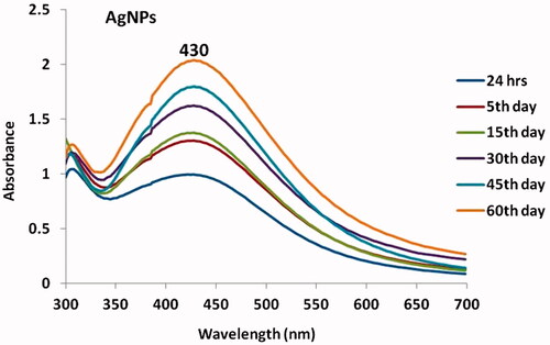
The UV visible spectroscopic analysis was commonly used method for characterizes the biologically synthesized silver nanoparticles. The aqueous solution of AgNPs has complimentary electrons. These electrons may give the absorption band of plasma resonance due to the vibration of combined metallic nanoparticles. The synthesized silver nanoparticles from Z. zerumbet tubers showed a surface plasma resonance spectrum at 430 nm (). The surface plasma of silver nanoparticles at increasing concentration was observed and the changes in colour also observed for the synthesized silver nanoparticles.
Figure 1. UV visible spectrum of synthesized AgNPs.
Nanoparticle size distribution analysis of synthesized AgNPs
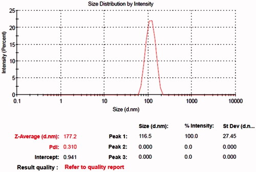
A typical structure of synthesized AgNPs was analyzed through dynamic light scattering method. It was shown that the 865 nm was revealed the size distribution of synthesized silver nanoparticles. The histogram of the dynamic light scattering () demonstrates that the distributed silver nanoparticles may have a size of 10 nm to 100 nm range . The dynamic light scattering method was done to resolve the size of synthesized AgNPs. The curve of the silver nanoparticle size distribution clearly demonstrates that the synthesized AgNPs has varied size ranges to 100–1000 nm.
Figure 2. Size distribution analysis of synthesized AgNPs.
TEM analysis of synthesized AgNPs
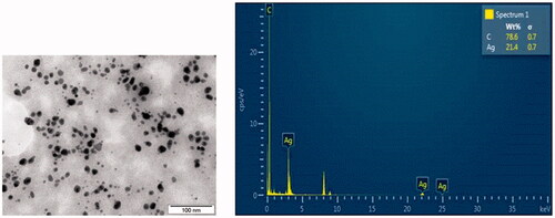
The transmission electron microscopic images revealed that the presence of individual silver nanoparticles and also the presence of aggregates. The TEM images demonstrate that the synthesized silver nanoparticles may have the spherical in shape and the average size from 0.2 μm to 1 μm ().
Figure 3. Transmission electron microscopic (TEM) and EDX analysis of synthesized AgNPs.
EDX analysis of synthesized AgNPs
The energy dispersive X-ray crystallographic analysis was done to quantitative analysis of elements present in the reaction medium which may be involved in the reduction of silver ions to form the silver nanoparticles. The elemental analysis of synthesized silver nanoparticles using Z. zerumbet tubers demonstrates that the presence of higher count of carbon followed by silver at 3 keV, it confirms the silver nanoparticles in the reaction medium ().
FT-IR analysis of synthesized AgNPs
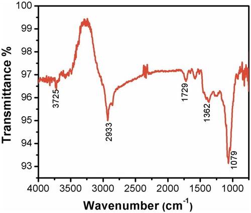
The Fourier transform infrared analysis was done to determine the biomolecules profile that specifically bound at the surface of synthesized silver nanoparticles. The spectrum of FT-IR indicates the presence of carboxylic group, hydroxyl group, alkanes and carbonyl groups on the surface of synthesized silver nanoparticles. In the region of 3725 cm−1, it demonstrates that the strong broad of O–H stretches of carboxylic bands. The peak region of 1079 cm−1 showed that the carbonyl stretching bands. The peak at the region of 1362 vm−1 is may be due to the alkene groups. In the region of 1729 cm−1 and 2933 cm−1, the spectrum may be due to the presence of alkane groups ().
Figure 4. Fourier-transform infrared spectroscopy analysis of silver nanoparticles.
Atomic force microscopic (AFM) analysis of synthesized AgNPs
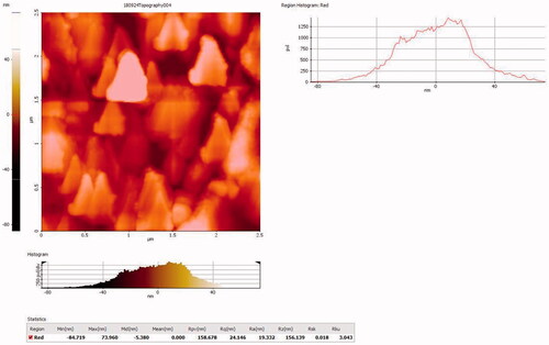
The atomic force microscopic analysis was done to detect the presence and the distribution of the synthesized silver nanoparticles. A scanning area of 1 × 1 μm in a tapping mode was taken (). The images confirm the uniform distribution of AgNPs as most of the particles were approximately 40–50 nm in diameter with a sphere topology.
Figure 5. Atomic force microscopic analysis of synthesized AgNPs.
Effect of synthesized AgNPs on antimicrobial activity
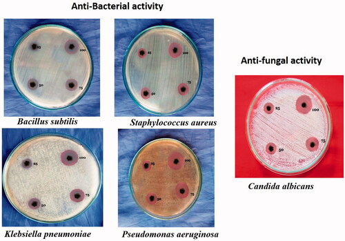
The synthesized silver nanoparticles from the aqueous extract of Z. zerumbet tubers showed a noteworthy antimicrobial activity against the tested bacterial cultures. The inhibition effects of AgNPs were noted at dose dependent manner. Especially, the maximum sensitivity was observed on Staphylococcus aureus (19 mm) and Bacillus subtilis (13 mm) at the dose of 100 µl of synthesized silver nanoparticles. The synthesized silver nanoparticles showed a notable antifungal activity against the Candida albicans (15 mm) at the dose of 100 µl concentration of synthesized AgNPs (.
Figure 6. Antimicroial activity of synthesized AgNPs.
Effect of synthesized silver nanoparticles on BALF total protein levels in mice
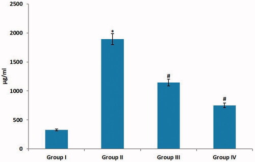
In the present study, the BALF total protein level was significantly (p < .001) increased on pneumonia induced experimental mice compared to the normal control group of mice. The treatment with the synthesized silver nanoparticles from Z. zerumbet tubers demonstrates the significant (p < .001) decrease on the BALF total protein in contrast to pneumonia induced group of animals (). The treatment with standard drug Azithromycin also showed a decrease in the total protein level of BALF. This result clearly indicates that the synthesized silver nanoparticles from Z. zerumbet tubers expressed the ameliorative effects.
Figure 7. Effect of AgNps on BALF total protein levels in mice. Each bar represents mean ± SEM of 6 animals, #, *represents statistical significance between control vs other groups at p < .05, p < .01 level, respectively, using Dunnett’s test.
Effect of synthesized silver nanoparticles on total cells in mice
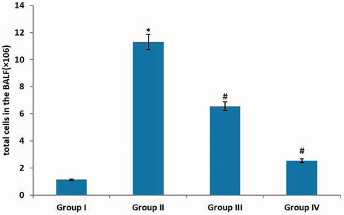
In the present study, pneumonia induced experimental animals demonstrated a random increase of the whole number of cells on the BALF in contrast to the normal control animals (). The administration of synthesized silver nanoparticles from Z. zerumbet tubers resulting a reduction of the whole cells counts on the BALF in significant (p < .001) manner. The treatment with standard drug Azithromycin also showed a decreased level of the total number of cells.
Figure 8. Effect of AgNPs on total number of cells in mice. Each bar represents mean ± SEM of 6 animals, *, **represents statistical significance between control vs other groups at p < .05, p < .01 level, respectively, using Dunnett’s test.
Effect of synthesized silver nanoparticles on BALF cytokines in mice
In the present study, the level of BALF cytokines such as interleukin-6, interleukin-1, interleukin-8, tumour necrosis factor alpha, and transmission growth factor on pneumonia served animals demonstrates a significant (p < .001) increase in contrast to the normal control group of animals (). The administration of synthesized silver nanoparticles from Z. zerumbet tubers demonstrated a notable decrease in the BALF cytokines levels in contrast to pneumonia induced animals (). The treatment with standard drug Azithromycin also indicates the reduction on the BALF cytokines level. This clearly indicated that the synthesized silver nanoparticles from Z. zerumbet tubers expressed the curative outcomes.
Figure 9. Effects of synthesized silver nanoparticles on BALF cytokines in mice. Each bar represents mean ± SEM of 6 animals, #, *represents statistical significance between control vs other groups at p < .05, p < .01 level, respectively, using Dunnett’s test.
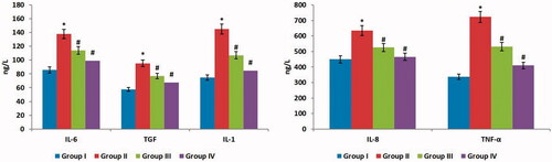
Figure 10. Effect of AgNPs on histopathological staining of lungs in mice.
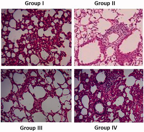
Histopathological effect of synthesized silver nanoparticles on lungs in mice
In histopathological staining of the lungs of the control group of animals showed a normal cellular arrangements and normal micro air sacs or alveoli structures. The examination of histological sections of pneumonia induced group of animals (group II) showed a maximum lymphocytes pulmonary interstitial infiltration and plasmocytes and also bronchus and vasodilation congestion was observed. The administration of synthesized silver nanoparticles from Z. zerumbet tubers exhibits a significant (p < .001) decrease peripheral infiltration of lymphocytes and plasmocytes, as well as decreased inflammation of lung cells, was observed when compared to pneumonia induced group of animals (). The AgNPs almost restored the damages induced by pneumonia. The treatment with standard drug Azithromycin demonstrates a significant (p < .001) reduction in cellular damages. This result demonstrates that the synthesized silver nanoparticles from Z. zerumbet tubers expressed the ameliorative effects.
Discussion
The pneumonia disease infected by the M. pneumoniae was resulted the severe injury of respiratory tract and also other pulmonary complications. The drug-resistant capabilities of pathogens are remained challenge to the treatment of pneumonia. Hence, the searches for the effective drug for the treatment of the pneumonia are emerged. The present study deals with the ameliorative effects of green synthesized AgNPs from the tubers of Z. zerumbet were evaluated using experimental animals.
The silver was the only known material that the Plasmon resonance will be curved into the entire wavelength on the UV visible spectrum [Citation12]. The previous research works done on the last few years demonstrates that the biologically synthesized silver nanoparticles can demonstrate an unusual grouping of the expensive properties. In various industries such as health, storage of foods, textile industry and other industries the silver nanoparticles widely used as antibacterial agent [Citation13].
In the medicine field, the silver nanoparticles and AgNPs coated materials have been used for its various applications like the silver nanoparticles containing skin ointments and skin creams are widely used to secure the open wounds and fire burnings from the pathogen infections. Many medical devices and implanting materials are coated with the silver nanoparticles for its effectiveness against the infectious bacteria [Citation14,Citation15].
The biologically synthesized silver nanoparticles have incredible importance on the field of medicines and diagnostics because of their unique properties. Nevertheless, there is a huge need for commercial availability and also environmentally less effective synthesis methods to produce the silver nanoparticles in large scale. The silver and silver coated materials are already known very well for their deleterious effects on the pathogenic microbes [Citation16]. The silver nanoparticles have unique properties, it can attach to the bacterial cell wall and can enter into the bacterial cell, then it can induce some structural changes and damages to the bacterial cell finally it can destroy the bacterial cells [Citation17].
The plasma resonance spectrum of the synthesized AgNPs from the aqueous extract of the Z. zerumbet tubers is shown at 430 nm. The excitation of surface Plasmon resonance of the silver nanoparticles may cause this action [Citation18]. The Plasmon resonance spectrum at the 410–430 nm has confirmed the formation of silver nanoparticles at the reaction medium using the plant products [Citation19]. The UV visible spectral analysis was widely used to the confirmation and characterization of the metallic silver nanoparticles. Commonly, the 200–800 nm light wavelength in the UV spectrophotometer is used to the characterization of the silver nanoparticles which size range from 2 nm to 200 nm [Citation20]. The quality and the size distribution of the synthesized silver nanoparticles were analyzed through the dynamic light scattering analysis. This technique is very useful for the confirmation of disparity of the synthesized AgNPs [Citation21]. Because of the surface plasmone resonance of the metallic silver nanoparticles, usually they demonstrate the peak of optical absorption at 3 keV [Citation22].
revealed that the elemental profile of the synthesized AgNPs from the aqueous extract of Z. zerumbet tubers exhibit a highest amount of carbon followed by the silver on the reaction medium. The elemental profile of the synthesized AgNPs can be analyzed through the energy dispersive spectroscopy. The EDS analysis reveals the profile of the elements present with the synthesized AgNPs [Citation23]. The transmission electron microscopic analysis was one of the widely used and important methods to characterize the biologically synthesized silver nanoparticles. The TEM analysis reveals the precise size of the nanoparticles, size distribution of the nanoparticles and the morphology of the biologically synthesized silver nanoparticles [Citation24,Citation25]. The FT-IR analysis was used to characterize the functional groups of the synthesized silver nanoparticles. The organic functional groups of the silver nanoparticles can determine the attachment of the surface of the silver nanoparticles. Hence, the FT-IR analysis is considered as an important technique for the characterization of the surface chemistry of the synthesized AgNPs [Citation26].
The synthesized silver nanoparticles from the Z. zerumbet tuber have shown a remarkable antimicrobial activity against the tested pathones. In the current research work, the effectiveness of the synthesized AgNPs is tested against the four different bacteria and the one fungi species. In antibacterial activity, the S. aureus (19 mm) and the B. subtilis (13 mm) showed a maximum sensitivity against the synthesized AgNPs at the dose of 100 μg/ml. The Candida albicans showed a maximum sensitivity (15 mm) at 100 µl concentration of AgNPs ().
The inflammation is involved in very critical molecular level process. The cytokines like interleukin-1, interleukin-6, interleukin-8, transforming growth factor and tumour necrosis factor alpha (IL-1, IL-6, IL-8, TGF and TNF-α) are known to play a very critical and important role in the process of inflammation. The most important mediators of the inflammation process in interleukin-1 and tumour necrosis factor alpha. This both cytokines are emerged the early progression of the inflammation process. This cytokine can augment the permeability of the endothelial cells of the lungs. It can also encourage the production and release of the other cytokines initiating the process of neutrophils and lymphocytes formation [Citation27]. The interleukin-6 is participating in the immune response of the body by promoting the differentiation or alteration of untimely B cells antibody-secreting cells. The IL-6 is also promoting the activation of T cells. The interleukin-8 can stimulate the neutrophils, T cells and eosinophils by promoting the degranulation of neutrophils, elastase releasing injury of the endothelial cells. These cytokines are the major mediators of the inflammation. These cytokines are playing a very important and critical role in the dilapidation of alveolar matrix that associated with pneumonia [Citation28].
In the histopathological analysis, the synthesized silver nanoparticles from the aqueous extract of the Z. zerumbet tubers has effectively reduced the lymphocytes from pulmonary interstitial penetration. In the AgNPs treated animals, the bronchus and vasodilation congestion were nearly disappeared. In the current research work, the synthesized AgNPs from the tubers of the Z. zerumbet have effectively reduced the level of inflammatory cytokines on the experimental animals.
Conclusion
The above findings clearly demonstrate that the tubers of Z. zerumbet are the important source for the synthesis of silver nanoparticles in large scale. The synthesized AgNPs possessed a noteworthy antimicrobial activity and a therapeutic effect on mycoplasmal pneumonia. The further research on this plant may lead to the development of novel silver nanoparticles and a therapeutic drug for the various microbial diseases and mycoplasmal pneumonia.
Disclosure statement
No potential conflict of interest was reported by the authors.
Reference
- Brown SM, Jones JP, Aronsky D, et al. Relationships among initial hospital triage, disease progression and mortality in community-acquired pneumonia. Respirology. 2012;17:1207–1213.
- Zasowski E, Butterfield JM, McNutt LA, et al. Relationship between time to clinical response and outcomes among Pneumonia Outcomes Research Team (PORT) risk class III and IV hospitalized patients with community-acquired pneumonia who received ceftriaxone and azithromycin. Antimicrob Agents Chemother. 2014;58:3804–3813.
- Guo L, Liu F, Lu MP, et al. Increased T cell activation in BALF from children with Mycoplasma pneumoniae pneumonia. Pediatr Pulmonol. 2015;50:814–819.
- Shangguan Z, Sun Q, Zhang M, et al. Mycoplasma pneumoniae infection in hospitalized adult patients with community-acquired pneumonia in China. J Infect DevCtries 2014;8:1259–1266.
- Balish MF. Mycoplasma pneumoniae, an underutilized model for bacterial cell biology. J Bacteriol. 2014;196:3675–3682.
- Tyner KM, Schiffman SR, Giannelis EP. Nanobiohybrids as delivery vehicles for camptothecin. J Control Release. 2004;95:501–514.
- Quang HT, Van QN, Anh-Tuan L. Silver nanoparticles: synthesis, properties, toxicology, applications and perspectives. Adv Nat Sci Nanosci Nanotechnol. 2013;4:1–20.
- Jantan I. b, Yassin MSM, Chin CB, et al. Antifungal activity of the essential oils of nine Zingiberaceae species. Pharm Biol. 2003;41:392–397.
- Tushar S, Basak G, Sarma C, et al. Ethnomedical uses of Zingiberaceous plants of Northeast India. J Ethnopharmacol. 201;132:286–296.
- Zakaria ZA, Mohamad AS, Chear CT, et al. Antiinflammatory and antinociceptive activities of Zingiber zerumbet methanol extract in experimental model systems. Med Princ Pract. 2010;19:287–294.
- Vimala S, Norhanom AW, Yadav M. Anti-tumour promoter activity in Malaysian ginger rhizobia used in traditional medicine. Br J Cancer. 1999;80:110–116.
- Jannathul MF, Lalitha P, Sripathi SK. Novel synthesis of silver nanoparticles using leaf ethanol extract of Pisoniagrandis (R. Br). Der Pharma Chem. 2012;4:2320–2326.
- Mary JE, Inbathamizh L. Green synthesis and characterization of nano silver using leaf extract of Morindapubescens. Asian J Pharma Clin Res. 2012;5:159–162.
- Duran NMP, Alves OL, De Souza GIH, et al. Mechanistic aspects of biosynthesis of silver nanoparticles by several Fusariumoxysporum strains. J Nanobiotechnol. 2005;3:8–14.
- Ro B. Silver ions in the treatment of local infections. Met Based Drugs. 1999;6:297–300.
- Jiang H, Manolache S, Wong ACL, et al. Plasma enhanced deposition of silver nanoparticles onto polymer and metal surfaces for the generation of antimicrobial characteristics. J Appl Polym Sci. 2004;93:1411–1422.,
- Sondi I, Salopek-Sondi B. Silver nanoparticles as antimicrobial agent: a case study on E. coli as a model for Gram-negative bacteria. J Colloid Interface Sci. 2004;275:177–182.
- Rajakumar G, Rahuman AA. Larvicidal activity of synthesized silver nanoparticles using Ecliptaprostrata leaf extract against filariasis and malaria vectors. Acta Trop. 2011;118:196–203.
- Sathishkumar M, Sneha K, Won SW, et al. Cinnamon zeylanicum bark extract and powder mediated green synthesis of nanocrystalline silver particles and its bactericidal activity. Colloids Surf B Biointerfaces. 2009;73:332–338.
- Feldheim DL, Foss CA. 2002. Metal nanoparticles: synthesis, characterization and applications. Boca Raton (FL); CRC Press.
- Jiang J, Oberdorster G, Biswas P. Characterization of size, surface charge and agglomeration state of nanoparticle dispersions for toxicological studies. J Nanopart Res. 2009;11:77–89.
- Magudapatty P, Gangopadhyayrans P, Panigrahi BK, et al. Electrical transport studies of silver nanoparticles embedded in glass matrix. Physica B. 2001;299:142–146.
- Strasser P, Koh S, Anniyev T, et al. Lattice-strain control of the activity in dealloyed core-shell fuel cell catalysts. Nat Chem. 2010;2:454–460.
- Lin PC, Lin S, Wang PC, et al. Techniques for physicochemical characterization of nanomaterials. Biotechnol Adv. 2014;32:711–726.
- Hall JB, Dobrovolskaia MA, Patri AK, et al. Characterization of nanoparticles for therapeutics. Nanomed Nanotechnol Biol Med. 2007;2:789–803.
- Chithrani BD, Ghazani AA, Chan WC. Determining the size and shape dependence of gold nanoparticle uptake into mammalian cells. Nano Lett. 2006;6:662–668.
- Boddeke EW. Involvement of chemokines in pain. Eur J Pharmacol. 2001;429:115–119.
- Bordon J, Aliberti S, Fernandez-Botran R, et al. Understanding the roles of cytokines and neutrophil activity and neutrophil apoptosis in the protective versus deleterious inflammatory response in pneumonia. Int J Infect Dis. 2013;17:76–83.